Abstract
Rejection caused by donor-specific antibodies (principally ABO and HLA antibodies) has become one of the major barriers to successful long-term transplantation. This review focuses on clinical outcomes in antibody-incompatible transplantation, the current state of the science underpinning clinical observations, and how these may be translated into further novel therapies. The clinical outcomes for allografts facing donor-specific antibodies are at present determined largely by the use of agents developed in the 20th century for the treatment of T-lymphocyte-mediated cellular rejection, such as interleukin-2 agents and anti-thymocyte globulin. These treatments are partially effective, because acute antibody-mediated rejection is mediated to a considerable extent by T lymphocytes. However these treatments are essentially ineffective in chronic antibody-mediated rejection. Future therapies for the prevention and treatment of antibody-mediated rejection are likely to fall into the categories of those that reduce antibody production, extracorporeal antibody removal and disruption of the effector arms of antibody-mediated tissue damage.
Keywords: ABO, antibody, antibody-mediated rejection, HLA, kidney transplant
INTRODUCTION
Antibodies directed against transplants are becoming recognized as a critical barrier to further improvements in the access of patients to transplantation and in the survival of allografts. This review will summarize the current results of transplantation in the presence of donor-specific antibodies (DSA), and the possible research pathways that will lead to the control and prevention of such antibodies and the effective treatment of antibody-mediated rejection (AMR), both acute (AAMR) and chronic (CAMR).
The modern era of renal transplantation began in the 1960s with the introduction of azathioprine. Within a few years the importance of blood group incompatibility and HLA-specific antibodies was recognized, and their presence essentially vetoed transplantation outside experimental settings [1]. A focus on T-lymphocyte-mediated cellular rejection over the next half century has resulted in a therapeutic toolkit that has eliminated the vast majority of graft losses from this cause in adherent patients. Ultimately the therapies required to prevent T-lymphocyte-mediated rejection proved relatively simple, namely effective multipoint targeting of the interleukin-2 pathway and lymphocyte deletion therapy. An international focus from clinicians, scientists and industry on developing new treatments for antibody-mediated rejection (AMR) arguably began only a decade ago, and we are currently in an exciting era characterized by a rapid series of new discoveries about anti-graft antibodies, their mechanisms of production and action and the treatment of antibody-mediated rejection.
CURRENT CLINICAL OUTCOMES
HLA antibodies
Donor-specific HLA antibodies may be preformed or develop de novo after a transplant. The current status of transplantation across preformed HLA antibodies (HLAi transplantation) is that acceptable graft outcomes can be achieved in living donor transplantation, so long as the pre-transplant complement dependent cytotoxic (CDC) crossmatch is negative [2]. However such transplants do require antibody screening and careful management. It is not enough simply to transplant across a negative CDC crossmatch without taking account of preformed HLA antibodies. In many patients with low pre-treatment levels of donor-specific HLA antibodies, successful engraftment may be achieved using standard immunosuppression of tacrolimus, mycophenolate, prednisolone and basiliximab. However, in cases with higher levels of DSA, for example where the flow cytometric (FC) crossmatch is positive, antibody removal and induction immunosuppression or therapies for antibody-mediated rejection are required [3–7].
As a generalization, current clinical outcomes seem to indicate a mortality and graft loss rate about twice that of ‘antibody-compatible’ transplantation in the first year, unless the CDC is positive when the graft loss rate is higher, rising to 50% at 5 years using CDC methodology where there is no enhancement with anti-human globulin [2], and 30% graft loss when the more sensitive technique using AHG is used [7]. Other adverse prognostic features that can be identified pre-transplant are DSA that are combinations of Class I and Class II, and DSA that bind the complement component C1q in microbead assays [7, 8].
Therapies used in such clinical series include antibody removal pre-transplantation (plasma exchange, plasmapheresis or immunoadsorption), cellular depleting therapies (anti-thymocyte globulin, rituximab, alemtuzumab), intravenous immunoglobulins and proteasome inhibitor therapy (bortezomib), but there is no consensus on which of these approaches is most effective, and randomized trials are awaited. Transplantation across preformed HLA antibodies is best performed with living donors where there is time to achieve effective antibody removal, and possibly the graft is better equipped to cope with the rigours of early post-transplant antibody assault [2–9].
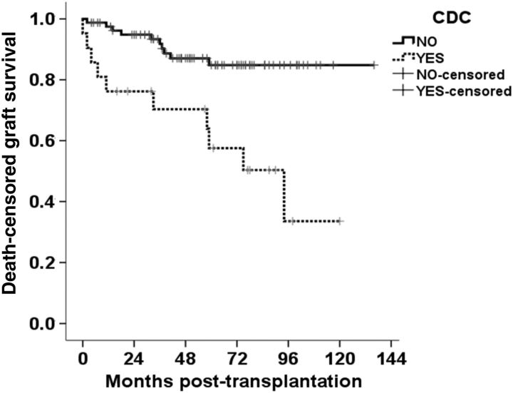
The outcomes in the face of CAMR due either to de novo HLA antibody production or persistent preformed DSA production are less encouraging [7–11]. For example one series showed a 10-year graft survival of <60% in those with de novo DSA, compared with >90% for those without DSA [12]. Rejection takes the form of glomerular basement membrane damage (transplant glomerulopathy), with proteinuria and progressive graft failure, usually over 2–3 years. There is no effective therapy for this condition, though we have seen it resolve durably if the DSA levels fall. Often transplant glomerulopathy may be associated with some active cellular infiltration in the peritubular capillaries and this may be temporarily amenable to therapy, but ultimately nearly every case of transplant glomerulopathy progresses to graft failure within 3–5 years (Figure 1).
FIGURE 1:
Graft survival at University Hospitals Coventry and Warwickshire in HLA antibody incompatible transplantation, for patients with pre-treatment complement dependent cytotoxic (CDC) positive crossmatch (n = 21) compared with those with DSA but with a negative CDC crossmatch (includes flow cytometric crossmatch positive and negative) (n = 91). Five- and 10-year death-censored graft survivals were 84.8% for the CDC-negative group, 57.5 and 33.6% respectively for the CDC-positive group.
ABO antibodies
Transplantation across ABO incompatibility (ABOi) generally produces excellent outcomes, and the experience in Japan over a period of decades indicates that outcomes are equivalent to ABO-compatible transplantation [13]. However, in the USA and in the UK, ABOi renal transplantation seems to have a slightly increased risk of severe AAMR which may result in graft loss, with catastrophic rejection progressing over a period of hours. This rejection may occur without any warning, and may indeed occur in those with very low pre-transplant antibody levels [14]. Further multicentre studies of this rare phenomenon (2–4% of grafts) are required.
ABOi transplantation does differ from HLAi transplantation in that chronic antibody-mediated rejection is not attributable to ABO antibodies. Biopsies may be ongoing staining for C4d on the peritubular basement membrane suggesting that there is some form of inflammation but the clinical outcome is not adversely affected. The significance of peritubular C4d differs from the same finding in HLAi transplantation, since the C4d deposition is a sign of CAMR. When CAMR is reported in ABOi grafts, this seems to be due to the concurrent presence of HLA antibodies [15].
Other antibodies
Non-HLA antibodies may be important players in AMR. Antibodies directed against the angiotensin receptor are of particular interest. These were first observed as a cause of sporadic AAMR associated with severe hypertension. Subsequent studies have extended their possible role to the causation of CAMR, and their presence has been associated with graft loss, independent of HLA antibodies [16, 17]. They may be auto- and allo-antibodies, and further work will define better the extent of their role in the causation of graft loss.
THE BIOLOGY OF ANTIBODY PRODUCTION AND REJECTION
Decades of focus on T-cell-mediated rejection and the lack of a clinical model of antibody incompatible transplantation mean that our understanding of the biology of antibody production, and the mechanisms of AMR are at a relatively early stage, but knowledge is accumulating rapidly.
Production of antibodies
Antibodies are synthesized by memory B lymphocytes and plasma cells. Both of these ultimately derive from immature cells of the B-lymphocyte lineage and receive T-cell help. It is possible that induction immunosuppression, enhanced beyond the level necessary to prevent most T-lymphocyte-mediated cellular rejection, may reduce the production of de novo HLA antibodies [18], and prospective and randomized studies are awaited.
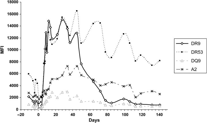
In HLAi transplantation, pre-formed donor-specific HLA antibodies may have been present for many years before the transplant, and detailed monitoring of their levels in the early post-transplant period may show large changes in their levels, with both rises and falls. Figure 2 shows a case with a heterogeneous response, the rates of post-transplant increases in DSA, the pre- to peak variation and the rate of fall of DSA showing variation. The rises and falls in HLA antibody levels post-transplant are not uniform across a range of patients, and there is no clear pattern governing the responses, though HLA Class II antibodies may show greater responses than Class I, with the profiles of HLA DQ and DP similar to HLA DR [19].
FIGURE 2:
HLA and ABO antibody levels pre- and post-transplant showing variation in antibody responses within the same patient, both in terms of rate of increase, magnitude of change from pre- to peak levels, and the rates of fall. The patient received a living donor transplant from his father. DSA levels were as shown in the legend. The CDC crossmatch was positive at a titre of 1 in 16, and there was additional ABO incompatibility, donor blood group A1 and recipient B. The recipient received plasma exchange pre-transplant, and post-transplant at Days 21 and 22 only. Immunosuppression was with prednisolone, tacrolimus, mycophenolate and basiliximab, with anti-rejection treatment including ATG (Days 1–15) and eculizumab (Days 24 and 31). The graft is still functioning at 5 years post-transplant, though with proteinuria and reduced function. DSA levels were measured by microbead assay (OneLambda). MFI levels were DR9—pre 2148, peak 14821 (Day 9), late 776; DR52—pre 5982, peak 12952 (Day 9), Day 141 8162; DQ9—pre 864, peak 1468 (Day 9), late 618; A2—pre 2774, peak 7258 (Day 45), late 2612. Follow-up at 2 years post-transplant showed little change in antibody levels from the Day 140 data shown here. Haemagglutination titres to blood group A1 were, IgG, pre 1, peak 128 (Day 10), late 4; IgM, pre 4, peak 128 (Day 10), late 32.
It is likely that insights can be gained from more detailed studies of the characteristics of the antibodies, for example the classes and IgG subclasses of the responses (Table 1). New antibody production is initially of the IgM class, and IgM donor-specific HLA antibodies have been associated with AAMR in the absence of equivalent IgG antibodies [20] and reported in de novo HLA antibody production [21]. HLA antibodies of the IgA class may also occur frequently, though their significance is not yet understood [26]. IgM antibody production may switch to IgG3 subclass, then to IgG1, IgG2 and ultimately IgG4. There is currently much interest in examining antibody subclasses in more detail [21], and we have observed some differences in subclass distribution, for example a higher incidence of IgG3 subclass in pregnancy-stimulated HLA antibodies and an association between pre-transplant IgG4 levels and subsequent AMR and graft survival [22].
Table 1.
HLA characteristics of antibody and antigen that might affect clinical outcomes
| Comment | Reference | |
|---|---|---|
| HLA antibody | ||
| Concentration | Not currently possible to measure antigen-specific concentration of antibody (see Figure 3) | |
| Antibody binding to HLA in solid phase assay (readout a combination of avidity and concentration) | Measureable by microbead assays | [2, 4] |
| Class | Mostly IgG, early reports of IgM mediated AMR and occurrence of IgA | [20, 21] |
| Subclass | Early studies show heterogeneity in responses | [21, 22] |
| Affinity | Measurement under investigation | |
| Glycosylation | Measurement under investigation | |
| Cellular binding | CDC and FC crossmatches Endothelial binding |
[2, 23] |
| Inhibitors | ||
| Soluble HLA | Early reports of methods to quantify | |
| HLA-E | Early reports of correlation with clinical outcomes | [24] |
| Idiotypic antibodies | Hard to measure with current tools but could be important | [25] |
| Immune complexes | ||
| HLA-sHLA immune complexes | Not currently measureable | |
One of the most remarkable observations in clinical practice is that DSA may disappear after transplantation. This has been observed in both ABO and HLA antibody-incompatible transplantation [13, 14, 19]. However, it seems likely that the mechanisms are different.
In HLA antibody-incompatible transplantation, many groups have noted that in some patients HLA antibodies may disappear after the transplant, even when sensitive microbead analysis is used. While some groups have attributed this to particular treatments administered, we have seen that this phenomenon is independent of any particular therapy over and above the standard immunosuppression of basiliximab, tacrolimus, mycophenolate and prednisolone [19]. The phenomenon does seem to be commoner when there is a vigourous antibody response post-transplant, and daily sampling shows that the fall in antibody levels occurs rapidly. This implies some form of ‘active’ removal of antibody from the circulation, and this is the subject of ongoing research. If it proved possible to induce the disappearance of DSA pre-transplant, that would be very useful. However, it is not clear whether this requires exposure to donor HLA (and if so in what form of delivery), as well as immunosuppression.
In ABOi transplantation, successful engraftment is often followed by the disappearance of antibody, but biopsies of the graft show ongoing C4d staining in peritubular capillaries, as if there is some persistent immunological stimulation, making it possible that some antibodies are being produced and then absorbed by the graft. It will be interesting to see what happens to ABO antibodies in patients who later lose their grafts from causes other than ABO-related rejection, and then have graft nephrectomy—will their ABO antibodies reappear or not? ABOi heart transplants in neonates may result in long-term tolerance to the transplanted ABO, but this could be a special case owing to the neonatal timing of the transplant.
Binding of antibodies to an allograft
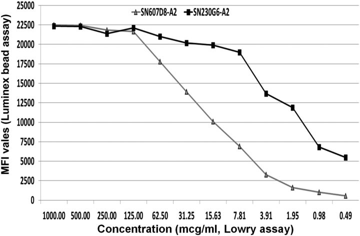
There is much current research examining how antibody binds to antigen at a molecular level, and then how that response evolves into clinical rejection, since the simple act of antibody binding to antigen alone does not seem to cause rejection. Although the development of microbead assays have enabled far better measurement of antibody levels than was previously possible, they do not measure the concentration of antibody, even in a semi-quantitative manner, but measure a combination of concentration and affinity, as shown in Figure 3. Table 1 lists some of the antibody characteristics that could be associated with clinical outcomes; we are still at a preliminary stage of the understanding of which of these are important, and which could be manipulated for therapeutic benefit.
FIGURE 3:
Dose response curve showing Luminex bead MFI value against concentrations of two human monoclonal HLA-A2-specific antibodies. The same concentrations of antibody may give markedly different MFI levels (e.g. <2000 and >10000, respectively, at 1.95 µg/mL).
HLA is profoundly polymorphic, and the binding of antibody to an HLA molecule is determined not by the whole HLA molecule, but by small areas of polymorphism within the protein structure known as epitopes. An epitope may be determined by as little as a single amino acid substitution in a critical area of the molecule, even though the binding footprint of an antibody molecule is much larger. Different HLA molecules are defined by a series of epitopes, many of which are shared between different HLA specificities. This is why an antibody generated against one HLA allele will bind to many others [27].
A better understanding of exactly how the antibody-antigen reaction develops may allow for the development of therapies that disrupt this interaction. Hence, research groups are studying antibody affinity for antigen, the atomic structure and biophysical properties of HLA, especially in relation to glycosylation as well as the amino acid backbone.
Clinical studies of the mechanisms of rejection are hampered by the inability of current techniques usefully to measure the binding of antibody to the graft [15]. It is not clear why this is the case, but the clinical classifications of antibody-mediated rejection depend on the detection of effector responses such as complement and cells, and not on whether there is any donor-specific antibody in the graft. A test that gave an accurate readout of the levels of donor-specific antibody bound to graft endothelium would give immediate and important insights into AMR.
Complement
It is widely acknowledged that activation of the complement system is a major driver of antibody-mediated tissue damage and subsequent transplant rejection. A good indicator of this is the CDC assay that correlates positively with risk of donor organ failure [2]. The presence of the complement protein fragment C4d in renal rejection is also a ‘smoking gun’ for activation of the complement cascade, which occurs downstream of C1q binding to clustered complexes of IgG and IgM antibodies on a cell surface. The antibody classes and subclasses typically found are IgG1 (HLA antibodies) and IgM (ABO antibodies), which are strong C1q binders and thus classical pathway activators. However, clinical investigation is made more difficult by the difficulties in measuring DSA in graft biopsies, and also by the observations that AAMR may occur in the absence of C4d deposition, and that C4d deposition may not reflect ongoing rejection; this could partly be due to endothelial synthesis of C4, so C4d should not necessarily be assumed to have been deposited on endothelium from circulating C4 [28].
Cellular response
The cellular aspect of acute antibody-mediated rejection is interesting, because although hyperacute rejection looks more like an innate response, with neutrophils and macrophages predominately, AAMR is characterized by a marked T-lymphocyte infiltration into the graft, visible using immunohistochemistry, and indistinguishable from T-cell-mediated cellular rejection at a molecular level [29, 30].
Response of endothelium
The predominant target of antibody-mediated rejection is vascular endothelium in the blood vessels (and glomeruli) of the allograft. Endothelial cells express ABO and HLA (classes I and II). Endothelium is not a passive victim of antibody binding and the rejection process, but has an adaptive response that may be protective to the graft. Indeed, clinical acute antibody-mediated rejection generally resolves in the presence of donor-specific antibody, and while this may be due to therapies which down-regulate the cellular responses, it is also possible that the graft becomes resistant to rejection [23, 28]. In the clinical setting, it is possible to measure vascular stress in the allograft by RNA analysis of renal biopsy material, the ‘molecular microscope’. This approach can also help characterize the T- and B-cell activity within the allograft [31].
In some cases, the antibody binds to endothelium but does not cause rejection, for example in ABOi transplantation, where staining for complement C4d in the peritubular capillaries of well-functioning grafts seems to indicate accommodation of the graft to antibody. In HLAi transplantation, by contrast, longer term C4d staining is likely to indicate low grade-AMR, though in some cases there does seem to be DSA present for many years without causing rejection or C4d staining on biopsy.
NEW THERAPIES
The successful treatment and ideally prevention of antibody-mediated rejection will require the development of new therapies that have specific new actions. It is not yet clear exactly which are the ideal targets, or whether there will be a solution as conceptually simple as the targeting of interleukin-2 in T-lymphocyte-mediated rejection. Therapies under consideration can be grouped into three main categories; (i) those that target the cells responsible for antibody production; (ii) those that target mediators of antibody damage, including complement; (iii) more effective removal of antibody using extracorporeal techniques.
Targeting the cells responsible for antibody production
Once the body has been programmed to produce the antibody in the long term, the cells responsible for this can be hard to target. For example, while bone marrow ablative therapy for haematological malignancies may appear to result in deletion of the host immune system, many antibody responses induced by prior vaccination may persist. We have seen HLA antibody production persist after chemotherapy and total lymphoid irradiation conditioning for cord cell transplantation.
There are some candidate therapies in use that could prove beneficial, but have not yet been tested in randomized trials. Rituximab (CD20 monoclonal antibody) and alemtuzumab (CD52 monoclonal antibody) are both capable of killing B lymphocytes, though do not have specific action against plasma cells, and both may spare memory B lymphocytes [18, 31, 32]. In the case of alemtuzumab, any benefits in clinical use might be due to deletion of effector leucocytes rather than effects on antibody production, and its use has even been reported to be associated with increases in the production of de novo HLA antibody levels in previously unsensitised transplant recipients [33]. In clinical use, neither agent can completely prevent increases in preformed HLA antibody levels after transplantation, though some data suggest that de novo HLA antibody production after transplantation may be reduced by prior administration of anti-CD20 therapy.
Anti-thymocyte globulin has potentially a very wide action, and may be particularly effective in dealing with cells in the effector arm of the rejection process, but again does not seem to completely prevent post-transplant synthesis of preformed DSA, although in a non-randomized study, its use was associated with less production of de novo HLA antibodies in the first 2 years post-transplant [33].
The cells that produce antibody could be targeted by means other than cytolytic therapies, and there are some candidate therapies available. Intravenous immunoglobulins (IVIg) have been used for some time. This product has a range of actions, but may down-regulate the production of HLA antibodies, and may be combined with anti-CD20 therapy [34]. Cytokine inhibition may be another route available, and studies are currently underway to evaluate the possible benefits of inhibitors of interleukin-6 and BAFF (B-cell activating factor). BAFF is an attractive target, as studies are emerging that associate BAFF with clinical outcomes in transplantation [35].
A further approach to reducing antibody production is targeting the protein synthetic capacity of plasma cells using proteasome inhibitors. Although these agents were initially conceived selectively to target malignant plasma cells, it is possible that they may impact on metabolically active cells producing DSA post-transplant, and uncontrolled studies have shown successful transplantation in the face of active immune responses [36, 37]. The effects of bortezomib on pre-transplant antibody production have been less encouraging, but randomized trials and the introduction of second generation proteasome inhibitors are welcomed with great interest.
Extracorporeal antibody removal
Most protocols for clinical antibody-incompatible transplantation involve extracorporeal antibody removal, at least in those with higher antibody levels. While the effects of antibody removal are partial and temporary with the current technology, it is generally believed to be beneficial [38, 39]. Randomized trials in the setting of preformed antibodies are lacking, but it does seem that removing antibodies to a level below the threshold of CDC crossmatch positivity prevents hyperacute rejection, and that plasma exchange is an effective therapy for AAMR.
Antibody removal therapies are constrained by several difficulties. These include the problem of providing long hours of therapy during times when there is a risk of bleeding; the pool of IgG outside the vascular compartment, which re-equilibrates only slowly with the vascular compartment; by rapid production of the antibody in the post-transplant period and by the inability of current therapies to selectively remove HLA antibodies, as all therapies remove total immunoglobulin, often together with other desirable blood components such as fibrinogen [39]. Similar constraints are also faced when total immunoglobulin removal methods are used in ABO-incompatible transplantation [40].
There is therefore a need for more selective therapies and modes of delivery that will allow removal of enough antibody at the times before and after transplant, and maybe soon after biopsy, and will impact on rejection at times when there is rapid antibody production (a doubling time of 12 h has been observed in some of our patients) [41]. The availability of large amounts of HLA protein has allowed the production of a selective HLA absorption column that indicates as proof of principle that HLA antibody removal therapy could be improved. Specific immunoadsorption therapy is already available to remove ABO antibodies [40, 42].
Mediators of antibody damage
Inhibition and modulation of complement activation is an attractive therapeutic route, with FDA-approved biopharmaceuticals already in clinical use for rare conditions such as hereditary angioedema (HA) and paroxysmal nocturnal haematuria (PNH). These include the drugs cinryze (C1 esterase inhibitor) for HA and eculizumab (anti-complement C5 monoclonal antibody) for PNH [43, 44]. These two agents act at different points within the complement activation cascade with potential to protect cells and tissues directly from aggressive lytic mechanisms and also broader immune activation. C1 esterase inhibitor is a protein that blocks the enzymatic function of the complement C1s component. This results in the proteolytic activation cascade being arrested upstream of powerful inflammatory and cytotoxic mechanisms such as C3/C4 opsonisation, anaphylatoxin release (a powerful leucocyte chemoattractant mechanism) and assembly of the membrane attack complex.
Eculizumab acts further downstream through functional blockade of the complement component C5, resulting in abrogated membrane attack complex formation and C5a anaphylatoxin release. Recruitment into an international randomized clinical trial of eculizumab in patients with pre-formed HLA antibodies has been completed, and results should be available in 2015. If a positive effect of this agent is shown, this will be pivotal in the future therapy of AMR, and possibly in the prevention of CAMR. If the randomized trial shows a marked reduction in AAMR rates in line with preliminary studies, there will be important questions to answer on the cost-effective application of the drug, and also whether its benefit will be extended to those with higher DSA levels who were not eligible for the current study.
SUMMARY
Antibody-incompatible transplantation has become a clinical reality in the 21st century, though graft survival rates are still suboptimal. Many patients with very high levels of preformed DSA, both HLA and ABO, are considered untransplantable with current technology. Treatment for CAMR when it leads to transplant glomerulopathy is almost completely ineffective. The understanding of the drivers of antibody production and mechanisms of AMR is progressing rapidly, and it is likely that combinations of new therapies targeting antibody production, complement and extracorporeal antibody removal will improve graft survival rates in the coming decades.
CONFLICT OF INTEREST STATEMENT
The authors are co-researchers with Pure Transplant Solutions in production of immunoadsorption column to remove HLA antibodies.
ACKNOWLEDGEMENTS
The authors would like to thank Professor Frans Claas and Dr Arend Mulder, University of Leiden, for the gift of monoclonal antibodies used in Figure 3.
REFERENCES
- 1.Patel R, Terasaki PI. Significance of the positive crossmatch test in kidney transplantation. New Engl J Med 1969; 280: 735–739 [DOI] [PubMed] [Google Scholar]
- 2.Higgins R, Lowe D, Hathaway M, et al. HLA antibody incompatible renal transplantation: excellent medium term outcomes with negative cytotoxic crossmatch. Transplantation 2011; 92; 900–906 [DOI] [PubMed] [Google Scholar]
- 3.Gupta A, Iveson V, Varagunam M, et al. Pretransplant donor-specific antibodies in cytotoxic negative crossmatch kidney transplants: are they relevant? Transplantation 2008; 85: 1200–1204 [DOI] [PubMed] [Google Scholar]
- 4.Lefaucheur C, Loupy A, Hill GS, et al. Preexisting donor-specific HLA antibodies predict outcome in kidney transplantation. J Am Soc Nephrol 2010; 21: 1398–1406 [DOI] [PMC free article] [PubMed] [Google Scholar]
- 5.Singh N, Djamali A, Lorentzen D, et al. Pretransplant donor-specific antibodies detected by single-antigen bead flow cytometry are associated with inferior kidney transplant outcomes. Transplantation 2010; 90: 1079–1084 [DOI] [PubMed] [Google Scholar]
- 6.Willicombe M, Brookes P, Santos-Nunez E, et al. Outcome of patients with preformed donor-specific antibodies following alemtuzumab induction and tacrolimus monotherapy. Am J Transplant 2011; 11: 470–477 [DOI] [PubMed] [Google Scholar]
- 7.Bentall A, Cornell LD, Gloor JM, et al. Five-year outcomes in living donor kidney transplants with a positive crossmatch. Am J Transplant 2013; 13: 76–85 [DOI] [PubMed] [Google Scholar]
- 8.Loupy A, Lefaucheur C, Vernerey D, et al. Complement-binding anti-HLA antibodies and kidney-allograft survival. N Engl J Med 2013; 369: 1215–1226 [DOI] [PubMed] [Google Scholar]
- 9.Tait BD, Süsal C, Gebel HM, et al. Consensus guidelines on the testing and clinical management issues associated with HLA and non-HLA antibodies in transplantation. Transplantation 2013; 95: 19–47 [DOI] [PubMed] [Google Scholar]
- 10.Kahwaji J, Najjar R, Kancherla D, et al. Histopathologic features of transplant glomerulopathy associated with response to therapy with intravenous immune globulin and rituximab. Clin Transpl 2014; 28: 546–553 [DOI] [PubMed] [Google Scholar]
- 11.Lefaucheur C, Loupy A, Vernerey D, et al. Antibody-mediated vascular rejection of kidney allografts: a population-based study. Lancet 2013; 381: 313–319 [DOI] [PubMed] [Google Scholar]
- 12.Wiebe C, Gibson IW, Blydt-Hansen TD, et al. Evolution and clinical pathologic correlations of de novo donor-specific HLA antibody post kidney transplant. Am J Transplant 2012; 12: 1157–1167 [DOI] [PubMed] [Google Scholar]
- 13.Tanabe K, Ishida H, Inui M, et al. ABO-incompatible kidney transplantation: long-term outcomes. Clin Transpl 2013: 307–312 [PubMed] [Google Scholar]
- 14.Montgomery JR, Berger JC, Warren DS, et al. Outcomes of ABO-incompatible kidney transplantation in the United States. Transplantation 2012; 93: 603–609 [DOI] [PMC free article] [PubMed] [Google Scholar]
- 15.Haas M, Sis B, Racusen LC, et al. Banff 2013 meeting report: inclusion of c4d-negative antibody-mediated rejection and antibody-associated arterial lesions. Am J Transplant 2014; 14: 272–283 [DOI] [PubMed] [Google Scholar]
- 16.Taniguchi M, Rebellato LM, Cai J, et al. Higher risk of kidney graft failure in the presence of anti-angiotensin II type-1 receptor antibodies. Am J Transplant 2013; 13: 2577–2589 [DOI] [PubMed] [Google Scholar]
- 17.Dragun D, Catar R, Kusch A, et al. Non-HLA-antibodies targeting Angiotensin type 1 receptor and antibody mediated rejection. Hum Immunol 2012; 73: 1282–1286 [DOI] [PubMed] [Google Scholar]
- 18.Jackson AM, Kraus ES, Orandi BJ, et al. A closer look at rituximab induction on HLA antibody rebound following HLA-incompatible kidney transplantation. Kidney Int 2015; 87: 409–416 [DOI] [PMC free article] [PubMed] [Google Scholar]
- 19.Higgins R, Lowe D, Hathaway M, et al. Rises and falls in donor specific and third party HLA antibody levels after antibody incompatible transplantation. Transplantation 2009; 87: 882–888 [DOI] [PubMed] [Google Scholar]
- 20.Bentall A, Tyan DB, Sequeira F, et al. Antibody-mediated rejection despite inhibition of terminal complement. Transpl Int 2014; 27: 1235–1243 [DOI] [PubMed] [Google Scholar]
- 21.Everly MJ, Rebellato LM, Haisch CE, et al. Impact of IgM and IgG3 anti-HLA alloantibodies in primary renal allograft recipients. Transplantation 2014; 97: 494–501 [DOI] [PubMed] [Google Scholar]
- 22.Lowe D, Higgins R, Zehnder D, et al. Significant IgG subclass heterogeneity in HLA-specific antibodies: implications for pathogenicity, prognosis, and the rejection response. Hum Immunol 2013; 74: 666–672 [DOI] [PubMed] [Google Scholar]
- 23.Naemi FM, Carter V, Kirby JA, et al. Anti-donor HLA class I antibodies: pathways to endothelial cell activation and cell-mediated allograft rejection. Transplantation 2013; 96: 258–266 [DOI] [PubMed] [Google Scholar]
- 24.Ravindranath MH, Pham T, Ozawa M, et al. Antibodies to HLA-E may account for the non-donor-specific anti-HLA class-Ia antibodies in renal and liver transplant recipients. Int Immunol 2012; 24: 43–57 [DOI] [PubMed] [Google Scholar]
- 25.Reed E, Hardy M, Benvenisty A, et al. Effect of antiidiotypic antibodies to HLA on graft survival in renal-allograft recipients. N Engl J Med 1987; 316: 1450–1455 [DOI] [PubMed] [Google Scholar]
- 26.Arnold ML, Heinemann FM, Horn P, et al. 16(th) IHIW: anti-HLA alloantibodies of the of IgA isotype in re-transplant candidates. Int J Immunogenet 2013; 40: 17–20 [DOI] [PubMed] [Google Scholar]
- 27.El-Awar N, Terasaki PI, Cai J, et al. Epitopes of HLA-A, B, C, DR, DQ, DP and MICA antigens. Clin Transpl 2009: 295–321 [PubMed] [Google Scholar]
- 28.Hamer R, Molostvov G, Lowe D, et al. Human leucocyte antigen-specific antibodies and gamma-interferon stimulate human microvascular and glomerular endothelial cells to produce complement factor C4. Transplantation 2012; 93; 867–873 [DOI] [PubMed] [Google Scholar]
- 29.Higgins R, Zehnder D, Chen K, et al. The histological development of acute antibody-mediated rejection in HLA antibody incompatible renal transplantation. Nephrol Dial Transplant 2010; 25: 1306–1312 [DOI] [PubMed] [Google Scholar]
- 30.Halloran PF, Reeve JP, Pereira AB, et al. Antibody-mediated rejection, T cell-mediated rejection, and the injury-repair response: new insights from the Genome Canada studies of kidney transplant biopsies. Kidney Int 2014; 85: 258–264 [DOI] [PubMed] [Google Scholar]
- 31.Loupy A, Lefaucheur C, Vernerey D, et al. Molecular microscope strategy to improve risk stratification in early antibody-mediated kidney allograft rejection. J Am Soc Nephrol 2014; 25: 2267–2277 [DOI] [PMC free article] [PubMed] [Google Scholar]
- 32.Vo AA, Choi J, Cisneros K, et al. Benefits of rituximab combined with intravenous immunoglobulin for desensitization in kidney transplant recipients. Transplantation 2014; 98: 312–319 [DOI] [PubMed] [Google Scholar]
- 33.Todeschini M, Cortinovis M, Perico N, et al. In kidney transplant patients, alemtuzumab but not basiliximab/low-dose rabbit anti-thymocyte globulin induces B cell depletion and regeneration, which associates with a high incidence of de novo donor-specific anti-HLA antibody development. J Immunol 2013; 191: 2818–2828 [DOI] [PubMed] [Google Scholar]
- 34.Jordan SC, Tyan D, Stablein D, et al. Evaluation of intravenous immunoglobulin as an agent to lower allosensitization and improve transplantation in highly sensitized adult patients with end-stage renal disease: report of the NIH IG02 trial. J Am Soc Nephrol 2004; 15: 3256–3262 [DOI] [PubMed] [Google Scholar]
- 35.Banham G, Prezzi D, Harford S, et al. Elevated pre-transplant soluble BAFF is associated with an increased risk of acute antibody-mediated rejection. Transplantation 2013; 96: 413–420 [DOI] [PMC free article] [PubMed] [Google Scholar]
- 36.Aubert O, Suberbielle C, Gauthe R, et al. Effect of a proteasome inhibitor plus steroids on HLA antibodies in sensitized patients awaiting a renal transplant. Transplantation 2014; 97: 946–952 [DOI] [PubMed] [Google Scholar]
- 37.Walsh RC, Brailey P, Girnita A, et al. Early and late acute antibody-mediated rejection differ immunologically and in response to proteasome inhibition. Transplantation 2011; 91: 1218–1226 [DOI] [PubMed] [Google Scholar]
- 38.Aklain E, Dinavahi R, Friedlander R, et al. Addition of plasmapheresis decreases the incidence of acute antibody-mediated rejection in sensitised patients with strong donor-specific antibodies. Clin J Am Soc Nephrol 2008; 3: 1160–1167 [DOI] [PMC free article] [PubMed] [Google Scholar]
- 39.Higgins RM, Lowe D, Hathaway M, et al. Double filtration plasmapheresis in antibody incompatible kidney transplantation. Ther Apher Dial 2010; 14; 392–399 [DOI] [PubMed] [Google Scholar]
- 40.Morath C, Becker LE, Leo A, et al. ABO-incompatible kidney transplantation enabled by non-antigen-specific immunoadsorption. Transplantation 2012; 93: 827–834 [DOI] [PubMed] [Google Scholar]
- 41.McMurtrey C, Lowe D, Buchli R, et al. Profiling antibodies to class II HLA in transplant patient sera. Hum Immunol 2014; 75: 261–270 [DOI] [PubMed] [Google Scholar]
- 42.Genberg H, Kumlien G, Wennberg L, et al. ABO-incompatible kidney transplantation using antigen-specific immunoadsorption and rituximab: a 3-year follow-up. Transplantation 2008; 85: 1745–1754 [DOI] [PubMed] [Google Scholar]
- 43.Stegall MD, Diwan T, Raghavaiah S, et al. Terminal complement inhibition decreases antibody-mediated rejection in sensitized renal transplant recipients. Am J Transplant 2011; 11: 2405–2413 [DOI] [PubMed] [Google Scholar]
- 44.Hamer R, Krishnan NS, Lowe D, et al. C5b-9 inhibitor (eculizumab) for antibody-mediated rejection in renal transplantation. Indian J Transpl 2011; 5: 6–8 [Google Scholar]


