

Use of metallic ink in Herculaneum scrolls
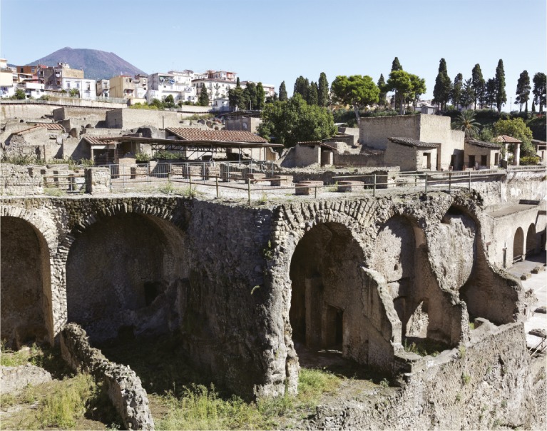
Herculaneum, with Mount Vesuvius in the background. Image courtesy of iStockphoto/GrahamMoore999.
Scholars of ancient scrolls hold that texts from antiquity, particularly Greek and Latin literary manuscripts produced until the fourth century AD, were largely written in carbon-based ink on papyri, the fibrous structure of which allowed scribes to jettison ruling lines. Emmanuel Brun et al. (pp. 3751–3754) used nondestructive synchrotron X-ray–based methods to chemically analyze barely visible black inscriptions on two nearly flat, multilayered papyrus fragments found at the Villa dei Papiri at Herculaneum in the mid-18th century and housed at the Institut de France in Paris. The introduction of metal in writing materials is generally dated to the fourth to fifth century AD, but the fragments’ high lead concentrations—around 84 µg/cm2 and 16 µg/cm2—suggest purposeful use of lead-containing ink, thus ruling out contamination from aqueducts, inkpots, or containers, and pushing back by several centuries the advent of metallic ink for literary inscription in the Greco-Roman period. Spots of concentrated lead likely correspond to the beginnings and ends of the scribes’ pen strokes on the scrolls. Letters on the fragments were bounded by naturally occurring horizontal lines of papyrus fibers that appear to have served as alignment guides for straight-line writing. The lines are likely signatures of cristobalite, a quartz-like mineral found in the papyrus plant. According to the authors, the findings might shape future analysis of unopened Herculaneum scrolls. — P.N.
High-resolution optical microscopy illuminates biofilms
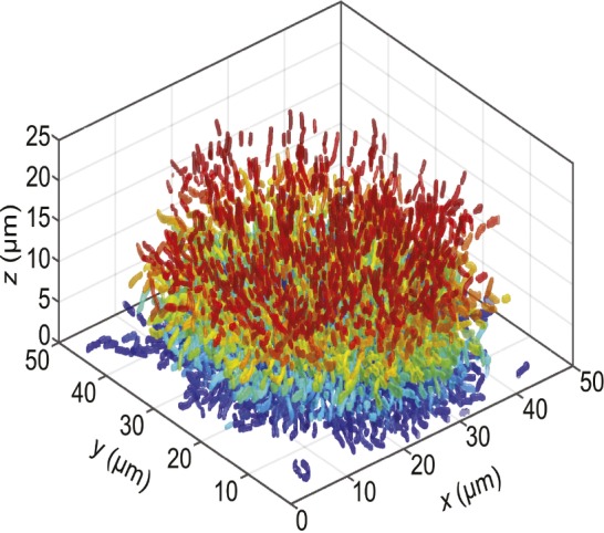
V. cholerae biofilm architecture at single-cell resolution.
Bacteria accrete in and adhere to surfaces in viscoelastic communities known as biofilms. Although human health concerns have driven extensive research into how these assemblages of antibiotic-resistant bacteria take hold and propagate, the cellular-level structure and morphology of biofilms remain largely unknown. Knut Drescher et al. (pp. E2066–E2072) used a custom-built confocal microscope to image the 3D position, size, and orientation of the thousands of individual cells that reside in Vibrio cholerae biofilms at different stages of growth. The investigation revealed four distinct growth phases in V. cholerae biofilms, each characterized by community-wide cellular reorganization and unique architecture. Transitions through these phases are driven primarily by the number of cells and the cell density in the growing community. The authors note that simple packing arguments alone do not fully explain the transitions by showing that mutant V. cholerae that lack particular biofilm matrix proteins produce drastically different biofilm architectures, compared with the corresponding wild-type strain. The findings demonstrate that optical imaging can illuminate biofilm formation, thus aiding modeling studies and suggesting strategies for inhibiting bacterial growth, according to the authors. — T.J.
Plant respiration and temperature response

Arctic vegetation in Alaska sampled for leaf respiration response to temperature.
During respiration, plants release carbon dioxide, a byproduct of the process of conversion of sugars into energy for cell maintenance and growth. Though plant respiration is a core component of the global carbon cycle, current climate models incorporate respiration using broad assumptions that fail to capture relationships that occur at small temporal and spatial scales. Mary Heskel et al. (pp. 3832–3837) quantified the relationship between temperature and leaf respiration rate based on 231 plant species in 7 different biomes. The authors demonstrate that whereas plant respiration rates increase with rising temperature, plant leaves become less sensitive to this effect as they warm. Notably, this deceleration in sensitivity is at odds with most terrestrial biosphere models, which assume that respiration increases exponentially with rising temperature. However, the analysis also reveals that the declining sensitivity to temperature appears to span multiple biomes and plant types, suggesting a global function that can be used to characterize the response. Accounting for this modified response, the authors present simulations showing that leaf respiration in cold biomes drops significantly, compared with other published studies aimed at addressing the shortcomings of exponential models. — T.J.
Food safety risk from Fukushima accident
Earthquake and tsunami damage to the Fukushima Dai-ichi nuclear power plant in March 2011 released large quantities of radioisotopes into the atmosphere and the Pacific Ocean. Since the accident, the government of Japan has sought to monitor radioisotope levels in potentially contaminated foods, but conflicting findings have done little to allay public concerns. Hiroshi Okamura et al. (pp. 3838–3843) present a statistical method that quantifies food contamination risk for specific species through time and space by tracking measurements of two cesium isotopes, 134Cs and 137Cs. Computing the probability that the sum of these isotopes exceeds a safety threshold, the authors found that the current overall risk of contamination is low, based on an evaluation of 1,646 combinations of aquatic species and locations. However, the authors caution that radioactive cesium levels remain relatively high in certain freshwater fish and crustaceans, compared with their marine counterparts. The model incorporates estimation techniques to help minimize confounders and biases in risk assessment. The findings offer a way to accurately assess aquatic food safety in the wake of the Fukushima accident, according to the authors. — T.J.
Virus evolution in the outback
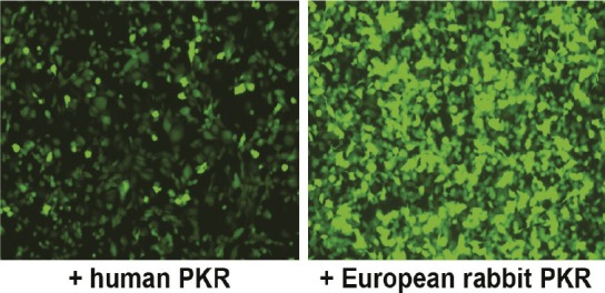
Human PKR restricts MYXV replication in congenic HeLa cells.
In Australia, where European rabbits are an invasive pest, the highly lethal, rabbit-specific myxoma virus (MYXV) was introduced in the 1950s to control the rabbit population. The rapid appearance of both attenuated virus and MYXV-resistant rabbits has provided researchers with a unique laboratory for studying host–virus coevolution in the wild. Chen Peng et al. (pp. 3855–3860) investigated the function of the MYXV protein M156, an ortholog of vaccinia virus K3, which enhances virulence by inhibiting the host antiviral protein kinase R (PKR). In both yeast and HeLa cell assays, the authors found that M156 expression inhibited rabbit PKR, but not PKR from humans or other mammals. Expression of human, but not rabbit, PKR enabled HeLa cells to suppress infection with MYXV. To test whether mutations in M156 affected MYXV virulence, the authors recreated a single amino acid substitution in a conserved region of M156 that was identified in 13 out of 24 Australian field isolates. The mutation abolished the ability of M156 to inhibit PKR and reduced viral gene expression and replication to levels seen in an M156 gene knockout background. According to the authors, species-specific inhibition of PKR by M156 may be responsible for the restricted host specificity of MYXV, and the M156 substitution may have contributed to the attenuation of MYXV since its introduction in Australia. — C.B.
Algal receptors and neuronal silencing

Simplified model shows the photocycle stages of ACRs.
Sensory photoreceptors called channelrhodopsins, which allow algae to move in response to light, have been harnessed for a powerful technique called optogenetics. By expressing these membrane proteins in genetically targeted neurons, researchers can use light to precisely control the activity of the neurons in freely moving animals. Oleg Sineshchekov et al. (pp. E1993–E2000) examined the light-absorbing and photochemical properties of a recently discovered family of natural channelrhodopsins that conduct negatively charged chloride ions, potentially enabling highly sensitive and efficient neuronal silencing. The authors expressed natural anion channelrhodopsins (ACRs) in yeast and human cells, exposed the proteins to laser flashes, and then measured changes in the absorption spectra and ion conductance. Similar to cation-conducting channelrhodopsins (CCRs) currently used for optogenetics, the ACRs transitioned through a sequence of photocycle stages, from the so-called K state to the O state, each of which was characterized by distinct flash-induced absorption changes. However, the ACR channels opened and conducted photocurrents during the L state, at an earlier photocycle stage than the CCRs. The findings show that ACRs and CCRs display fundamental differences in ion conduction and photochemistry, confirming that these proteins form distinct families of channelrhodopsins. According to the authors, analysis of the molecular mechanisms underlying the function of ACRs could help optimize the proteins for optogenetics or gene therapy. — J.W.