Significance
The common belief has been that no metal is present in Greco-Roman inks. In this work, we show that lead is present in the ink of two Herculaneum papyrus fragments. The concentration found is very high and not to be explained merely by contamination. The metal found in these fragments deeply modifies our knowledge of Greek and Latin writing in antiquity. Moreover, these concentration values allow the optimization of future computed tomography experiments on still-unrolled Herculaneum scrolls to enable the recovery of texts in the only surviving ancient Greco-Roman library. The possibility of using additional material to trace down ruled lines guiding the scribes' writing along straight lines is also addressed. We demonstrate that no additional material was used for this goal.
Keywords: paleography, papyrology, Herculaneum papyri, ruled lines, metallic ink
Abstract
Writing on paper is essential to civilization, as Pliny the Elder remarks in his Natural History, when he describes the various types of papyri, the method of manufacturing them, and all that concerns writing materials in the mid-first century AD. For this reason, a rigorous scientific study of writing is of fundamental importance for the historical understanding of ancient societies. We show that metallic ink was used several centuries earlier than previously thought. In particular, we found strong evidence that lead was intentionally used in the ink of Herculaneum papyri and discuss the possible existence of ruled lines traced on the papyrus texture. In addition, the metallic concentrations found in these fragments deliver important information in view of optimizing future computed tomography (CT) experiments on still-unrolled Herculaneum scrolls to improve the readability of texts in the only surviving ancient Greco-Roman library.
The development of alphabetic writing is one of the crucial steps in the history of Western civilization (1). Starting from the earliest examples, the writing that Greeks imported from Phoenicia is characterized by a quite regular layout, with letters evenly written between imaginary parallel lines (2). Capital letters, initially found in Greek manuscripts, then in Latin documents, and later in all of the languages based on Roman scripts (such as most Western and Central European languages, as well as many languages from other parts of the world), are based on this bilinear characteristic (3). If ruling lines is a well-established practice for writing on parchment supports in the Middle Ages (4), it is generally recognized that they were not necessary in papyri, the fibrous structure of which has been deemed sufficient for enabling the good alignment of written lines, even if their horizontal spacing was sometimes marked by a series of vertical dots. Eric G. Turner (3) argued that some material, no longer visible, was perhaps used for ruled lines on papyri; however, this claim cannot be supported by material evidence in the present state of paleographical knowledge (3, 5).
The same historical considerations apply to the chemical composition of the inks used in antiquity. Pliny the Elder carefully describes the carbon-based ink used in his time, which was obtained from smoke from wood burnt in furnaces, without any deliberate addition of metal (1). In the case of the most ancient manuscripts, and particularly the literary papyri both in Greek and Latin, it has been assumed that the ink used for writing was carbon-based, at least until the fourth to fifth centuries AD (4, 6, 7). The occasional use of metallic ink before this period has been known: it is reported to be used for writing secret messages in the second century BC (8), and Pliny remarks that papyrus soaked in tannin turns black after contact with a solution of iron salt (1). Moreover, Wagner et al. demonstrate the use of metal in inks in an ancient Egyptian papyrus (9), although none of those special inks has ever been mentioned in a Greco-Roman calligraphic context. However, a metallic iron-gall mixture was definitely elaborated and then adopted as a new writing ink for parchments beginning from around 420 AD (10, 11), because for this different kind of support, another, more adherent, ink was required. Thereafter, metallic inks became the standard for parchments in late antiquity and for most of the Middle Ages (4, 7).
In this work, we study the chemical composition of papyrus fragments carbonized by Mount Vesuvius' eruption in 79 AD and found in the Villa dei Papiri at Herculaneum between 1752 and 1754. Some fragments from the villa were analyzed on several occasions (12–14). In ref. 14, no metal was found, contrary to in refs. 12, 13, in which Seales et al. found lead and strontium in the ink at relatively high concentrations. Unfortunately, the analysis, which was made on several spots of 0.5 mm, did not include any elemental mapping. In this work, we address the issues left open in ref. 14 by a careful and detailed chemical and structural analysis of two fragments, using nondestructive synchrotron X-ray based techniques. The two multilayered fragments that were analyzed are of different sizes and come from the Institut de France’s collection in Paris, which is composed of six Herculaneum papyri donated by the King of Naples to Napoleon Bonaparte in 1802.
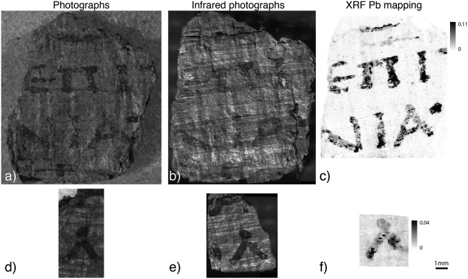
To resolve the chemical composition of the ink and papyrus texture in the fragments, a scanning X-ray fluorescence (XRF) experiment was performed at the ID21 beamline of the European Synchrotron Radiation Facility (ESRF), Grenoble, France. Via this technique, the identification of the principal constituent elements of the ink and papyrus support was possible. Both fragments are close to being flat, made of several layers, and on one surface, black letters are (barely) visible (Fig. 1 A and D). They were scanned using X-ray beams of different sizes, from a submillimeter down to a micrometric scale. The details of the different acquisition parameters are described in the Supporting Information.
Fig. 1.
Comparison of visible light photographs (A and D), infrared microscopy images (B and E), and lead distribution maps obtained by XRF (C and F) for both of the examined Herculaneum papyrus fragments. XRF maps were normalized by the incident flux and are in arbitrary units.
Results
Fig. 1 shows a comparison between photographs taken in visible light, infrared (IR) microscopy, and XRF mapping obtained at the M-edge of lead for both fragments. The photograph in visible light was taken using a standard digital single-lens reflex camera. For the IR setup, we illuminated the samples with an IR light of 940 nm wavelength and captured the reflected image (details in Supporting Information). XRF maps were obtained by raster scanning the samples using a 50-µm pinhole beam and 50-µm steps. The maps of the different elements were extracted using the PyMCA code (15).
In Fig. 1, the letters are clearly readable both under visible light and in IR photographs: in the larger fragment, the letter contours are sharper in the visible light range (Fig. 1A), whereas in the smaller fragment, the contrast is slightly enhanced in the IR photograph (Fig. 1E), a well-known feature that is widely exploited by specialists working on Herculaneum papyri (16). The XRF maps highlight letters especially in the larger sample; this means that for Herculaneum fragments that are difficult to read, the use of spectrometric techniques sensitive to lead may render the writing more clearly.
Thanks to an iterative Monte Carlo simulation (17), the mass concentration of lead in the letters can be estimated to be 84 ± 5 µg/cm2 for the larger sample and 16 ± 5 µg/cm2 for the smaller one. These fairly high concentrations of lead cannot be attributed only to lead contamination of water from Roman aqueducts (18) or from a copper inkpot or a bronze container (Supporting Information).
This level of concentration implies that a lead-bearing material was intentionally introduced in the ink production process. This would deeply modify our knowledge about the ancient Greek and Roman inks, as it is generally admitted that metal was not introduced before the fourth to fifth century AD.
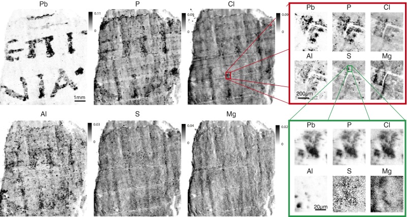
For a deeper evaluation of the chemical composition of the ink, we present in Fig. 2 the different elemental maps obtained for the larger sample. The red and green frames delimit XRF maps obtained at different resolutions, 10 and 1 µm, respectively, on small written zones identified by the color rectangles in the images.
Fig. 2.
Micro-XRF images of elemental distributions at low resolution (Left, 50-µm beam and mesh scan step size) and high resolution in the red frame (10-µm beam and mesh scan step size) and at very high resolution in the green frame (1-µm beam and mesh scan step size). Maps were normalized to the incident flux and are in arbitrary units.
From Fig. 2, it is clear that lead is the best discriminating element between ink and papyrus. At low resolution, lead (Pb), phosphorus (P), chlorine (Cl), and aluminum (Al) seem well colocalized, whereas the codistribution of sulfur (S) is difficult to see, and magnesium (Mg) does not show any correlation with the ink. At very high resolution (green frame inset), Al is not colocalized with Pb; Cl, Pb, and P seem strongly correlated, even if a high concentration of P can be found in other places.
Let us note that places with a greater concentration of Pb indicate the points where the scribe began and ended individual pen strokes, leaving a small ink blob that is not clearly visible to the eye.
The presence of Pb in the ink may be explained by several hypotheses. Pb could have been used as a pigment; for instance, lead sulfide or lead white (a mixture of lead carbonate and lead hydroxyl-carbonate) were frequently used in ancient times as pigments for cosmetic products (19). In addition, Pb could originate from a binding medium in the ink: Pb compounds such as litharge (PbO) have been extensively used in paintings, as they speed up the process of oil drying. Pliny does describe the use of minium (red lead) in books, but only for writing specific letters. These hypotheses are being tested by further experiments and will be discussed elsewhere (20).
The reason why no contrast based on the Pb concentration was observed in the phase contrast experiment reported in ref. 21 is possibly because of a different chemical composition of the ink. Individual scribes concocted their own inks, and one can expect variations in the materials they used. Moreover, the papyri in the Herculaneum library were written over a period of more than 3 centuries, and thus their composition and fabrication are likely to have evolved.
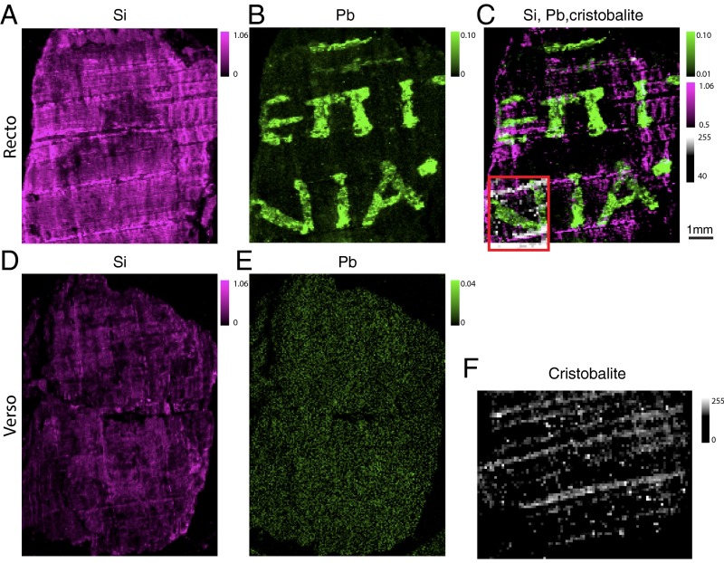
Some horizontal lines delimiting the height of letters were observed in the samples and appear in the Cl, S, and Mg maps because of the contrast with the papyrus support. Diverse experimental means were used to ascertain that the lines are formed of the fibers of the papyrus itself, and thus were used as guides by the scribes, rather than being deliberately introduced in a process of ruling. The general belief is that ruled lines were only traced on papyrus at the beginning of the third century AD (6). In Fig. 3, we present additional silicon (Si) XRF maps along with Pb XRF maps for both recto and verso of the larger sample, as well as X-ray diffraction (XRD) images in transmission geometry, which reveal the presence of cristobalite, a mineral for which the formula is the same as that of quartz (i.e., SiO2). The XRD experiments were performed at the ID11 beamline of the ESRF on a portion of the larger sample (details in the Supporting Information).
Fig. 3.
XRF and XRD maps obtained for the larger fragment. (A) Si XRF maps for the recto of the sample and (D) for its verso, (B and E) Pb XRF maps, (C) XRD cristobalite map superposed on the Pb XRF map, and (F) XRD cristobalite map on the portion of the sample defined by the red region of interest in C. The maps are normalized to the incident flux and are in arbitrary units.
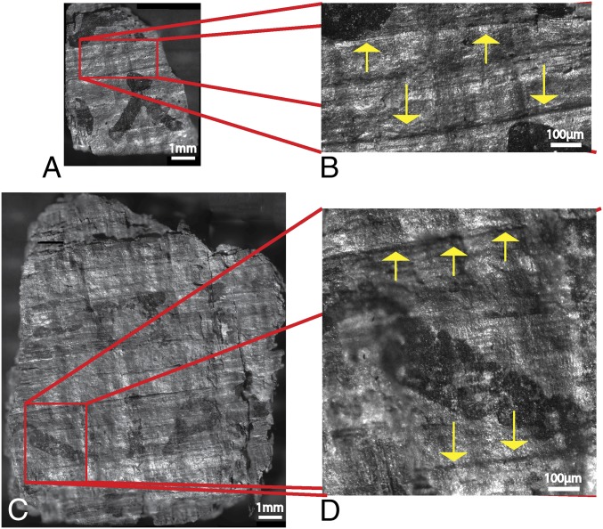
The XRD cristobalite map, obtained in the volume through the full thickness of the multilayered papyrus fragment, shows mainly horizontal lines (the images shown in the figures are slightly tilted) that match perfectly the observed Si XRF lines. The distance between two of these lines coincides with the size of letters in the lower row, whereas papyrus fibers are spaced more tightly. Moreover, one can find neither horizontal lines on the verso of the sample nor vertical lines on both sides of the sample. Note that because the XRD measurements were made in transmission mode, additional horizontal lines appear as a result of the sample consisting of several papyrus sheets. In addition, some lines are evident in the IR images shot at 1-µm resolution on both samples, as shown in Fig. 4. The selected zone for the larger fragment has to be compared with the red frame zone in Fig. 3C. Cristobalite and the infrared visible lines coincide.
Fig. 4.
Close-up in the microscopic IR image of the smaller fragment (A and B) and of the larger one (C and D). The close-up areas are labeled by red rectangles, and the ruled lines are indicated by yellow arrows.
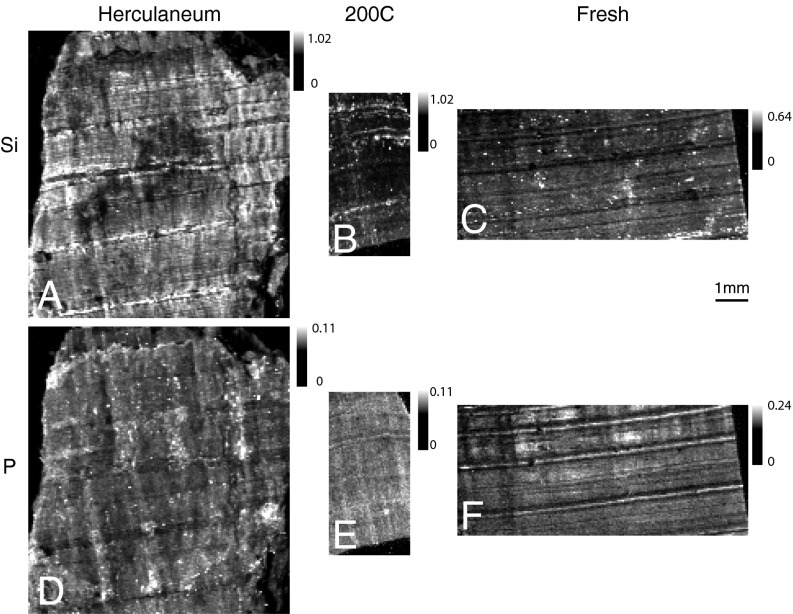
To confirm that the lines we found belong to the natural papyrus fibrous pattern, an additional XRF experiment was performed on two samples of modern papyrus: one fresh and one carbonized in an oven at 200 °C in vacuum. Fig. 5 presents Si and P maps for both modern samples and the larger ancient fragment. It is clear that horizontal lines are visible in all of the six images, meaning the lines in the fragments are probably the signature of cristobalite (22) naturally produced by the papyrus plant. The Si lines are more striking on increasing the temperature, whereas the opposite occurs with the P map because of the carbonization.
Fig. 5.
XRF maps of the three papyrus fragments: Si maps (A–C) and P maps (D–F). The larger fragment is shown in A and D, the papyrus sheet carbonized at 200 °C in vacuum is shown in B and E, and a fresh (modern) papyrus sheet is shown in C and F.
As a consequence, the visible lines in the Herculaneum fragments appear to be natural as well (5).
Discussion
To conclude, we have demonstrated that in the ink present on two Herculaneum fragments there is a high concentration of lead, and that the scribes used straight and thick horizontal papyrus fibers to guide the writing of letters in straight lines. The finding of metal in the ink radically modifies our knowledge of Greek and Latin writing in antiquity and might strongly influence the analysis of unopened papyrus rolls (21) and other ancient manuscripts. In our previous study (21), where X-ray phase contrast tomography was applied to unrolled Herculaneum scrolls, we first assumed that the ink was carbon-based. However, its actual chemical composition can strongly influence the choice of the imaging technique to be used and the wavelengths to be selected. Both of these findings may have some effect on other archaeological studies. For instance, Canevali et al. (23) analyzed black powders found in Pompeii's excavations to verify whether these powders were remainders of ink or make-up. Despite the presence of metals such as lead, they rejected different powders as candidates for writing inks primarily according to the archaeologists' assumption that at those times, in the Greco-Roman world, carbon-based inks without lead compounds were used exclusively.
The results of the present study push back by several centuries the introduction of metal into ink during the Greco-Roman period. The concentrations of metal found in the ink of the fragments we investigated provide evidence of an intentional practice and will aid in optimizing future tomographic experiments on undisclosed Herculaneum scrolls. More extensive research using other fragments from Herculaneum (whenever available) must be undertaken to assess the extent of presence of lead in the inks.
Description of the Samples
Two small fragments of six Herculaneum papyri from the Institut de France’s collection, a gift from the King of Naples to Napoleon Bonaparte in 1802, were analyzed. Found in the box “Objet 59,” they belong to one (or two) of these six rolls, and unfortunately cannot be more precisely identified. The larger of the two fragments measures ∼0.9 × 1.2 cm, and the smaller one is ∼0.5 × 0.8 cm. Both samples have an average thickness of about 0.3 mm and consist of two or more layers of papyrus with potential hidden writing.
Infrared Acquisition
The used infrared setup consists of an infrared extended source composed of an array of 4 × 4 IR light-emitting diodes (total dissipated power, 15 W) emitting at a wavelength of 940 nm and illuminating the samples. Images were captured in reflection mode using a BASLER piA400-17gm CCD camera equipped with a near-infrared 5× Mitutoyo microscope objective. The CCD camera is composed of an array of 2,456 × 2,048 pixels at 3.45-µm pitch.
XRF Acquisition
The XRF microbeam measurements were performed on the X-ray microscopy beamline ID21 at the ESRF, Grenoble, France. The primary beam energy was tuned by a Si(111) monochromator. Two energies were used: 2.48 and 3 keV (below and above the Pb M4,5-edges). The elemental maps for Mg and S are obtained at the lower energy, whereas all the others are extracted at the higher energy. At low resolution, the beam spot size on the sample was defined by a 50-µm pinhole for the larger fragment and a 100-µm pinhole for the smaller sample. The beam flux on the sample was continuously recorded by a pin diode to normalize the intensity variations to the fluctuating ring current. An average beam flux of 3.7 × 10⁹ ph/s and 6.9 × 10⁹ ph/s was obtained during the measurements on the larger and the smaller fragments, respectively. The samples were scanned across the X-ray beam in regular steps (38 × 31 100-µm steps for the smaller fragment and 160 × 175 50-µm steps for the larger one), and a XRF spectrum was recorded at each pixel. Additional measurements at higher spatial resolution were performed (see the high-resolution images of Fig. 2). The first dataset (red frame) was acquired by scanning the samples with a beam focused to ∼0.3 × 0.6 µm2 in 10-µm steps, whereas the second dataset was recorded with 1-µm steps.
XRD Acquisition
The microbeam XRD experiment was performed on the ID11 beamline at the ESRF, Grenoble, France. The primary beam energy was tuned by a Laue-Laue monochromator and set to 50 KeV. The transmitted diffraction patterns were recorded using a FReLoN CCD camera with fiber optic taper and an effective pixel size of 50 µm. The 2D images were processed using the pyFAI (24) program to yield 1D diffraction patterns. A blind deconvolution of the diffraction patterns was performed using the projected gradient method for nonnegative matrix factorization (25) to identify features in the data that were inspected and matched against crystallographic phase entries in the Inorganic Crystal Structural Database from FIZ-Karlsruhe (26).
Demonstration of the Intentional Use of Pb
For the time being it is difficult to know when and where the manuscripts were written (79 AD at the latest). It is therefore impossible to know precisely the concentration of lead in the water used for its dilution, and an educated guess is thus mandatory.
Nowadays, the European Community estimates that water is polluted if the concentration of lead is in excess of 10 µg/L (50 µg/L before 1983). According to Delile et al., water in the early Roman Empire was mostly 40 times more polluted than normal water (18). Here, we assume that the concentration of Pb in water used for diluting the ink was 2,000 µg/L, which would probably have induced lead poisoning.
The lead concentrations observed in both fragments are estimated to be as high as 16 µg/cm2 and 85 µg/cm2 for the smaller and the bigger fragments, respectively. If we assume than the water used for diluting the soot/arabic gum mixture was just “tap water,” this means that the total amount of water in 1 cm2 of ink would be 42 mL in the bigger sample and 8 mL in the smaller sample.
In addition, this would signify that for a papyrus roll of a 15 × 0.15 m (i.e., 22,500 cm2), assuming that 40% of it is covered by ink, 432 L of water should have been used (i.e., approximately three big bathtubs). This is definitely unrealistic.
Acknowledgments
The European Synchrotron Radiation Facility (ESRF) is gratefully acknowledged for granting in-house X-ray beam time. We thank C. Jarnias for his help during the infrared microscopy experiment and the Institut de France, as well as the “Commission des Bibliothèques,” for generously lending the papyrus samples, and F. Topin for the fruitful scientific discussion. We are greatly indebted to the referees for their very invaluable recommendations, and especially their critical comments about natural cristobalite. P.T.’s work is funded by a PhD grant from the Agency for Innovation by Science and Technology.
Footnotes
The authors declare no conflict of interest.
This article is a PNAS Direct Submission. R.J. is a guest editor invited by the Editorial Board.
This article contains supporting information online at www.pnas.org/lookup/suppl/doi:10.1073/pnas.1519958113/-/DCSupplemental.
References
- 1.Eichholz DE, Jones WHS, Rackham H. Pliny the Elder, Natural History. Cambridge, MA: Harvard University Press; 1938. [Google Scholar]
- 2.Cavallo G, Maehler H. Hellenistic Bookhands. Walter De Gruyter; Berlin: 2008. [Google Scholar]
- 3.Turner EG. In: Greek Manuscripts of the Ancient World. 2nd ed Parsons PJ, editor. London: University of London, Institute of Classical Studies; 1987. [Google Scholar]
- 4.Thompson EM. An Introduction to Greek and Latin Palaeography. Clarendon; Oxford: 1912. [Google Scholar]
- 5.Cavallo G. Reviewed work: Greek Manuscripts of the Ancient World by Eric G. Turner. Gnomon. 1974;46:147–152. [Google Scholar]
- 6.Thompson EM. Handbook of Greek and Latin Paleography. Kessinger Publishing; Whitefish, Montana: 2007. [Google Scholar]
- 7.Bischoff B. Latin Palaeography: Antiquity and the Middle Ages. Cambridge University Press; Cambridge: 1990. [Google Scholar]
- 8.Graux C. L’encre à base métallique dans l’antiquité. Revue de Philolologie. 1880;IV:82–85. [Google Scholar]
- 9.Wagner B, et al. Analytical approach to the conservation of the ancient Egyptian manuscript ‘Bakai Book of the Dead’: A case study. Mikrochim Acta. 2007;159:101–108. [Google Scholar]
- 10. Martianus Capella (1977) The Marriage of Philology and Mercury; trans Stahl WH, Johnson R, Burge EL (Columbia Univ Press, New York)
- 11.Dick A. 1925. Martianus Capella (Leipzig, Teubner)
- 12.Seales WB, Delattre D. Virtual Unrolling of Carbonized Herculaneum Scrolls: Research Status (2007-2012) Cronache Ercolanesi. 2013;43:191–208. [Google Scholar]
- 13.Seales WB. 2009. Lire sans détruire les papyrus carbonisés d'Herculanum, Comptes rendus des séances de l'Académie des Inscriptions et Belles-Lettres 153(2):907–923.
- 14.Störmer F, Lorentzen I, Fosse B, Capasso M, Kleve K. Ink in Herculaneum. Cronache Ercolanesi. 1990;20:183–184. [Google Scholar]
- 15.Solé VA, Papillon E, Cotte M, Walter P, Susini J. A multiplatform code for the analysis of energy-dispersive X-ray fluorescence spectra. Spectrochim Acta B At Spectrosc. 2007;62(1):63–68. [Google Scholar]
- 16.Chabries DM, Booras SW, Bearman GH. Imaging the past: Recent applications of multispectral imaging technology to deciphering manuscripts. Antiquity. 2003;77:359–372. [Google Scholar]
- 17.Schoonjans T, Solé VA, Vincze L, del Rio MS, Appel K, Ferrero C. A general Monte Carlo simulation of energy-dispersive X-ray fluorescence spectrometers - Part 6. Quantification through iterative simulations. Spectrochim Acta B At Spectrosc. 2013;82(1):36–41. [Google Scholar]
- 18.Delile H, Blichert-Toft J, Goiran JP, Keay S, Albarède F. Lead in ancient Rome’s city waters. Proc Natl Acad Sci USA. 2014;111(18):6594–6599. doi: 10.1073/pnas.1400097111. [DOI] [PMC free article] [PubMed] [Google Scholar]
- 19.Walter P, et al. Making make-up in ancient Egypt. Nature. 1999;397:483–484. [Google Scholar]
- 20.Tack P, et al. February 8, 2016. Tracking the ink composition on Herculaneum papyrus scrolls: Identification, localization, quantification and speciation of lead by X-ray based techniques and Monte Carlo simulations. Sci Rep, 10.1038/srep20763.
- 21.Mocella V, Brun E, Ferrero C, Delattre D. Revealing letters in rolled Herculaneum papyri by X-ray phase-contrast imaging. Nat Commun. 2015;6:5895. doi: 10.1038/ncomms6895. [DOI] [PubMed] [Google Scholar]
- 22.Dollase W. Reinvestigation of the structure of low cristobalite. Zeitschrift für Krist. 1965;121(5):369–377. [Google Scholar]
- 23.Canevali C, et al. A multi-analytical approach for the characterization of powders from the Pompeii archaeological site. Anal Bioanal Chem. 2011;401(6):1801–1814. doi: 10.1007/s00216-011-5216-8. [DOI] [PubMed] [Google Scholar]
- 24.Ashiotis G, et al. The fast azimuthal integration Python library: PyFAI. J Appl Cryst. 2015;48(Pt 2):510–519. doi: 10.1107/S1600576715004306. [DOI] [PMC free article] [PubMed] [Google Scholar]
- 25.Pedregosa F, et al. Scikit-learn: Machine Learning in Python. J Mach Learn Res. 2011;12:2825–2830. [Google Scholar]
- 26. Introduction to ICSD Web. Available at https://icsd.fiz-karlsruhe.de/search. Accessed July 2015.


