



Abstract
Study Objectives:
To investigate the relationship between K-complexes (KCs) and cardiac functioning.
Methods:
Forty healthy adolescents aged 16–22 y (19 females) participated in the study. Heart rate (HR) fluctuations associated with spontaneous and evoked KCs were investigated on two nights, one with (event-related potential night) and one without auditory tones presented across the night.
Results:
There was a clear biphasic cardiac response to evoked and spontaneous KCs, with an initial acceleration in HR followed by a deceleration (P < 0.001). HR acceleration occurred immediately to KCs in response to tones presented in the first third of the interbeat interval, but was delayed a beat when the tone occurred later in the cardiac cycle (P < 0.05). Sex differences were also evident. Pretone baseline HR was higher, and the magnitude of the HR response was blunted and delayed, in female compared to male adolescents (P < 0.001). Also, pretone baseline HR was lower when a tone elicited a KC compared to when it did not (P < 0.001), suggesting that KCs are possibly more likely to be elicited by external stimuli in states of reduced cardiac activation.
Conclusions:
The strict dependency observed between KCs and cardiac control indicates a potential role of KCs in modulating the cardiovascular system during sleep. Sex differences in the KC-cardiac response indicate the sensitivity of this measure in capturing sex differences in cardiac regulatory physiology.
Citation:
de Zambotti M, Willoughby AR, Franzen PL, Clark DB, Baker FC. K-complexes: interaction between the central and autonomic nervous systems during sleep. SLEEP 2016;39(5):1129–1137.
Keywords: autonomic nervous system, cardiovascular, heart rate, K-complex, sex differences, sleep
Significance.
This study shows a reciprocal interaction between the K-complex, a hallmark of NREM sleep, and cardiac activation, suggesting that K-complexes play a role in cardiovascular homeostasis. An understanding of the relationship between the K-complex and peripheral physiology, such as autonomic nervous system functioning, may help elucidate the functional role of K-complexes. Future research should focus on clarifying the relationship between the cardiovascular state as a predictor of K-complex occurrence and whether this relationship unravels in the presence of clinical conditions such as hypertension.
INTRODUCTION
The K-complex (KC) is a specific pattern of electrical brain activity that appears both spontaneously or in response to an external stimulus.1 The KC is characterized by a positive-negative-positive waveform lasting at least half a second. When KCs are averaged, these peaks are seen as P200, N550, and P900 components in the averaged event related potential (ERP), with the N550 and P900 components typically largest over frontal electrode sites. Because the N550 amplitude can be in excess of 100 μV, it is thought to reflect the synchronized activity of a large number of cortical neurons1 and has been hypothesized to reflect cortical integrity.2 Despite our knowledge of the neurophysiological generation of the KC, our understanding of the functional role of the KC remains limited. Although research3–6 suggests that the KC is sleep protective, there is still ongoing debate about its sleep protective role, and whether it reflects an arousal response or is a marker of sleep depth.7–9
Although many studies have focused on central nervous system (CNS) mechanisms involved in the generation of KCs, little attention has been paid to the relationship between the KC and peripheral autonomic physiology. Nevertheless, several elegant experiments, starting back in the 1950s, provide important results. Ackner and Pampiglione10,11 showed a temporal relationship between KCs and peripheral vasoconstriction, measured using a finger plethysmograph, leading them to suggest that both phenomena were related to “spontaneous fluctuations in the levels of alertness or sleep.”
Johnson and Lubing12 reported that autonomic nervous system (ANS) responses (myocardial contraction and peripheral vasoconstriction) were greater when KCs were elicited by tones than when they occurred spontaneously. However, a later experiment by Johnson and Karpan13 found ANS changes related to both spontaneous and evoked KCs were the same. Following the intuition of Johnson and Lubing,12 Berg et al.14 showed that cardiac responses (a biphasic response with tachycardia followed by bradycardia) were only associated with trials where KCs were elicited. Interestingly, Fruhistorfer et al.15 showed that spontaneous KCs were most likely to occur 200–300 msec and 600–800 msec after the onset of the P wave in the electrocardiogram (ECG), suggesting a relationship between spontaneous KCs and the cardiac cycle. However, theoretical interpretation is difficult in this study because no distinction was made between KCs and “vertex sharp waves” which are faster, theta frequency waveforms maximal over the vertex rather frontal scalp.16
Later research used ANS measures as indices of arousal associated with a KC. Many of these studies used microneurography to measure muscle sympathetic activity (MSNA) in addition to measuring heart rate (HR) and blood pressure (BP). They typically found that KCs were associated with abrupt increases in sympathetic activity, tachycardia (an increase in HR) and an increase in BP.17–21
Even if provocative, these early studies had several limitations: KCs were poorly characterized, the majority of the results were descriptive rather than quantitative, techniques were limited, sample sizes were small (the majority of the studies included fewer than 10 participants) and, in several studies, data were collected in drug induced states or during sleep deprivation.
More recently, Monstad and Guilleminault22 found a significant relationship between KC incidence and Mayer waves (a spontaneous oscillation in BP with a frequency of approximately 0.1 Hz) in eight volunteers (32–71 y). Spontaneous KCs were more likely to occur while BP was decreasing and evoked KCs were more likely to be elicited if the tone was presented in the downward slope of the Mayer wave (i.e., while BP was decreasing). KCs were also associated with a significant rise in mean BP: BP increased (after approximately 6 to 8 sec) by 2.4 mmHg following an evoked KC and by 9.7 mmHg following a spontaneous KC. Similarly, HR also increased following evoked and spontaneous KCs, peaking 2 to 6 sec after the KC. A relationship between KCs and BP regulation during sleep was also suggested by Tank et al.23 By analyzing seven healthy participants (22–41 y), the authors showed that single large KCs were preceded by a baroreflex-mediated increase in MSNA in 55% of the cases and were associated with a 22.5% increase in MSNA, a 5.2% increase in systolic BP and a 6.5% increase in diastolic BP.
The current study aims to extend previous research into cardiovascular control associated with KCs by investigating the magnitude and temporal dynamics of HR changes associated with spontaneous and tone-evoked KCs in a large sample of healthy male and female adolescents. Previous studies focused on adult populations and, to our knowledge, none investigated potential sex differences in cardiac responses to a KC. Two advantages of studying young healthy individuals (16–22 y) are that it allows investigation of the KC when it is most prominent (higher amplitude and more likely to be elicited to a tone), compared to older individuals,24 and it reduces the confounding effect of age on the interaction between the KC and heart rate. Based on previous literature, we hypothesized that there would be a biphasic cardiac response to the KC, with an initial acceleration followed by a deceleration in HR, and that responses would be of a similar magnitude for both spontaneous and evoked KCs. Based on the hypothesis that KCs play a role in cardiovascular control during sleep,22 we also hypothesized differences in the baseline cardiac state during tones that elicited a KC, compared to those that did not (lower pretone HR when tones elicit a KC).
Finally, to account for sex differences in cardiovascular physiology, such as a higher resting HR in women than in men,25–28 we included a mixed sample of male and female adolescents to enable an investigation of potential sex differences in cardiac responses to a KC.
METHODS
Participants
Forty adolescents, 19 females (11 Caucasian; mean ± standard deviation [SD]: age, 18.5 ± 1.3 y; body mass index [BMI]: 23.1 ± 5.1 kg/m2) and 21 males (17 Caucasian; mean ± SD: age, 18.9 ± 1.3 y; BMI: 22.6 ± 3.6 y kg/m2), between 16 and 22 years old were included in this analysis. They were participating in a special project about sleep conduced at SRI International and University of Pittsburgh as part of a large ongoing (2012–2017) multisite longitudinal study of adolescents (National Consortium on Alcohol and NeuroDevelopment in Adolescence, NCANDA). A full description of NCANDA methodology and sample characteristics at baseline has been provided in the literature.29 They were recruited through distribution of school and community fliers (SRI International) or random digit dialing (Pittsburgh). Data describing the characteristics of the sample were obtained from data release version: NCANDA_DATA_00010_V2.
Briefly, all participants had a phone interview and in-person screening session including the Semi-Structured Assessment for the Genetics of Alcoholism.30 None of the participants had severe medical conditions (e.g., heart disease, epilepsy, traumatic brain injury) or current/past major Diagnostic and Statistical Manual of Mental Disorders, Fourth Edition31 Axis I disorders (e.g., major depression, generalized anxiety disorders). None of the participants showed evidence of sleep disordered breathing, periodic limb movement disorder, or narcolepsy, as assessed by a clinical sleep evaluation. None of the participants currently used medication known to affect sleep and cardiovascular systems.
The study was approved by the Institutional Review Boards at SRI International and University of Pittsburgh. Adult participants consented to participate and minors provided written assent along with consent from a parent/legal guardian. Participants and parents were compensated for participation.
Procedures
Participants had two randomized overnight polysomnography (PSG) recordings at SRI International or University of Pittsburgh sites in sound-attenuated, temperature-controlled bedrooms. On one night, tones were presented throughout the night to elicit KCs (event-related potential - ERP night); on the other night, tones were not presented (sleep architecture, Arch night). Twenty-one participants (12 males and 9 females) had their ERP night recorded first. Participants chose their own lights-out and lights-on times. Each night, a breath alcohol test (S75 Pro, BACtrack Breathalyzers, San Francisco, CA, USA) and urine drug test (10 Panel iCup drug test kit, Instant Technologies, Inc.) confirmed the absence of recent alcohol or drug use.
Evoked Related Potential Protocol
A standard sleep ERP protocol was used.32 Auditory stimuli of 1,000 Hz were presented binaurally at 80 dB for 50 msec (2 msec rise and fall time) using Compumedics NeuroScan Stim software (Compumedics Ltd, Abbotsford, Victoria, Australia) or Eprime (Psychology Software Tools, Inc, Pittsburgh, PA) through E-A-RTONE 3A insert earphones (3M Auditory Systems, Indianapolis, IN, USA) with a random interstimulus interval of 15–30 sec. Tones were first presented after about 30 min of stable nonrapid eye movement (NREM) sleep and continued to be presented during NREM sleep throughout the night. Tones were halted during major arousals or awakenings, and re-started once stable sleep returned. All participants reported normal hearing and confirmed they could hear the tones before going to bed.
Assessment of Polysomnographic Sleep and ECG
Standard PSG was performed using the Compumedics Grael HD-PSG system (Compumedics, Abbotsford, Victoria, Australia). The electroencephalogram (EEG) (FP1, FP2, F3, F4, FC3, FC4, C3, C4, CP3, CP4, P3, P4, O1, O2 referenced to the contra-lateral mastoids), submental electromyogram and bipolar electrooculogram were recorded according to American Academy of Sleep Medicine (AASM) rules.33 The EEG was sampled at 256 Hz and filtered at 0.3–30 Hz. Sleep was scored according to AASM criteria.33 The ECG was collected simultaneously at 256 Hz through the Compumedics system using Ag/AgCl Meditrace surface spot electrodes in a modified lead II Einthoven configu-ration (electrodes were placed under the right clavicle and on the lower left abdomen within the rib cage frame).
Evoked and Spontaneous K-Complexes
EEG data were analyzed using the EEGLAB34 toolbox, the ERPLAB35 toolbox, and custom processing scripts for MATLAB (MathWorks, Natick, MA, USA).36 For both the Arch and ERP nights, the EEG was re-referenced to the average mastoid and filtered using a zero-phase shift finite impulse response filter (with 6 dB cutoffs at 0.15 and 30.15 Hz).
On ERP nights, KCs were visually identified in N2 sleep from F3 and C3 and average KC waveforms from F3 were created according to established criteria.32,37 Briefly, epochs starting 2,000 msec before to 3,000 msec after the presentation of the tone were extracted and baseline corrected to the 1,000 msec prestimulus period. Epochs were categorized as KC+ (KC elicited by the presentation of the tone) if a negative peak occurred 400–900 msec following tone presentation or as KC- if the tone did not elicit a KC. Mean KC waveforms were created by averaging all the KC+ trials for each participant and mean N550 latency (msec) and amplitude (μV) were calculated for each participant.
On the Arch nights, spontaneous KCs (KCSP) were identified using a semi-automated method. The EEG at electrodes F3 and C3 was visually inspected for single-instance KCSP which were then automatically checked to ensure they were more than 10s apart from each other. The N550 peak was detected at F3 and a dummy trigger position calculated based on the mean N550 latency for each participant (derived from the evoked KC condition). The inclusion of the dummy trigger was necessary for analogous processing of KC+ and KCSP data. Amplitude of the N550 component (μV) for KCSP was also calculated.
Heart Rate Changes in Response to Tone (or Dummy Trigger) Presentation
R-wave peaks in the ECG were automatically detected using a customized Matlab algorithm and checked for accuracy by visual inspection. Interbeat intervals (IBIs, msec) were calculated as the difference between the R-wave peaks.
On the ERP nights, IBI durations were calculated for the IBI in which the tone was presented (IBI0), the five previous IBIs (IBI−5 to IBI−1) and the subsequent 12 IBIs (IBI1 to IBI12). In order to compare the Arch night to the ERP night, the same procedure was used for data collected on the Arch night using the dummy triggers. Figure 1 shows a schematic representation of the three conditions analyzed: EEG KC+, EEG KC-, and EEG KCSP. The mean of IBI−4 to IBI−2 was used as a baseline for comparison with post-tone IBIs.
Figure 1.

Representation of the interbeat intervals (IBIs) analyzed for each of the K-complex (KC) conditions (KC+, KC-, and KCSP). For the KCSP condition, the estimated time of tone presentation (dummy tone) was calculated based on the averaged tone-to-N550 peak latency of KC+ trials on the event-relate potential nights. Electroencephalographic (EEG) traces for KC+, KC- and KCSP conditions are displayed with the simultaneous registration of the electrocardiogram (ECG). Pretone (IBI-5-to-IBI-1), tone (IBI0), and posttone (IBIs 1-to-12) IBIs are marked. IBI-4-to-IBI-2 were used as a baseline.
Data Analyses
K-complex characteristics: N550 latency and amplitude. For the KC+ condition, a paired t-test was used to compare mean N550 latency in males and females. A KC Condition (KC+ and KCSP) × Sex (males and females) repeated-measures analysis of variance (ANOVA) was used to investigate differences in N550 mean amplitude.
Characterization of the biphasic cardiac response to evoked K-complexes. The magnitude of the peak in cardiac acceleration was measured as the percentage difference between the shortest posttone IBI (i.e., fastest HR, between IBI0 and IBI5) and the baseline measure (mean of IBI−4-to-IBI−2). The magnitude of the peak in deceleration following the first acceleration was measured as the percentage difference between the longest posttone IBI (i.e., slowest HR, between IBI5 and IBI12) and the baseline measure. If the maximum deceleration was observed in the final IBI (IBI12), that trial was removed from the analysis, because it was impossible to determine whether the peak deceleration had been reached (number of removed trials: mean ± SD: 6.6 ± 5.3). In addition, three males were further excluded from the analysis of the “deceleration response” due to having a low number of trials (< 10). The posttone IBI positions in which the cardiac acceleration and subsequent deceleration reached their peaks were also computed. Sex differences in the magnitude of acceleration and deceleration and for the IBI position in which they occurred were tested using independent t-tests.
Assessment of the nature of the cardiac response: K-complex-or tone-dependent? We used a repeated-measures ANOVA to compare the magnitude of the peak in cardiac acceleration as a function of KC+, KC- and KCSP conditions.
Effect of tone position within the cardiac cycle on the temporal dynamics and magnitude of the cardiac acceleration. On ERP nights, tones randomly fell between two consecutive R waves. In order to test whether the position of tone within the cardiac cycle affects timing and magnitude of the cardiac response, all KC+ trials were classified based on whether the tone fell in the first (T1), second (T2), or third (T3) part of the cardiac cycle. Repeated-measures ANOVAs were used to determine differences in the magnitude of the peak in cardiac acceleration and post-tone IBI position in which the cardiac acceleration reached its peak, according to cardiac cycle time (T1, T2, and T3).
Assessment of pretone baseline cardiac activity: are K-complexes more likely to be elicited in a specific cardiac state? Differences in baseline pretone cardiac activity were analyzed according to KC Condition (KC+ and KC-) and Sex (male and female) using a repeated measure ANOVA.
All analyses were performed based on the averaged cardiac evoked response for each participant. IBIs (msec) were used to analyze the evoked response and then were converted to HR (bpm). In ANOVA models including more than two within levels, the Huynh-Feldt correction was applied to correct for sphericity and Tukey post hoc tests were used to analyze significant interactions. The value P < 0.05 was considered significant in all analyses.
RESULTS
K-Complex Characteristics: N550 Latency and Amplitude
In the KC+ condition, N550 latency was similar in male (693.6 ± 60.1 msec) and female (679.3 ± 58.3 msec) adolescents. N550 amplitude was higher in KCSP (−282.9 ± 65.6 μV) than KC+ (−228.2 ± 84.4 μV) conditions (F1,38 = 59.19, P < 0.001) and was similar in males and females (P = 0.418).
Characterization of the Biphasic Cardiac Response to the evoked K-Complexes
The cardiac response to evoked KCs occurring in artifact-free N2 sleep was characterized by a strong cardiac acceleration following by a deceleration (Figure 2) with fluctuations exceeding baseline levels by more than 5% in both males (HR in bpm, mean ± SD: pretone baseline, 54.5 ± 4.9 bpm; peak in acceleration; 59.0 ± 5.4 bpm, peak in deceleration, 53.3 ± 3.5 bpm) and females (HR in bpm, mean ± SD, bpm: pretone baseline, 63.5 ± 6.4 bpm; peak in acceleration; 67.2 ± 7.9 bpm, peak in deceleration, 60.3 ± 6.4 bpm). The peak in acceleration occurred, on average, between the second and third posttone IBI (IBI2-IBI3) whereas the subsequent deceleration peaked, on average, between IBI7 and IBI10.
Figure 2.
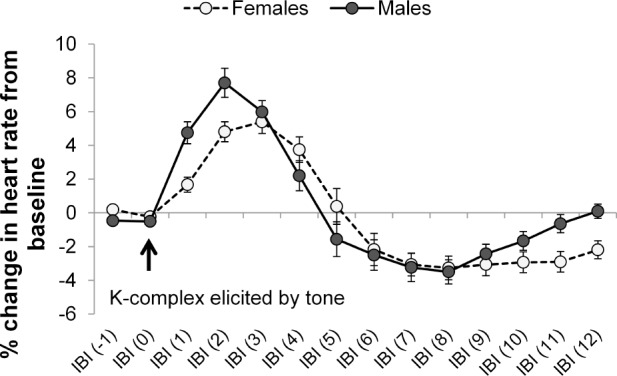
Representation of the beat-to-beat dynamics of the biphasic cardiac response to evoked KCs occurring in artifact-free N2 sleep in males (N = 18) and females (N = 19). Each point reflects the percentage change in heart rate from the pretone baseline level. Vertical bars represent standard errors. IBI = interbeat interval.
The peak in cardiac acceleration occurred earlier (P < 0.05) and was of a greater magnitude (P < 0.05) in males compared to females. The peak in cardiac deceleration was also delayed (P < 0.001), but did not differ in magnitude, in females compared to males. See Table 1 for details.
Table 1.
Magnitude of the peak in cardiac acceleration (baseline to peak, %) and subsequent deceleration (baseline to peak, %) and interbeat interval position in which the peaks in acceleration and deceleration occurred in response to evoked K-complexes.

Considering that the higher number of trials in females may confound the results, we re-ran all the analyses after randomly reducing the numbers of trials selected in females by one-third to match the number of trials selected in males. All results remain unchanged.
Dependence of the Cardiac Response on the Occurrence of K-Complexes
ANOVA revealed a KC Condition main effect for the magnitude of the peak in cardiac acceleration (F2,78 = 43.50, P < 0.001). Post hoc analysis indicated that the cardiac response was similar for K+ and KSP condition, which were both significantly greater than the K- condition (P < 0.001), indicating a dependence of the cardiac response on the occurrence of KCs and not on the presence of a tone (Figure 3).
Figure 3.
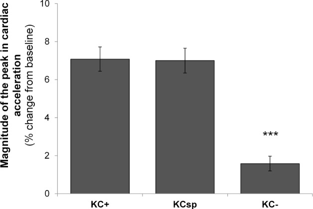
Magnitude of the peak in cardiac acceleration (baseline to peak, %) in response to K-complexes evoked by tones (KC+), tones that did not elicit a K-complex (KC-), and spontaneous K-complexes (KCSP). Vertical bars represent standard errors. ***P < 0.001 compared to KC+ and KCSP conditions.
Temporal Dynamics and Magnitude of Cardiac Acceleration as a Function of the Period of the Cardiac Cycle during which the Tone Occurred
The ANOVA revealed a Tone Position main effect (F2,78 = 16.17, P < 0.001) for the IBI position in which the peak in acceleration occurred (Figure 4). Post hoc analyses indicated that when the tone was presented in the first period (T1) of the cardiac cycle, the peak in acceleration occurred, on average, earlier than when the tone was presented in the second (T2; P = 0.020) and third (T3; P < 0.001) period of the cardiac cycle. Similarly, when the tone was presented in T2, the posttone peak in acceleration occurred, on average, earlier that when it was presented in T3 (P < 0.001). In contrast, no significant effect of Tone Position emerged for the magnitude of the peak in cardiac acceleration, indicating that the latency to the peak in cardiac acceleration but not magnitude of the acceleration is dependent on tone position.
Figure 4.
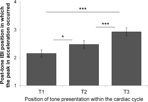
IBI position in which the peak in acceleration occurred as a function of the period within the cardiac cycle in which the tone was presented. Vertical bars represent standard errors. *P < 0.05; ***P < 0.001. IBI = interbeat interval.
Sex Differences and K-Complex-Dependent Pretone Cardiac Activity
ANOVA revealed KC Condition (F1,38 = 32.70, P < 0.001) and Sex (F1,38 = 26.16, P < 0.001) main effects for the baseline pretone HR. Baseline HR was higher in females compared to males. In both males and females, baseline HR was lower when KCs were elicited compared to when they were not elicited by tones (Figure 5).
Figure 5.
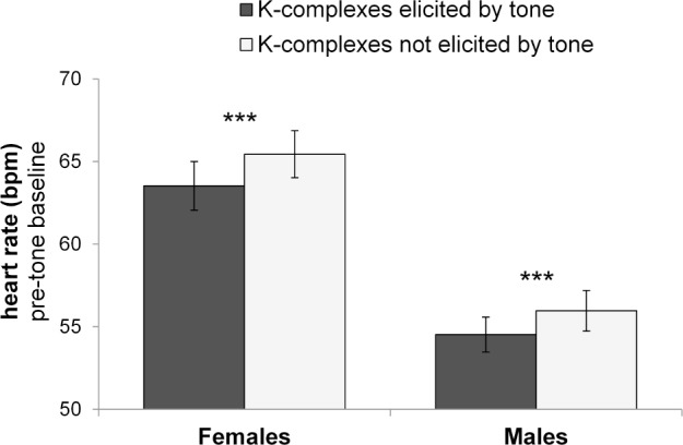
Baseline pretone cardiac activity (heart rate, bpm) when K-complexes are elicited (KC+) or not (KC-) by tones in both males and females. Vertical bars represent standard errors. Asterisk represent significant differences (P < 0.001) between KC+ and KC- conditions, and between sexes.
DISCUSSION
The main purpose of the study was to characterize the magnitude and temporal dynamics of heart rate fluctuations associated with the occurrence of KCs to gain insight into the functional significance of the KC and the associated autonomic cardiac response. The main findings can be summarized as follows: (1) heart rate showed a clear biphasic pattern in association with KCs, with a marked acceleration followed by a more gradual deceleration; this response was absent to tones unaccompanied by a KC; (2) baseline heart rate was slower when KCs were elicited compared to when they were not elicited by tones, suggesting that KCs are more likely to be evoked in states of lower cardiac activation; (3) the KC-dependent heart rate acceleration was delayed when the tones were presented later in the cardiac cycle; (4) heart rate was faster and the cardiac response to KCs was blunted and delayed in females compared to males, suggesting sex differences in the KC-cardiac relationship.
We replicated and extended previous research8,14,38 by showing that both spontaneous and evoked KCs are associated with a clear biphasic cardiac response of tachycardia followed by bradycardia lasting ∼12 cardiac cycles (Figure 2). Tones that were not accompanied by a KC lacked a cardiac response, and strongly suggest that this biphasic cardiac response is linked with the KC itself. These results show that spontaneous and evoked KCs are associated with an increase in heart rate of > 5%, with the increase being maximal in the first two cardiac cycles following the IBI in which the tone was presented (Figure 2). In our study we were able to compare KC+, KC-, and KSP conditions within the same sample. The use of a sample of healthy adolescents allowed us to investigate EEG and cardiac functioning free from confounding effects of age and age-related diseases.39–41 Also, the analysis of single-instance KCs in arousal free trials selected in stable N2 NREM sleep allowed us to investigate cardiac responses to KCs free from other manifestations of cortical synchronization or desynchronization that may be initiated through a different mechanism and could affect cardiac control differently.
Autonomic cardiac modulation associated with KCs has typically been cited as evidence that the KC represents an arousal response associated with tachycardia and increased sympathetic nerve activity.17–21,23 Sforza and colleagues8 compared the HR pattern across microarousals, phase transitory activation, and bursts of KCs and delta waves, and suggested that bursts of KCs and delta waves reflect subcortical (or autonomic) arousal and are part of a continuum on the arousal spectrum, which progresses from brainstem to cortical levels. Although it seems clear that KCs are accompanied by ANS changes, the role of KCs and the exact nature of the relationship between KCs and ANS responses are less clear. Research shows that spontaneous and evoked KCs are more likely to occur in synchrony with Mayer blood pressure waves22 with KCs predominantly occurring during a decrease in blood pressure; in the same study, spontaneous KCs were also preceded by a decline in heart rate (0.3 ± 2.4 bpm) over a 20-sec period prior to the KC.22 Heart rate oscillation (tachycardia followed by bradycardia) during NREM sleep also precedes increases in EEG delta power.42 Tank et al.23 found that a large proportion of single KCs (55%) were preceded by a decrease in blood pressure or a baroreflex-mediated increase in MSNA, suggesting that the baroreceptor input may modulate KCs. Similarly, our data show that the pretone baseline heart rate in the KC+ was lower compared with the KC- condition, supporting the existence of a meaningful relationship between the ANS and KC generation. These results are consistent with the notion that KCs are more likely to be elicited when the ANS is in a more deactivated state.
Our results also show that cardiac responses were sensitive to when in the cardiac cycle the tone was presented. In trials where KCs were elicited, the cardiac response peaked earlier if the tone was presented in the first period of the cardiac cycle; if the tone was presented in the second and third period of the cycle, the peak in heart rate acceleration was delayed. While for measurement reasons, R-R intervals are used to determine interbeat intervals, it is the smaller P wave of the ECG that reflects atrial contraction following firing of the sinoatrial node (SA) that kicks off the cardiac electrical cycle. Assuming a HR of 60 bpm, (IBI of 1,000 msec) the P wave occurs roughly 200 msec before the R wave. Given these assumptions, the first third of an R-R interval starts around 800 msec before, the second third starts around 466 msec before and the last third of an R-R interval starts around 133 msec before SA node firing.
Given that the change in HR is dependent on KCs themselves and not on the tones used to evoke them, then the eliciting event should coincide with the first electrical manifestation of the KC. The initial positive deflection (P200) that occurs approximately 200 msec after tone presentation is a candidate, but tones that do not produce a KC still produce a P200 (P2), and thus it is considered a component independent of KC production.43 Although it was proposed that the N350 component might reflect a KC trigger,44 subsequent work led to the conclusion that this is unlikely to be the case.16 Clearly, however, there is some triggering event after the P200 component that is involved in KC generation. For the sake of argument, we can assume that this is around 250 msec after the tone onset. Thus, for a tone to generate a KC before SA node firing, it needs to occur at least 250 msec before the following P wave and thus 450 msec before the following R wave. This means that all of the tones presented in the first third of the R-R interval have time to generate a KC before SA node firing and thus have an immediate effect on HR. Tones presented in the final third will produce a KC after the SA node has fired but could still influence firing of the SA node in the next cycle. Those presented in the middle third might or might not produce a KC before the SA node has fired, and thus could influence the HR immediately, or have the effect delayed by a cycle. This is the pattern observed in Figure 4.
Cardiac fluctuations associated with KCs seem to contribute together with other major oscillatory rhythms (e.g., cardiac acceleration and deceleration with the inspiratory and expira-tory phases of respiration) to the phenomenon known as heart rate variability (HRV). HRV techniques allow decomposing the overall variability of the heart period into specific frequency bands reflecting sympathetic and vagal influence on the myocardium. Any oscillation falling within the HRV total power spectrum ranging from 0 to 0.5 Hz contributes to the power (msec2) in the very low (0.003–0.05Hz; controversially interpreted to reflect thermoregulation and the renin-angiotensin-aldosterone system), low (0.05–0.15 Hz; representing a mixture of sympathetic and parasympathetic rhythms and includes the 0.1-Hz frequency of Mayer waves) or high frequency (0.15–0.4Hz; corresponding to respiratory sinus arrhythmia and considered an index of vagal cardiac control) bands.45 HRV is known to vary across sleep stages with increased high and decreased low frequency power observed during NREM sleep compared to wake and REM sleep.46,47 Also, there is growing evidence supporting the relationship between cortical EEG synchronization and high HRV.48–51 Reduced variability in cardiac cycle period and, in particular a shift in sympathovagal balance toward sympathetic dominance during sleep, is related to several diseases involving ANS dysfunction.52–54 The investigation of KC associated cardiac fluctuations may provide new insight into the effect of this source of variability in the HRV spectrum and help to clarify the influence of processes involved in cortical synchronization on cardiac activity.
Although males and females showed similar EEG KC features, there were marked sex differences in baseline HR and in the magnitude and temporal dynamic of the cardiac response to evoked KCs. HR was faster during the pretone baseline and the cardiac response to evoked KCs was blunted and delayed in females compared to males. These results suggest sex differences in cardiac ANS control, or possibly sex differences in cardiac reactivity to a stimulus (in this case, the KC). Male adolescents and adults have a lower HR than female adolescents and adults,25–28 which is attributed to differences in body composition and physical fitness as well as differences in intrinsic electrophysiological properties of the sinus node and autonomic nervous system.55,56 Sex differences in ANS modulation have also been shown in response to stress with conflicting results showing greater increases in HR,57 greater reduction in vagal-HRV measures,58 or blunted59 HR responses in females compared to males. It is tempting to speculate that a blunted cardiac response to the evoked KC, as seen here in female adolescents, could be interpreted as a smaller arousal response and might play a role in protecting sleep. Based on objective PSG measures, women have better sleep, with less wakefulness and light N1 sleep, and a better sleep efficiency overall compared to men across a wide age range.60 Further studies are needed to explore sex differences in the KC-autonomic relationship and whether these differences have functional importance.
In our study, some limitations need to be considered: (1) our findings represent relationships between KCs and cardiac control in healthy adolescents. Whether this relationship is maintained in younger children, across older age, or in disease conditions remains to be clarified; (2) we did not measure cardiovascular indices such as BP, cardiac output, or indices of peripheral vasoconstriction/vasodilatation reflecting vascular tone. Additional studies need to investigate the functional role of KCs with regard to cardiovascular physiology; (3) we show a similar cardiac response for KCSP and KC+ conditions; however, the N550 amplitude was higher in KCSP than KC+ conditions, probably related to the process of selection of trials (during the ERP night all KCs generated in response to a tone are averaged whereas for the Arch night, only isolated and well-defined KCs were selected and averaged). Following that, the potential dependency between KC features and aspects of the cardiac response to KCs needs to be further investigated.
From a theoretical point of view, accounting for the similarity between KCs and slow waves, we may consider KCs as a part of a continuum of reactive sleep EEG elements (vertex waves, KCs, slow waves, and phase A1 of cyclic alternating pattern, which includes KCs and slow wave groups).61 The reciprocal interactions between the ascending reticular activating system and the ventrolateral preoptic nucleus, responsible for the regulation of wake states, may also be implicated in maintaining the stability of the cardiovascular system during sleep. Thus, we may expect that different reactive sleep EEG elements have different effects on cardiovascular regulation such as an arousal-type response in sensory evoked potentials or vertex waves and a shifting to a more sleep-type response in reactive sleep slow waves. Further investigation of cardiovascular measures in response to different reactive sleep EEG elements as well as a function of homeostatic and circadian processes may help clarify the nature of KCs as a single event or as part of a continuum of sleep EEG elements.
Overall, these results suggest that KCs are not a limited CNS phenomenon, and rather interact with other systems such as the ANS in a complex manner. They raise the intriguing possibility that the KC may play a role in regulating HR and maintaining cardiovascular homeostasis, opening a new line of research into their functional role in sleep.
DISCLOSURE STATEMENT
This was not an industry supported study. This study was supported by the National Consortium on Alcohol and NeuroDevelopment in Adolescence (NCANDA); grants: AA021690 (DBC), and AA021696 (IMC+FCB). The authors have indicated no financial conflicts of interest.
ACKNOWLEDGMENTS
The authors thank Justin Greco, Lena Kardos, Rebecca Carr, David Sugarbaker, David Dresser, Stephanie Claudatos, Sarah Inkelis, and Sarah Gratzmiller for their effort in the data collection process. The content is solely the responsibility of the authors and does not necessarily represent the official views the National Institutes of Health.
REFERENCES
- 1.Colrain IM. The K-complex: a 7-decade history. Sleep. 2005;28:255–73. doi: 10.1093/sleep/28.2.255. [DOI] [PubMed] [Google Scholar]
- 2.Colrain IM, Sullivan EV, Rohlfing T, et al. Independent contributions of cortical gray matter, aging, sex and alcoholism to K-complex amplitude evoked during sleep. Sleep. 2011;34:787–95. doi: 10.5665/SLEEP.1050. [DOI] [PMC free article] [PubMed] [Google Scholar]
- 3.Forget D, Morin CM, Bastien CH. The role of the spontaneous and evoked k-complex in good-sleeper controls and in individuals with insomnia. Sleep. 2011;34:1251–60. doi: 10.5665/SLEEP.1250. [DOI] [PMC free article] [PubMed] [Google Scholar]
- 4.Nicholas CL, Trinder J, Colrain IM. Increased production of evoked and spontaneous K-complexes following a night of fragmented sleep. Sleep. 2002;25:882–7. [PubMed] [Google Scholar]
- 5.De Gennaro L, Ferrara M, Bertini M. The spontaneous K-complex during stage 2 sleep: is it the ‘forerunner’ of delta waves? Neurosci Lett. 2000;291:41–3. doi: 10.1016/s0304-3940(00)01366-5. [DOI] [PubMed] [Google Scholar]
- 6.Peszka J, Harsh J. Effect of sleep deprivation on NREM sleep ERPs and related activity at sleep onset. Int J Psychophysiol. 2002;46:275–86. doi: 10.1016/s0167-8760(02)00115-0. [DOI] [PubMed] [Google Scholar]
- 7.Jahnke K, von Wegner F, Morzelewski A, et al. To wake or not to wake? The two-sided nature of the human K-complex. NeuroImage. 2012;59:1631–8. doi: 10.1016/j.neuroimage.2011.09.013. [DOI] [PubMed] [Google Scholar]
- 8.Sforza E, Jouny C, Ibanez V. Cardiac activation during arousal in humans: further evidence for hierarchy in the arousal response. Clin Neurophysiol. 2000;111:1611–9. doi: 10.1016/s1388-2457(00)00363-1. [DOI] [PubMed] [Google Scholar]
- 9.Halász P, Terzano M, Parrino L, Bódizs R. The nature of arousal in sleep. J Sleep Res. 2004;13:1–23. doi: 10.1111/j.1365-2869.2004.00388.x. [DOI] [PubMed] [Google Scholar]
- 10.Ackner B, Pampiglione G. Some relationships between peripheral vasomotor and EEG changes. J Neurol Neurosurg Psychiatry. 1957;20:58–64. doi: 10.1136/jnnp.20.1.58. [DOI] [PMC free article] [PubMed] [Google Scholar]
- 11.Pampiglione G, Ackner B. The effects of repeated stimuli upon EEG and vasomotor activity during sleep in man. Brain. 1958;81:64–74. doi: 10.1093/brain/81.1.64. [DOI] [PubMed] [Google Scholar]
- 12.Johnson LC, Lubin A. The orienting reflex during waking and sleeping. Electroencephalogr Clin Neurophysiol. 1967;22:11–21. doi: 10.1016/0013-4694(67)90004-1. [DOI] [PubMed] [Google Scholar]
- 13.Johnson LC, Karpan WE. Autonomic correlates of the spontaneous K-complex. Psychophysiology. 1968;4:444–52. doi: 10.1111/j.1469-8986.1968.tb02785.x. [DOI] [PubMed] [Google Scholar]
- 14.Berg WK, Jackson JC, Graham FK. Tone intensity and rise-decay time effects on cardiac responses during sleep. Psychophysiology. 1975;12:254–61. doi: 10.1111/j.1469-8986.1975.tb01287.x. [DOI] [PubMed] [Google Scholar]
- 15.Fruhistorfer H, Partanen J, Lumio J. Vertex sharp waves and heart action during the onset of sleep. Electroencephalogr Clin Neurophysiol. 1971;31:614–7. doi: 10.1016/0013-4694(71)90078-2. [DOI] [PubMed] [Google Scholar]
- 16.Bastien CH, Crowley KE, Colrain IM. Evoked potential components unique to non-REM sleep: relationship to evoked K-complexes and vertex sharp waves. Int J Psychophysiol. 2002;46:257–74. doi: 10.1016/s0167-8760(02)00117-4. [DOI] [PubMed] [Google Scholar]
- 17.Okada H, Iwase S, Mano T, Sugiyama Y, Watanabe T. Changes in muscle sympathetic nerve activity during sleep in humans. Neurology. 1991;41:1961–6. doi: 10.1212/wnl.41.12.1961. [DOI] [PubMed] [Google Scholar]
- 18.Shimizu T, Takahashi Y, Suzuki K, et al. Muscle nerve sympathetic activity during sleep and its change with arousal response. J Sleep Res. 1992;1:178–85. doi: 10.1111/j.1365-2869.1992.tb00035.x. [DOI] [PubMed] [Google Scholar]
- 19.Hornyak M, Cejnar M, Elam M, Matousek M, Wallin BG. Sympathetic muscle nerve activity during sleep in man. Brain. 1991;114:1281–95. doi: 10.1093/brain/114.3.1281. [DOI] [PubMed] [Google Scholar]
- 20.Xie A, Skatrud JB, Puleo DS, Morgan BJ. Arousal from sleep shortens sympathetic burst latency in humans. J Physiol. 1999;515:621–8. doi: 10.1111/j.1469-7793.1999.621ac.x. [DOI] [PMC free article] [PubMed] [Google Scholar]
- 21.Heald S, Siebers RW, Maling TJ. The k-complex vasoconstrictor response: evidence for central vasomotor downregulation in borderline hypertension. J Hypertens Suppl. 1989;7:S28–S29. doi: 10.1097/00004872-198900076-00011. [DOI] [PubMed] [Google Scholar]
- 22.Monstad P, Guilleminault C. Cardiovascular changes associated with spontaneous and evoked K-complexes. Neurosci Lett. 1999;263:211–3. doi: 10.1016/s0304-3940(99)00142-1. [DOI] [PubMed] [Google Scholar]
- 23.Tank J, Diedrich A, Hale N, et al. Relationship between blood pressure, sleep K-complexes, and muscle sympathetic nerve activity in humans. Am J Physiol Regul Integr Comp Physiol. 2003;285:R208–R214. doi: 10.1152/ajpregu.00013.2003. [DOI] [PubMed] [Google Scholar]
- 24.Colrain IM, Crowley KE, Nicholas CL, et al. Sleep evoked delta frequency responses show a linear decline in amplitude across the adult lifespan. Neurobiol Aging. 2010;31:874–83. doi: 10.1016/j.neurobiolaging.2008.06.003. [DOI] [PMC free article] [PubMed] [Google Scholar]
- 25.Scholle S, Wiater A, Scholle HC. Normative values of polysomnographic parameters in childhood and adolescence: cardiorespiratory parameters. Sleep Med. 2011;12:988–96. doi: 10.1016/j.sleep.2011.05.006. [DOI] [PubMed] [Google Scholar]
- 26.Hedger-Archbold K, Sorensen ST, Goodwin JL, Quan SF. Average heart rates of Hispanic and Caucasian adolescents during sleep: longitudinal analysis from the TuCASA cohort. J Clin Sleep Med. 2014;10:991–5. doi: 10.5664/jcsm.4034. [DOI] [PMC free article] [PubMed] [Google Scholar]
- 27.Krishnan B, Jeffery A, Metcalf B, et al. Gender differences in the relationship between heart rate control and adiposity in young children: a cross-sectional study (EarlyBird 33) Pediatr Diabetes. 2009;10:127–34. doi: 10.1111/j.1399-5448.2008.00455.x. [DOI] [PubMed] [Google Scholar]
- 28.Liu K, Ballew C, Jacobs DR, Jr, et al. Ethnic differences in blood pressure, pulse rate, and related characteristics in young adults. The CARDIA study. Hypertension. 1989;14:218–26. doi: 10.1161/01.hyp.14.2.218. [DOI] [PubMed] [Google Scholar]
- 29.Brown SA, Brumback T, Tomlinson K, et al. The National Consortium on Alcohol and NeuroDevelopment in Adolescence (NCANDA): a multi-site study of adolescent development and substance use. J Stud Alcohol Drugs. 2015;76:895–908. doi: 10.15288/jsad.2015.76.895. [DOI] [PMC free article] [PubMed] [Google Scholar]
- 30.Bucholz KK, Cadoret R, Cloninger CR, et al. A new, semi-structured psychiatric interview for use in genetic linkage studies: a report on the reliability of the SSAGA. J Stud Alcohol. 1994;55:149–58. doi: 10.15288/jsa.1994.55.149. [DOI] [PubMed] [Google Scholar]
- 31.American Psychiatric Association. Washington, DC: American Psychiatric Association Press; 2000. Diagnostic and statistical manual of mental disorders, 4th ed. text rev. [Google Scholar]
- 32.Colrain IM, Crowley KE, Nicholas CL, Padilla M, Baker FC. The Impact of Alcoholism on Sleep Evoked Delta Frequency Responses. Biol Psychiatry. 2009;66:177–84. doi: 10.1016/j.biopsych.2008.10.010. [DOI] [PMC free article] [PubMed] [Google Scholar]
- 33.Iber C, Ancoli-Israel S, Chesson A, Quan SF for the American Academy of Sleep Medicine. Westchester, IL: American Academy of Sleep Medicine; 2007. The AASM manual for the scoring of sleep and associated events: rules, terminology and technical specifications, 1st ed. [Google Scholar]
- 34.Delorme A, Makeig S. EEGLAB: an open source toolbox for analysis of single-trial EEG dynamics including independent component analysis. J Neurosci Methods. 2004;134:9–21. doi: 10.1016/j.jneumeth.2003.10.009. [DOI] [PubMed] [Google Scholar]
- 35.Lopez-Calderon J, Luck SJ. ERPLAB: an open-source toolbox for the analysis of event-related potentials. Frontiers in human neuroscience. 2014;8:213. doi: 10.3389/fnhum.2014.00213. [DOI] [PMC free article] [PubMed] [Google Scholar]
- 36.Willoughby AR, de Zambotti M, Baker FC, Colrain IM. Partial K-Complex Recovery Following Short-Term Abstinence in Individuals with Alcohol Use Disorder. Alcohol Clin Exp Res. 2015;39:1417–24. doi: 10.1111/acer.12769. [DOI] [PMC free article] [PubMed] [Google Scholar]
- 37.Crowley KE, Trinder J, Kim Y, Carrington M, Colrain IM. The effects of normal aging on sleep spindle and K-complex production. Clin Neurophysiol. 2002;113:1615–22. doi: 10.1016/s1388-2457(02)00237-7. [DOI] [PubMed] [Google Scholar]
- 38.Hord DJ, Lubin A, Johnson LC. The evoked heart rate response during sleep. Psychophysiology. 1966;3:47–54. doi: 10.1111/j.1469-8986.1966.tb02679.x. [DOI] [PubMed] [Google Scholar]
- 39.Carrier J, Land S, Buysse DJ, Kupfer DJ, Monk TH. The effects of age and gender on sleep EEG power spectral density in the middle years of life (ages 20-60 years old) Psychophysiology. 2001;38:232–42. [PubMed] [Google Scholar]
- 40.Reardon M, Malik M. Changes in heart rate variability with age. Pacing Clin Electrophysiol. 1996;19:1863–6. doi: 10.1111/j.1540-8159.1996.tb03241.x. [DOI] [PubMed] [Google Scholar]
- 41.Umetani K, Singer DH, McCraty R, Atkinson M. Twenty-four hour time domain heart rate variability and heart rate: relations to age and gender over nine decades. J Am Coll Cardiol. 1998;31:593–601. doi: 10.1016/s0735-1097(97)00554-8. [DOI] [PubMed] [Google Scholar]
- 42.Togo F, Cherniack NS, Natelson BH. Electroencephalogram Characteristics of Autonomic Arousals During Sleep in Healthy Men. Clin Neurophysiol. 2006;117:2597–603. doi: 10.1016/j.clinph.2006.07.314. [DOI] [PMC free article] [PubMed] [Google Scholar]
- 43.Crowley KE, Colrain IM. A review of the evidence for P2 being an independent component process: age, sleep and modality. Clin Neurophysiol. 2004;115:732–44. doi: 10.1016/j.clinph.2003.11.021. [DOI] [PubMed] [Google Scholar]
- 44.Bastien C, Campbell K. The evoked K-complex: all-or-none phenomenon? Sleep. 1992;15:236–45. doi: 10.1093/sleep/15.3.236. [DOI] [PubMed] [Google Scholar]
- 45.Cacioppo JT, Tassinary LG, Berntson GG. Handbook of psychophysiology. 3rd ed. New York, NY: Cambridge University Press; 2007. [Google Scholar]
- 46.Trinder J. Cardiac activity and sympathovagal balance during sleep. Sleep Med Clin. 2007;2:199–208. [Google Scholar]
- 47.Trinder J, Kleiman J, Carrington M, et al. Autonomic activity during human sleep as a function of time and sleep stage. J Sleep Res. 2001;10:253–64. doi: 10.1046/j.1365-2869.2001.00263.x. [DOI] [PubMed] [Google Scholar]
- 48.Yang CC, Lai CW, Lai HY, Kuo TB. Relationship between electroencephalogram slow-wave magnitude and heart rate variability during sleep in humans. Neurosci Lett. 2002;329:213–6. doi: 10.1016/s0304-3940(02)00661-4. [DOI] [PubMed] [Google Scholar]
- 49.Kuo TB, Yang CC. Scatterplot analysis of EEG slow-wave magnitude and heart rate variability: an integrative exploration of cerebral cortical and autonomic functions. Sleep. 2004;27:648–56. doi: 10.1093/sleep/27.4.648. [DOI] [PubMed] [Google Scholar]
- 50.Miyashita T, Ogawa K, Itoh H, et al. Spectral analyses of electroencephalography and heart rate variability during sleep in normal subjects. Auton Neurosci. 2003;103:114–20. doi: 10.1016/s1566-0702(02)00259-x. [DOI] [PubMed] [Google Scholar]
- 51.Rothenberger SD, Krafty RT, Taylor BJ, et al. Time-varying correlations between delta EEG power and heart rate variability in midlife women: the SWAN Sleep Study. Psychophysiology. 2015;52:572–84. doi: 10.1111/psyp.12383. [DOI] [PMC free article] [PubMed] [Google Scholar]
- 52.Vanoli E, Adamson PB, Pinna GD, Lazzara R, Orr WC. Heart rate variability during specific sleep stages. A comparison of healthy subjects with patients after myocardial infarction. Circulation. 1995;91:1918–22. doi: 10.1161/01.cir.91.7.1918. [DOI] [PubMed] [Google Scholar]
- 53.Bonnet MH, Arand DL. Heart rate variability in insomniacs and matched normal sleepers. Psychosom Med. 1998;60:610–5. doi: 10.1097/00006842-199809000-00017. [DOI] [PubMed] [Google Scholar]
- 54.Thayer JF, Yamamoto SS, Brosschot JF. The relationship of autonomic imbalance, heart rate variability and cardiovascular disease risk factors. Int J Cardiol. 2010;141:122–31. doi: 10.1016/j.ijcard.2009.09.543. [DOI] [PubMed] [Google Scholar]
- 55.Eyre EL, Duncan MJ, Birch SL, Fisher JP. The influence of age and weight status on cardiac autonomic control in healthy children: a review. Auton Neurosci. 2014;186:8–21. doi: 10.1016/j.autneu.2014.09.019. [DOI] [PubMed] [Google Scholar]
- 56.Larsen JA, Kadish AH. Effects of gender on cardiac arrhythmias. J Cardiovasc Electrophysiol. 1998;9:655–64. doi: 10.1111/j.1540-8167.1998.tb00950.x. [DOI] [PubMed] [Google Scholar]
- 57.Kudielka BM, Buske-Kirschbaum A, Hellhammer DH, Kirschbaum C. Differential heart rate reactivity and recovery after psychosocial stress (TSST) in healthy children, younger adults, and elderly adults: the impact of age and gender. Int J Behav Med. 2004;11:116–21. doi: 10.1207/s15327558ijbm1102_8. [DOI] [PubMed] [Google Scholar]
- 58.Li Z, Snieder H, Su S, et al. A longitudinal study in youth of heart rate variability at rest and in response to stress. Int J Psychophysiol. 2009;73:212–7. doi: 10.1016/j.ijpsycho.2009.03.002. [DOI] [PMC free article] [PubMed] [Google Scholar]
- 59.Steiner H, Ryst E, Berkowitz J, Gschwendt MA, Koopman C. Boys' and girls' responses to stress: affect and heart rate during a speech task. J Adolesc Health. 2002;30:14–21. doi: 10.1016/s1054-139x(01)00387-1. [DOI] [PubMed] [Google Scholar]
- 60.Redline S, Kirchner HL, Quan SF, Gottlieb DJ, Kapur V, Newman A. The effects of age, sex, ethnicity, and sleep-disordered breathing on sleep architecture. Arch Intern Med. 2004;164:406–18. doi: 10.1001/archinte.164.4.406. [DOI] [PubMed] [Google Scholar]
- 61.Halász P. The K-complex as a special reactive sleep slow wave-a theoretical update. Sleep Med Rev. 2015;29:34–40. doi: 10.1016/j.smrv.2015.09.004. [DOI] [PubMed] [Google Scholar]