Abstract
Study Design Retrospective study of a prospectively maintained database.
Objective Our aim was to retrospectively review management and outcomes of patients with low-grade hangman's fractures, specifically looking at differences in outcomes between collars and halo immobilization. We also studied fracture patterns and their treatment outcomes.
Methods Forty-one patients with hangman's fractures were identified from 105 patients with axis fractures between 2007 and 2013. Typical hangman's fractures were defined as traumatic spondylolisthesis of the axis causing a bilateral pars interarticularis fracture. Fractures involving the posterior cortex of C2 on one or both sides or an asymmetrical pattern were defined as atypical.
Results There were 41 patients with a mean age of 59 years, with 13 (31.7%) typical and 28 (68.2%) atypical fractures. There were 22 (53.6%) type 1 fractures, 7 (41.4%) type 2 fractures, and 2 (4.9%) type 2a fractures in this series. Cervical collars were used to manage 11 patients (27% of all patients with hangman's fractures) and halo orthosis was used in 27 (65.8%). Three (7.3%) patients underwent surgical fixation of the fracture. Bony union was achieved in all patients on radiologic follow-up. Permanent neurologic deficit occurred in one patient due to associated injuries. Neck pain and stiffness were reported more commonly in the atypical group, but this finding was not statistically significant.
Conclusions The majority of hangman type fractures can be treated nonoperatively. We found no difference in outcomes between a rigid collar or halo immobilization for treatment of low-grade fractures. Radiologic follow-up is essential to identify cases of nonunion.
Keywords: hangman's fracture, atypical hangman's, traumatic spondylolisthesis, C2, cervical spine trauma, halo
Introduction
In 1913 Wood-Jones described a bilateral fracture-dislocation of the axis that separated the posterior arch from the vertebral body.1 The term hangman's fracture was first used in 1965 to define a similar fracture of the C2 neural arch without damage to the odontoid process and with or without forward listhesis of the C2 vertebral body upon the C3 vertebral body.2 Most authors define hangman's fracture as bilateral fractures of the pars interarticularis.3 4 5 6 7
Atypical hangman's fractures are those involving the posterior aspect of the vertebral body, on one or both sides, as opposed to the neural arch.8 Asymmetrical as opposed to atypical hangman's fractures have been more recently defined as a fracture of the pars interarticularis on one side of the neural arch plus another fractured component, either the posterior cortex of the C2 body or the posterior elements of the neural arch, on the other side.6
Hangman's fractures are one of the most frequent types of high cervical spine injury, accounting for 20 to 22% of all axis fractures.4 5 9 This injury results from cervical hyperextension and axial loading most commonly occurring during road traffic accidents (RTAs) and falls.2 3 5 10
Management guidelines in the literature are based on level III evidence. A recent review concluded that external immobilization is recommended as the initial management of traumatic spondylolisthesis, with surgical stabilization and fusion reserved for cases of severe angulation of C2 on C3, disruption of the C2–C3 disk space, and/or inability to achieve or maintain fracture alignment with external immobilization.11 Most authors suggest nonoperative management for stable fracture types.3 5 12 13 14
There is a paucity of literature looking directly at atypical hangman's fractures. Only three case series have recorded the fracture type as a distinct subtype of hangman's fracture.6 8 15 Other authors acknowledge this fracture type but classify it within a broader context of miscellaneous axis fractures or axis body fractures.12 16
Materials and Methods
The study was conducted in an adult neurosurgical regional tertiary referral center in the United Kingdom with a catchment population of ∼3.5 million (The Walton Centre, Liverpool) between 2007 and 2013. A prospectively collected clinical database of 105 patient records coded as “axis fractures” was used to identify patients. We reviewed all the radiologic records. Inclusion criteria were all patients with hangman's fractures; there were no exclusion criteria. The study was evaluated and approved by an internal review board.
The clinical notes were retrospectively reviewed for information on the mechanism of injury, associated injury, comorbidities, presentation, management, and follow-up in terms of the clinical and radiologic outcomes. The clinical outcomes included management complications, pain scores, and reported neck stiffness. The radiologic outcomes were independently reviewed by a neuroradiologist; bony union was assessed on follow-up computed tomography (CT) or plain radiographs.
Fractures were reviewed and classified by an independent radiologist. The modified Effendi and Francis classification for hangman's fractures was applied (Table 1), and displacement and angulation of the C2 body on the first cervical spine CT recorded. All patients had follow-up imaging (cervical CT or lateral radiographs), which were used to assess bony union, final displacement, and angulation. Atypical hangman's fractures patterns include a coronally orientated fracture line through the body of C2 and oblique fractures through one side of the C2 body with contralateral fracture of posterior elements of the axis ring (either the pars or the lamina). The criteria for union was obliteration of the fracture line with cortical continuity or bridging of fracture by callus, trabeculae, or bone.
Table 1. Hangman's fracture classification.
| Classification | Definition | Mechanism |
|---|---|---|
| Effendi | ||
| Type I | Isolated hairline fracture of ring of axis | Axial loading and hyperextension |
| Type II | Displacement of anterior fragment and abnormal disk below axis | Further hyperextension and rebound flexion |
| Type III | Displacement of anterior fragment and locked facet at C2–C3 | Flexion and rebound extension |
| Levine and Edwards | ||
| Type I | Nondisplaced fracture (<3 mm) | Hyperextension and axial loading |
| Type II | Significant angulation (>11 degrees) and translation (>3 mm) | Hyperextension, axial loading and rebound flexion |
| Type IIa | Very severe angulation without translation | Flexion-distraction |
| Type III | Severe angulation and displacement with facet dislocation | Flexion-compression |
Statistical analysis was performed using the Fisher exact (two-tailed) test, with significance set at p < 0.05.12 13
Results
Clinicopathologic Characteristics and Mechanism of Injury
Forty-one (18 male and 23 female) patients with a mean age of 59 years were included. The most common mechanism of injury was falls, accounting for 23 (56%) injuries. RTAs were also common, causing 14 (34%) injuries. The incidence of falls and RTAs was similar for both typical and atypical fractures.
Associated injuries (Table 2) were reported in 21 (51%) cases. Fifteen patients (37%) had an associated spinal fracture, 14 in the cervical spine and 1 in the thoracic spine (the mechanism of injury in this group was falls). Four had multiple spinal fractures. Eight patients suffered head trauma, 5 with minor head injuries and 3 with major head injuries. Rib fractures occurred in 2 patients. Trauma to the chest or abdomen occurred in 6 (15%) patients.
Table 2. Associated injuries.
| Associated injury | n |
|---|---|
| Multiple spinal level fractures | 4 |
| Cervical fracture | 14 (7 odontoid peg fractures) |
| Thoracic fracture | 1 |
| Rib fracture | 2 |
| Manubrium fracture | 1 |
| Hip fracture | 1 |
| Scapular fracture | 1 |
| Major head injury | 3 |
| Minor head injury | 5 |
| Blunt abdominal trauma | 1 |
The neurologic signs and symptoms were reported in 5 (12%) patients on admission; 4 were transient either in the form of paresthesia or monoparesis. One patient suffered a dense hemiparesis, which only recovered partially.
Diagnostic Work-up
On presentation, all patients had a cervical CT scan with sagittal reconstructions. Twenty-eight (68%) fractures were atypical and 13 (32%) of fractures showed the typical bilateral pars interarticularis fracture.
Levine-Edwards type 1 fractures were seen in 22 patients (53.6% of all hangman's fractures), 8 typical and 13 atypical. There were 17 type 2 fractures (41.5% of all hangman's fractures), 4 typical and 13 atypical. Type 2a fractures were identified in 2 (4.9%) patients, 1 typical and 1 atypical. There were no type 3 fractures.
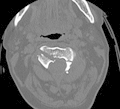
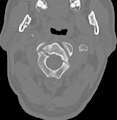
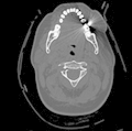
Of the atypical fractures, 12 fractures (43% of the atypical fractures in the series) involved the C2 body on one side and a fracture of the contralateral posterior element. In these cases, the fracture pattern was through one side of the vertebral body obliquely and another fracture through either the pars (n = 10) or lamina (n = 2). The remaining 16 (57% of the atypical group) atypical fractures showed a coronally orientated fracture through the body of C2 anterior to the pars interarticularis, which sometimes left the ring of the axis intact. The possible fracture patterns and nomenclature are illustrated in Table 3.
Table 3. Fracture classification.
| Fracture type | Description | Sagittal view | Axial view |
|---|---|---|---|
| Coronally orientated (type 1) | Coronally orientated fracture line through the body of C2, which may or may not leave the ring of the axis intact |

|

|
| Unilateral oblique body fracture with contralateral pars fracture (type 2a) | Unilateral oblique fracture through the C2 body extending into the canal, with contralateral fracture of the pars interarticularis |

|

|
| Unilateral oblique body fracture with contralateral lamina fracture (type 2b) | Unilateral oblique fracture through the C2 body with contralateral fracture of the lamina |

|

|
| Typical hangman's fracture | Bilateral fracture through the pars interarticularis of C2 with or without forward listhesis of the C2 body |

|

|
Management
Six patients with typical fractures were managed by immobilization in a halo and 5 with hard cervical collars. Twenty-one patients with atypical fractures were managed with halos and 6 with collars. Two patients developed pin site infections while in a halo and required a change of management to a collar for the remainder of treatment (Table 4).
Table 4. Summary of management.
| Levine-Edwards type | Typical | Atypical | Total (n) | ||
|---|---|---|---|---|---|
| n | Treatment (n) | n | Treatment (n) | ||
| 1 (n = 22) | 8 | Halo (3) Collar (4) Surgery (1) |
14 | Halo (11) Collar (2) Surgery (failed halo treatment) (1) |
Halo (14) Collar (6) Surgery (2) |
| 2 (n = 17) | 4 | Halo (3) Collar (1) |
13 | Halo (9) Collar (4) |
Halo (12) Collar (5) |
| 2a (n = 2) | 1 | Surgery (1) | 1 | Halo (1) | Halo (1) Surgery (1) |
| 3 (n = 0) | – | – | – | – | – |
| Total (n = 41) | 13 | Halo (6) Collar (5) Surgery (2) |
28 | Halo (21) Collar (6) Surgery (1) |
Halo (27) Collar (11) Surgery (3) |
Halo immobilization was used more frequently than collars to treat both type 1 and type 2 fracture types. Collars were used if a patient was elderly or deemed unable to tolerate a halo or if collar use was the surgeon's preferred management strategy. The mean age of patients treated with a collar was 67.6 (range 33 to 85). The mean age of patients managed with halos was 54.9 (range 19 to 76).
Thirteen patients (62% of type 1 fractures) with type 1 fractures were managed with halo immobilization, 6 (28.6% of type 1 fractures) were managed with collars, and 1 (4.8% of type 1 fractures) required surgery. Twelve (71%) type 2 fractures were managed in halos and 5 (29%) in collars. One type 2a fracture was managed in a halo and 2 (66%) required surgery (Table 4).
The mean duration of management using a halo was 117.5 days (range 70 to 186). The mean duration of management using a cervical collar was 124.1 days (range 84 to 170).
Three patients underwent surgical fixation. The first patient had an asymmetrical atypical hangman's fracture (vertebral body and pars type 2a). On initial CT scan, the fracture was a Levine type 1. The subject was initially treated with a halo; follow-up imaging showed a progressive C2–C3 subluxation. A C1–C3 posterior fixation was performed 8 days after the initial presentation. The remaining 2 patients had surgery due to associated cervical fractures. One patient had a typical hangman's fracture with a burst fracture of the body of C3 with retropulsion. She was managed nonoperatively in a halo. Follow-up imaging at 3 months revealed nonunion at C3, and the patient underwent a C2–C4 posterior fixation. The final patient had a typical hangman's fracture with a comminuted C2 body fracture including an anteriorly displaced teardrop fragment. He was initially managed in a halo. At 3 months, there were no signs of fusion of the C2 body fracture, and he therefore underwent a C1–C3 posterior fixation.
Outcomes
The mean duration of follow-up was 9.4 months (range 3 to 25 months).
There were 5 (18%) cases of infection occurring at the pin sites, which led to the discontinuation of halo management in 2 cases. The management strategy was changed to collars in these patients.
Pain was assessed according to the visual analog scale at last follow-up. Five patients in total (12.2% of all hangman's fractures) were still experiencing moderate to severe pain at that time. Pain and stiffness were more common with atypical fractures, but this finding did not reach statistical significance, with 4 in the atypical group (14% of atypical fractures) reporting moderate or severe pain compared with 1 patient in the typical group (p > 0.999). Stiffness was reported in 11 patients with atypical fractures (39% of all atypical fractures) compared with 4 in the typical group (31% of all typical fractures; p = 0.734).
All fractures in our series apart from the three patients who underwent surgical fixation discussed previously had documented bony union on follow-up CT or plain radiographs, which were typically performed at 4 to 6 weeks, after removal of the halo or collar and at final follow-up. The criteria for union were obliteration of the fracture line with cortical continuity or bridging of fracture by callus, trabeculae, or bone.
Fracture union rates in grade I and II hangman's fractures were not significantly different between halo immobilization and collars (p > 0.999).
Five patients (12%) had focal neurologic signs on admission. Four of these patients recovered completely. The remaining patient was left with a permanent left-sided hemiparesis, which was likely the result of a second cervical spine injury (an unstable and retropulsed comminuted C3 fracture).
Three patients underwent surgical fixation following management with a halo, 1 for developing a progressive listhesis of C2–C3 and 2 for nonunion of associated cervical fractures. Bony union was otherwise achieved in all patients managed nonoperatively.
Discussion
This retrospective analysis aimed at analyzing the patterns of hangman's fractures and auditing our treatment methods. We sought to establish whether treatment failures were more likely with cervical collars compared with halo immobilization.
Mechanism of Injury and Presentation
Hangman's fractures are usually caused by RTAs, falls, and occasionally diving or athletic accidents.2 A blow to the face results in hyperextension with axial loading of the cervical spine, leading to the anterior ligaments becoming stretched as well as compression of the posterior aspect of the bony facets. This force results in fracture of the pars interarticularis and separation of the body from the neural arch.2 3 12 13 A variety of mechanisms for the production of coronally orientated C2 body fractures were described by Benzel including extension or hyperextension with axial load, flexion with axial load, and flexion with distraction.16
In other series, RTAs are the commonest cause, accounting for 50 to 79% of injuries.6 12 13 14 15 Falls were the main mechanism of hangman's fractures (both typical and atypical), accounting for 56% of injuries, whereas RTAs accounted for 33%.
The mean age of patients in this study (59 years) was higher than reported by other authors (31.2 to 40 years),3 5 6 13 14 15 16 which may explain the higher incidence of falls as the mechanism of injury.
The associated injuries seen were similar to those reported in other studies. Trauma to the head and face was reported in 11 to 79% of patients, which compares with 19% in this study.3 12 14 Secondary cervical fractures in association with hangman's fractures are reported in the literature to range between 8 and 32% with odontoid fractures described in 5 to 6%.3 13 In our series secondary cervical fractures were seen in 14 patients (34%), and 7 (17%) of these were odontoid peg fractures. Fractures of the thoracic and lumbar spine are less frequent than associated cervical spine injuries, occurring in 0 to 11% of patients in other case series and in 2% in this study.3 12 13 17
In our series, 5 patients (12%) had focal neurologic signs on admission. Four of these patients recovered completely. The 1 patient (3%) who did not recover completely had a burst fracture of C3 with a retropulsed fragment and intramedullary signal change, in addition to a bilateral C2 pars fracture. These rates of neurologic deficit are similar to those observed by others. The quoted incidence of neurologic signs on admission is 3 to 13% and a permanent deficit occurs in 0 to 5% of patients.3 12 14 18 This low incidence of neurologic deficit occurs due to the body of the axis moving forward and enlarging the spinal canal and intervertebral foramen.14 15 Atypical fractures may compress the spinal cord against the posterior cortex of the C2 body, causing a higher rate of neurologic deficit.15 In our series, 3 of the 4 patients with associated neurologic deficits at presentation had atypical fractures.
Radiology
The classical radiologic appearance of a true hangman's fracture is a bilateral fracture of the pars interarticularis (isthmus) with or without forward listhesis of the C2 body. In modern use, the term has come to include alternatives to the classic fracture pattern. Due to the rotational component associated with this type of extension injury, bilateral fractures of the pars are rarely symmetrical.12
Our preferred terminology is typical and atypical hangman's fracture. Atypical hangman's fracture patterns include a coronally orientated fracture line through the body of C2 and oblique fractures through one side of the C2 body with contralateral fracture of posterior elements of the axis ring (either the pars or the lamina). Our recommended fracture nomenclature can be seen in Table 3.
Several grading systems have been proposed previously; the most widely accepted is the Effendi (modified by Levine) classification. In our view, this system is also applicable to atypical hangman's fractures.12 13
The distribution of Effendi fracture type varies between studies (Table 5). Our high incidence of atypical fractures (68%) compared with other studies (18 to 54%)6 8 15 may be due to the use of CT imaging, which gives a more detailed and clear picture of the anatomy, or due to the fact that older studies have looked at typical hangman's fractures only.13 14 There was no standard protocol for vertebral artery imaging for this cohort of patients in our institution.
Table 5. Summary of literature.
| Type 1 (%) | Type 2 (%) | Type 2a (%) | Type 3 (%) | Atypical (%) | Treatment, n (%) | |
|---|---|---|---|---|---|---|
| Effendi et al (1981)12 (n = 131) | 85 (65%) | 37 (28%) | – | 9 (6%) | – | Brace, 80 (65%) Surgery, 42 (34%) |
| Levine and Edwards (1985)13 (n = 52) | 15 (29%) | 29 (56%) | 3 (6%) | 5 (9%) | – | Collar, 10 (19%) Halo, 32 (62%) Surgery, 3 (6%) |
| Burke and Harris (1989)8 (n = 62) | 13 (21%) | 35 (56%) | – | 3 (5%) | 11 (18%) | – |
| Green et al (1997)4 (n = 74) | 53 (72%) | 20 (27%) | – | 1 (1%) | – | Halo, 56 (76%) SOMI, 6 (8%) Collar, 3 (4%) Surgery, 7 (9%) |
| Müller et al (2000)39 (n = 39) | 10 (26%) | 29 (74%) | – | – | – | Halo, 18 (46%) Collar, 12 (31%) Minerva, 1 (3%) Surgery, 8 (21%) |
| Ramieri et al (2010)41 (n = 16) | 11 (69%) | 5 (31%) | – | – | – | Halo, 11 (69%) SOMI, 5 (31%) |
| Samaha et al (2010)6 (n = 24) | – | – | – | – | 13 (54%) | Minerva, 15 (63%) Surgery, 9 (38%) |
| Vaccaro et al (2002)30 (n = 31) | – | 27 (87%) | 4 (13%) | – | – | Traction + halo, 31 (100%) |
| Moon et al (2002)42 (n = 42) | – | – | – | – | – | Cervical orthosis, 20 (48%) Surgery, 22 (52%) |
| Al-Mahfoudh et al, this study (n = 41) | 21 (51%) | 17 (41%) | 3 (7%) | 0 (0%) | 28 (68%) | Halo, 27 (66%) Collar, 11 (27%) Surgery, 3 (7%) |
The incidence of vertebral artery injury (VAI) associated with blunt cervical spine injury ranges from 10.5 to 88%.19 In a recent meta-analyses, a statistically significant association between blunt cerebrovascular injury (BCVI) and two screening criteria, cervical spine injury and thoracic injury, was established. Patients with cervical spine injury had a fivefold greater likelihood of BCVI compared with those patients without cervical spine injury.20
A predominance of certain fracture patterns in the cervical spine in patients with VAIs has been reported. These include transverse foramen fractures (8 to 37%) and C1 to C3 body fractures (31 to 36%).21 22 In one study specifically correlating C2 fractures to VAI, 17.8% of patients with C2 fractures were found to have VAI. There was a correlation of VAI with specific fracture patterns, including traumatic spondylolisthesis of axis and a greater degree of angulation, in addition to C2 fractures with comminuted fractures involving the foramen transversarium.19
Conventional catheter cerebral angiography is the gold standard for screening but carries risk for iatrogenic injury, stroke, and death. Complication rates with catheter angiography have been reported up to 4%.22 Sixteen-slice computed tomography angiography (CTA) is now an established alternative; it is noninvasive and it has become a routine screening tool for BCVI. A major advantage of utilizing screening CTA in the emergency setting (relative to digital subtraction angiogram) is the reduced time to diagnosis with BCVI, which has been reported to be up to 12-fold, with a consequent reduction in stroke rate by up to fourfold.23
Several studies and guidelines indicate that a 16-slice CTA is an acceptable modality based on the data from several reports.20 22 24 25 Historically, however, 16-slice magnetic resonance angiography and CTA scanners have poor sensitivity compared with conventional angiography.22
Heparin has been associated with better overall neurologic outcome in patients with VAI.26 27 Specifically for symptomatic cases, antithrombotic therapy for most asymptomatic VAIs is still controversial, and there is a lack of class I evidence to support any strong guidelines for treatment.22
Overall, the literature points to a higher incidence of VAI in cervical spine trauma, specifically with fractures involving the transverse foramen and high cervical fractures. In addition, there is some evidence to support improved outcomes in patients with VAI who receive anticoagulation. Altogether, this evidence probably supports the view that all patients with hangman's fractures should undergo imaging to exclude VAIs. Sixteen-slice CTA is probably the modality of choice, although angiography is still considered the gold standard.
Management
Most authors agree that the majority of hangman's fractures can be managed nonoperatively.3 4 18 28 29 30 31 32 33 34 The indications for primary surgical management differ between authors and are usually applicable only to unstable fractures; however, definitions of stability vary. Effendi type 1 fractures are usually considered stable and type 3 fractures are considered unstable, but there is no consensus regarding the stability of type 2 or type 2a fractures.10 17 35 The instability of a type 2 fracture depends on the disk or ligamentous damage, which can be evaluated by anterior movement and angulation of the dens. Angulation of greater than 20 degrees signifies damage to the posterior ligaments and C2–C3 disk space.36
Nonoperative management of hangman's fractures involves external immobilization with variations of cervical collar and halo ring orthosis.3 37
Immobilization with cervical collars or cervicothoracic orthosis has been used to successfully treat fractures with up to 6-mm displacement. They provide a more comfortable form of treatment and do not put the patient at risk of the complications associated with a halo. Collar management is indicated in type 1 and some stable type 2 fractures.35 38 39
Halo orthosis provides the most rigid form of nonsurgical immobilization and is the most commonly used form of nonoperative therapy. Halos are used in fractures where the level of instability warrants the increased discomfort and risk of complications but also allows mechanical traction to be externally applied for reduction.40 Complications of halo management are reported to occur in 12 to 36% of cases and can include pin site infection, subdural abscesses, increased risk of falls, and respiratory problems.3 41 Displaced (Effendi type 2) fractures can be reduced with 5 to 15 lbs prior to application of the halo vest and jacket.30 Management with halo orthosis has been reported to produce good results in type I, IIA, and IIB fractures.35 41 In a systematic review by Li et al, 20 of the included publications (62.5%) advocated conservative management for all types of hangman's fractures.35 Of the remaining 12 publications in this review, 11 suggested that conservative treatment was suitable only for some stable fractures. The method of conservative treatment in most articles in this systematic review was tong traction used in the earliest stage, with halo immobilization strongly recommended when Levine-Edwards type IIa and III fractures were managed conservatively. Nonrigid external fixation was only used in some type I and Levine-Edwards type II fractures, often supplemented with rigid immobilization.35 41
In our study, the union rates in low-grade hangman's fractures treated with halos and those treated with collars were comparable. As such and given the higher complications associated with halo immobilization, collars may be a more favorable management option especially when a halo is not tolerated.
In this study, nonoperative management was successful in all but 3 (7%) patients treated with a halo, which was due to nonunion of another cervical fracture in 2 and progression of C2–C3 subluxation in the third patient. This result is comparable to other studies that have emphasized nonoperative management.3 5 Nonoperative management fails in around 5% of total cases but may fail in up to 50% of type 2a and type 3 fractures.3 4 14 35 Therefore, close follow-up is required with frequent radiologic assessment.
Surgical management of hangman's fractures has historically been reserved for Effendi type 3 fractures, patients with nonunion after 3 months in a halo, and some patients with second fractures of the cervical spine.3 4 12 13 14 38 The consensus appears to be early surgical intervention for patients with significant displacement and angulation at C2–C3.6 36 42 43
Surgical intervention is unnecessary in the majority of Effendi type 1 and type 2 fractures, unless nonoperative management fails. Type 2a and 3 fractures might be candidates for primary surgical stabilization and fusion.34 If surgery is deemed necessary, the posterior approach allows direct access to the C2–C3 facet joints for reduction (necessary in type 3 fractures) and correction of local kyphosis, but requires considerable muscle dissection followed by C1–C3 or C2–C3 instrumented fusion.7 35 44 The anterolateral approach with autologous bone graft allows for a C2–C3 diskectomy and fusion if traumatic disk herniation compromises the spinal cord. Plate and screws may be used.45 46 A combined approach may be indicated in highly unstable fractures that require posterior reduction and fixation with additional anterior disk removal and C2–C3 fusion. Direct pars fixation with bilateral C2 screws conducted from a posterior approach has been discussed as a motion-preserving alternative surgical method for hangman's fractures that have limited disk and ligament injury.7 47 48 49 50 In the systematic review, the fusion rate of type 3 fractures was similar for those treated posteriorly (39%) and anteriorly (43%).35
Atypical Hangman's Fractures
Atypical hangman's fractures, which are asymmetrical or involve the C2 body, were first noted by Effendi et al in 1981 but have not been included in any traumatic spondylolisthesis classification system (Effendi, Francis, Levine, Roy-Camille) and are not included in several large case series.3 4 5 12 13 14 30 41
Coronally oriented vertical fractures of the C2 body were described as type 1 C2 body fractures by Benzel.16 The unilateral oblique body fractures with contralateral pars or lamina fracture we described have not been included in any classification system.
There are no separate grading systems for atypical hangman's fractures although both Starr and Eismont and Samaha et al commented that typical traumatic spondylolisthesis classifications (Levine and Roy-Camille, respectively) can be applied.6 13 15
Symmetry of the fracture may not affect outcomes and therefore it has been suggested that treatment strategies should be the same.6 13 15 Nonoperative treatment has also been advocated for most cases of vertical C2 body fractures.51 Others have suggested that atypical fractures may have greater instability and that obtaining closed reduction is difficult due to interposition of soft tissue between the atypical fragments.8
In our series, 1 of 28 (3%) atypical hangman's fractures required surgical fixation. Therefore, conducting a primarily nonoperative approach to management similar to typical hangman's fracture seems appropriate. Extrapolating meaningful clinical guidelines for a small subset of patients remains difficult, however. We believe a unified nomenclature system will be useful to identify which fracture patterns in the atypical group are more likely to fail nonoperative management.
We concede the weaknesses associated with a single-center retrospective study. The treatment modality of collar or halo fixation for low-grade fractures may have been influenced by individual surgeon choice, which may well represent the pragmatic state with managing these fractures. We also acknowledge the low number of patients in the different treatment arms and therefore note that firm conclusions cannot be made based on this study alone.
Conclusions
We classify the different fracture patterns of atypical hangman's fractures and propose applying the same grading systems as for typical hangman's fractures. A unified nomenclature system for fracture subtypes will aid in developing evidence-based management strategies. Nonoperative management of these fractures is effective in most cases of low-grade fracture with surgical management being reserved for higher-grade variants and those that show nonunion or a progression after nonoperative management. Cervical hard collars may be an appropriate management alternative for low-grade hangman's fractures with a lower complication rate than halo immobilization. Neurologic deficit is rare in all types of hangman's fracture.
Footnotes
Disclosures Rafid Al-Mahfoudh, none Christopher Beagrie, none Ele Woolley, none Rasheed Zakaria, none Mark Radon, none Simon Clark, none Robin Pillay, none Martin Wilby, none
References
- 1.Wood-Jones F. The ideal lesion produced by judicial hanging. Lancet. 1913;181(4462):53. [Google Scholar]
- 2.Schneider R C, Livingston K E, Cave A J, Hamilton G. “Hangman's fracture” of the cervical spine. J Neurosurg. 1965;22(2):141–154. doi: 10.3171/jns.1965.22.2.0141. [DOI] [PubMed] [Google Scholar]
- 3.Coric D, Wilson J A, Kelly D L Jr. Treatment of traumatic spondylolisthesis of the axis with nonrigid immobilization: a review of 64 cases. J Neurosurg. 1996;85(4):550–554. doi: 10.3171/jns.1996.85.4.0550. [DOI] [PubMed] [Google Scholar]
- 4.Greene K A, Dickman C A, Marciano F F, Drabier J B, Hadley M N, Sonntag V K. Acute axis fractures. Analysis of management and outcome in 340 consecutive cases. Spine (Phila Pa 1976) 1997;22(16):1843–1852. doi: 10.1097/00007632-199708150-00009. [DOI] [PubMed] [Google Scholar]
- 5.Ferro F P, Borgo G D, Letaif O B, Cristante A F, Marcon R M, Lutaka A S. Traumatic spondylolisthesis of the axis: epidemiology, management and outcome. Acta Ortop Bras. 2012;20(2):84–87. doi: 10.1590/S1413-78522012000200005. [DOI] [PMC free article] [PubMed] [Google Scholar]
- 6.Samaha C, Lazennec J Y, Laporte C, Saillant G. Hangman's fracture: the relationship between asymmetry and instability. J Bone Joint Surg Br. 2000;82(7):1046–1052. doi: 10.1302/0301-620x.82b7.10408. [DOI] [PubMed] [Google Scholar]
- 7.Suchomel P, Hradil J. Berlin, Germany: Springer; 2011. Fractures of the ring of axis (Hangman type fractures) pp. 179–196. [Google Scholar]
- 8.Burke J T, Harris J H Jr. Acute injuries of the axis vertebra. Skeletal Radiol. 1989;18(5):335–346. doi: 10.1007/BF00361422. [DOI] [PubMed] [Google Scholar]
- 9.Hadley M N, Dickman C A, Browner C M, Sonntag V K. Acute axis fractures: a review of 229 cases. J Neurosurg. 1989;71(5 Pt 1):642–647. doi: 10.3171/jns.1989.71.5.0642. [DOI] [PubMed] [Google Scholar]
- 10.White A A III, Panjabi M M. The clinical biomechanics of the occipitoatlantoaxial complex. Orthop Clin North Am. 1978;9(4):867–878. [PubMed] [Google Scholar]
- 11.Ryken T C, Hadley M N, Aarabi B. et al. Management of isolated fractures of the axis in adults. Neurosurgery. 2013;72 02:132–150. doi: 10.1227/NEU.0b013e318276ee40. [DOI] [PubMed] [Google Scholar]
- 12.Effendi B, Roy D, Cornish B, Dussault R G, Laurin C A. Fractures of the ring of the axis. A classification based on the analysis of 131 cases. J Bone Joint Surg Br. 1981;63-B(3):319–327. doi: 10.1302/0301-620X.63B3.7263741. [DOI] [PubMed] [Google Scholar]
- 13.Levine A M, Edwards C C. The management of traumatic spondylolisthesis of the axis. J Bone Joint Surg Am. 1985;67(2):217–226. [PubMed] [Google Scholar]
- 14.Francis W R, Fielding J W, Hawkins R J, Pepin J, Hensinger R. Traumatic spondylolisthesis of the axis. J Bone Joint Surg Br. 1981;63-B(3):313–318. doi: 10.1302/0301-620X.63B3.7263740. [DOI] [PubMed] [Google Scholar]
- 15.Starr J K, Eismont F J. Atypical hangman's fractures. Spine (Phila Pa 1976) 1993;18(14):1954–1957. doi: 10.1097/00007632-199310001-00005. [DOI] [PubMed] [Google Scholar]
- 16.Benzel E C. Conservative treatment of neural arch fractures of the axis: computed tomography scan and X-ray study on consolidation time. World Neurosurg. 2011;75(2):229–230. doi: 10.1016/j.wneu.2010.09.036. [DOI] [PubMed] [Google Scholar]
- 17.Govender S, Charles R W. Traumatic spondylolisthesis of the axis. Injury. 1987;18(5):333–335. doi: 10.1016/0020-1383(87)90055-6. [DOI] [PubMed] [Google Scholar]
- 18.Fielding J W, Francis W R Jr, Hawkins R J, Pepin J, Hensinger R. Traumatic spondylolisthesis of the axis. Clin Orthop Relat Res. 1989;(239):47–52. [PubMed] [Google Scholar]
- 19.Ding T, Maltenfort M, Yang H. et al. Correlation of C2 fractures and vertebral artery injury. Spine (Phila Pa 1976) 2010;35(12):E520–E524. doi: 10.1097/BRS.0b013e3181cd98b6. [DOI] [PubMed] [Google Scholar]
- 20.Franz R W, Willette P A, Wood M J, Wright M L, Hartman J F. A systematic review and meta-analysis of diagnostic screening criteria for blunt cerebrovascular injuries. J Am Coll Surg. 2012;214(3):313–327. doi: 10.1016/j.jamcollsurg.2011.11.012. [DOI] [PubMed] [Google Scholar]
- 21.Cothren C C, Moore E E, Ray C E Jr, Johnson J L, Moore J B, Burch J M. Cervical spine fracture patterns mandating screening to rule out blunt cerebrovascular injury. Surgery. 2007;141(1):76–82. doi: 10.1016/j.surg.2006.04.005. [DOI] [PubMed] [Google Scholar]
- 22.Fassett D R, Dailey A T, Vaccaro A R. Vertebral artery injuries associated with cervical spine injuries: a review of the literature. J Spinal Disord Tech. 2008;21(4):252–258. doi: 10.1097/BSD.0b013e3180cab162. [DOI] [PubMed] [Google Scholar]
- 23.Eastman A L Muraliraj V Sperry J L Minei J P CTA-based screening reduces time to diagnosis and stroke rate in blunt cervical vascular injury J Trauma 2009673551–556., discussion 555–556 [DOI] [PubMed] [Google Scholar]
- 24.Payabvash S, McKinney A M, McKinney Z J, Palmer C S, Truwit C L. Screening and detection of blunt vertebral artery injury in patients with upper cervical fractures: the role of cervical CT and CT angiography. Eur J Radiol. 2014;83(3):571–577. doi: 10.1016/j.ejrad.2013.11.020. [DOI] [PubMed] [Google Scholar]
- 25.Biffl W L Egglin T Benedetto B Gibbs F Cioffi W G Sixteen-slice computed tomographic angiography is a reliable noninvasive screening test for clinically significant blunt cerebrovascular injuries J Trauma 2006604745–751., discussion 751–752 [DOI] [PubMed] [Google Scholar]
- 26.Miller P R Fabian T C Bee T K et al. Blunt cerebrovascular injuries: diagnosis and treatment J Trauma 2001512279–285., discussion 285–286 [DOI] [PubMed] [Google Scholar]
- 27.Miller P R Fabian T C Croce M A et al. Prospective screening for blunt cerebrovascular injuries: analysis of diagnostic modalities and outcomes Ann Surg 20022363386–393., discussion 393–395 [DOI] [PMC free article] [PubMed] [Google Scholar]
- 28.Hadley M N, Browner C, Sonntag V K. Axis fractures: a comprehensive review of management and treatment in 107 cases. Neurosurgery. 1985;17(2):281–290. doi: 10.1227/00006123-198508000-00006. [DOI] [PubMed] [Google Scholar]
- 29.Seljeskog E L, Chou S N. Spectrum of the hangman's fracture. J Neurosurg. 1976;45(1):3–8. doi: 10.3171/jns.1976.45.1.0003. [DOI] [PubMed] [Google Scholar]
- 30.Vaccaro A R, Madigan L, Bauerle W B, Blescia A, Cotler J M. Early halo immobilization of displaced traumatic spondylolisthesis of the axis. Spine (Phila Pa 1976) 2002;27(20):2229–2233. doi: 10.1097/00007632-200210150-00009. [DOI] [PubMed] [Google Scholar]
- 31.Bucholz R D, Cheung K C. Halo vest versus spinal fusion for cervical injury: evidence from an outcome study. J Neurosurg. 1989;70(6):884–892. doi: 10.3171/jns.1989.70.6.0884. [DOI] [PubMed] [Google Scholar]
- 32.Elliott J M Jr, Rogers L F, Wissinger J P, Lee J F. The hangman's fracture. Fractures of the neural arch of the axis. Radiology. 1972;104(2):303–307. doi: 10.1148/104.2.303. [DOI] [PubMed] [Google Scholar]
- 33.Pepin J W, Hawkins R J. Traumatic spondylolisthesis of the axis: hangman's fracture. Clin Orthop Relat Res. 1981;(157):133–138. [PubMed] [Google Scholar]
- 34.Sherk H H, Howard T. Clinical and pathologic correlations in traumatic spondylolisthesis of the axis. Clin Orthop Relat Res. 1983;(174):122–126. [PubMed] [Google Scholar]
- 35.Li X F, Dai L Y, Lu H, Chen X D. A systematic review of the management of hangman's fractures. Eur Spine J. 2006;15(3):257–269. doi: 10.1007/s00586-005-0918-2. [DOI] [PMC free article] [PubMed] [Google Scholar]
- 36.Marton E, Billeci D, Carteri A. Therapeutic indications in upper cervical spine instability. Considerations on 58 cases. J Neurosurg Sci. 2000;44(4):192–202. [PubMed] [Google Scholar]
- 37.Cosan T E, Tel E, Arslantas A, Vural M, Guner A I. Indications of Philadelphia collar in the treatment of upper cervical injuries. Eur J Emerg Med. 2001;8(1):33–37. doi: 10.1097/00063110-200103000-00007. [DOI] [PubMed] [Google Scholar]
- 38.Ryken T C, Hadley M N, Aarabi B. et al. Management of isolated fractures of the axis in adults. Neurosurgery. 2013;72(3) 02:132–150. doi: 10.1227/NEU.0b013e318276ee40. [DOI] [PubMed] [Google Scholar]
- 39.Müller E J, Wick M, Muhr G. Traumatic spondylolisthesis of the axis: treatment rationale based on the stability of the different fracture types. Eur Spine J. 2000;9(2):123–128. doi: 10.1007/s005860050222. [DOI] [PMC free article] [PubMed] [Google Scholar]
- 40.Garfin S R, Rothman R H. Philadelphia, PA: JB Lippincott; 1983. Traumatic spondylolisthesis of the axis (Hangman's fracture) pp. 223–232. [Google Scholar]
- 41.Ramieri A, Domenicucci M, Landi A, Rastelli E, Raco A. Conservative treatment of neural arch fractures of the axis: computed tomography scan and X-ray study on consolidation time. World Neurosurg. 2011;75(2):314–319. doi: 10.1016/j.wneu.2010.09.004. [DOI] [PubMed] [Google Scholar]
- 42.Moon M S Moon J L Moon Y W Sun D H Choi W T Traumatic spondylolisthesis of the axis: 42 cases Bull Hosp Jt Dis 2001. –2002;60261–66. [PubMed] [Google Scholar]
- 43.Suchomel P, Hradil J, Barsa P. et al. [Surgical treatment of fracture of the ring of axis - “hangman's fracture”] Acta Chir Orthop Traumatol Cech. 2006;73(5):321–328. [PubMed] [Google Scholar]
- 44.Salmon J H. Fractures of the second cervical vertebra: Internal fixation by interlaminar wiring. Neurosurgery. 1977;1(2):125–127. doi: 10.1227/00006123-197709000-00007. [DOI] [PubMed] [Google Scholar]
- 45.Ying Z, Wen Y, Xinwei W. et al. Anterior cervical discectomy and fusion for unstable traumatic spondylolisthesis of the axis. Spine (Phila Pa 1976) 2008;33(3):255–258. doi: 10.1097/BRS.0b013e31816233d0. [DOI] [PubMed] [Google Scholar]
- 46.Xu H, Zhao J, Yuan J, Wang C. Anterior discectomy and fusion with internal fixation for unstable hangman's fracture. Int Orthop. 2010;34(1):85–88. doi: 10.1007/s00264-008-0658-0. [DOI] [PMC free article] [PubMed] [Google Scholar]
- 47.ElMiligui Y, Koptan W, Emran I. Transpedicular screw fixation for type II Hangman's fracture: a motion preserving procedure. Eur Spine J. 2010;19(8):1299–1305. doi: 10.1007/s00586-010-1401-2. [DOI] [PMC free article] [PubMed] [Google Scholar]
- 48.Boullosa J L Colli B O Carlotti C G Jr Tanaka K dos Santos M B Surgical management of axis' traumatic spondylolisthesis (hangman's fracture) Arq Neuropsiquiatr 200462(3B):821–826. [DOI] [PubMed] [Google Scholar]
- 49.Dalbayrak S, Yilmaz M, Firidin M, Naderi S. Traumatic spondylolisthesis of the axis treated with direct C2 pars screw. Turk Neurosurg. 2009;19(2):163–167. [PubMed] [Google Scholar]
- 50.Borne G M, Bedou G L, Pinaudeau M. Treatment of pedicular fractures of the axis. A clinical study and screw fixation technique. J Neurosurg. 1984;60(1):88–93. doi: 10.3171/jns.1984.60.1.0088. [DOI] [PubMed] [Google Scholar]
- 51.German J W Hart B L Benzel E C Nonoperative management of vertical C2 body fractures Neurosurgery 2005563516–521., discussion 516–521 [DOI] [PubMed] [Google Scholar]


