

Abstract
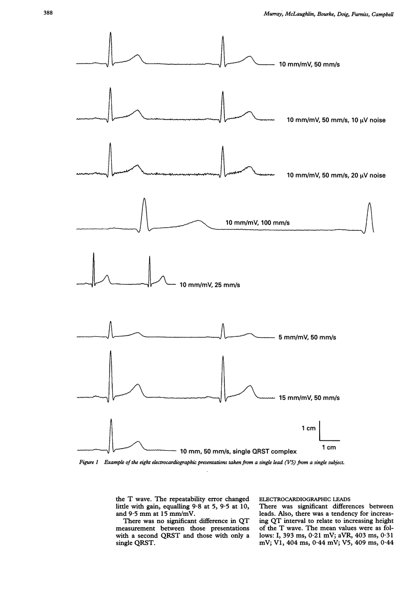
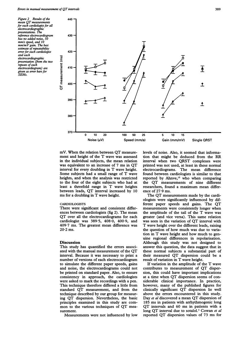
OBJECTIVE--To quantify the errors associated with manual measurement of QT intervals and to determine the source of the errors. DESIGN--A randomised study of QT measurement by four cardiologists of electrocardiograms plotted on paper in presentations with different noise levels, paper speeds, amplifier gains, and with and without a second QRST complex to indicate the RR interval. SUBJECTS--Four electrocardiograph leads (I, aVR, V1, V5) recorded in eight healthy people relaxing in a semirecumbent position. MAIN OUTCOME MEASURES--Manual measurement of QT interval in 512 electrocardiograms (eight subjects x four leads x eight presentations x two repeats) by each of four cardiologists. RESULTS--QT intervals measured were significantly longer with greater amplifier gain: by 8 ms for a doubling of gain (p < 0.005), equivalent to a doubling of T wave height. QT intervals measured were significantly longer at slower paper speeds: by 11 ms when paper speed was reduced from 100 to 50 mm/s (p < 0.001) and by 16 ms when speed was further reduced from 50 to 25 mm/s (p < 0.001). Neither the presence of noise nor the presence of a second QRST complex altered the mean QT measurements. There were consistent differences in the measurements between cardiologists, amounting to a maximum mean difference of 20 ms. CONCLUSIONS--Manual measurement of QT interval is significantly affected by the paper speed used to plot the electrocardiogram and by electrocardiogram gain, and hence also T wave amplitude. Manual QT measurement also differed consistently with different cardiologists.
Full text
PDF
Selected References
These references are in PubMed. This may not be the complete list of references from this article.
- Ahnve S. Errors in the visual determination of corrected QT (QTc) interval during acute myocardial infarction. J Am Coll Cardiol. 1985 Mar;5(3):699–702. doi: 10.1016/s0735-1097(85)80396-x. [DOI] [PubMed] [Google Scholar]
- Campbell R. W., Gardiner P., Amos P. A., Chadwick D., Jordan R. S. Measurement of the QT interval. Eur Heart J. 1985 Nov;6 (Suppl 500):81–83. doi: 10.1093/eurheartj/6.suppl_d.81. [DOI] [PubMed] [Google Scholar]
- Cowan J. C., Hilton C. J., Griffiths C. J., Tansuphaswadikul S., Bourke J. P., Murray A., Campbell R. W. Sequence of epicardial repolarisation and configuration of the T wave. Br Heart J. 1988 Nov;60(5):424–433. doi: 10.1136/hrt.60.5.424. [DOI] [PMC free article] [PubMed] [Google Scholar]
- Day C. P., McComb J. M., Campbell R. W. QT dispersion in sinus beats and ventricular extrasystoles in normal hearts. Br Heart J. 1992 Jan;67(1):39–41. doi: 10.1136/hrt.67.1.39. [DOI] [PMC free article] [PubMed] [Google Scholar]
- Day C. P., McComb J. M., Campbell R. W. QT dispersion: an indication of arrhythmia risk in patients with long QT intervals. Br Heart J. 1990 Jun;63(6):342–344. doi: 10.1136/hrt.63.6.342. [DOI] [PMC free article] [PubMed] [Google Scholar]
- Franz M. R., Bargheer K., Rafflenbeul W., Haverich A., Lichtlen P. R. Monophasic action potential mapping in human subjects with normal electrocardiograms: direct evidence for the genesis of the T wave. Circulation. 1987 Feb;75(2):379–386. doi: 10.1161/01.cir.75.2.379. [DOI] [PubMed] [Google Scholar]
- Hii J. T., Wyse D. G., Gillis A. M., Duff H. J., Solylo M. A., Mitchell L. B. Precordial QT interval dispersion as a marker of torsade de pointes. Disparate effects of class Ia antiarrhythmic drugs and amiodarone. Circulation. 1992 Nov;86(5):1376–1382. doi: 10.1161/01.cir.86.5.1376. [DOI] [PubMed] [Google Scholar]
- Laguna P., Thakor N. V., Caminal P., Jané R., Yoon H. R., Bayés de Luna A., Marti V., Guindo J. New algorithm for QT interval analysis in 24-hour Holter ECG: performance and applications. Med Biol Eng Comput. 1990 Jan;28(1):67–73. doi: 10.1007/BF02441680. [DOI] [PubMed] [Google Scholar]
- Merri M., Benhorin J., Alberti M., Locati E., Moss A. J. Electrocardiographic quantitation of ventricular repolarization. Circulation. 1989 Nov;80(5):1301–1308. doi: 10.1161/01.cir.80.5.1301. [DOI] [PubMed] [Google Scholar]
- Mirvis D. M. Spatial variation of QT intervals in normal persons and patients with acute myocardial infarction. J Am Coll Cardiol. 1985 Mar;5(3):625–631. doi: 10.1016/s0735-1097(85)80387-9. [DOI] [PubMed] [Google Scholar]
- O'Donnell J., Lovelace D. E., Knoebel S. B., McHenry P. L. Behavior of the terminal T wave during exercise in normal subjects, patients with symptomatic coronary artery disease and apparently healthy subjects with abnormal ST segment depression. J Am Coll Cardiol. 1985 Jan;5(1):78–84. doi: 10.1016/s0735-1097(85)80087-5. [DOI] [PubMed] [Google Scholar]
- Schwartz P. J., Wolf S. QT interval prolongation as predictor of sudden death in patients with myocardial infarction. Circulation. 1978 Jun;57(6):1074–1077. doi: 10.1161/01.cir.57.6.1074. [DOI] [PubMed] [Google Scholar]
- Somberg J., Tepper D., Wynn J. Prolonged repolarization: a historical perspective. Am Heart J. 1985 Feb;109(2):395–398. doi: 10.1016/0002-8703(85)90625-8. [DOI] [PubMed] [Google Scholar]