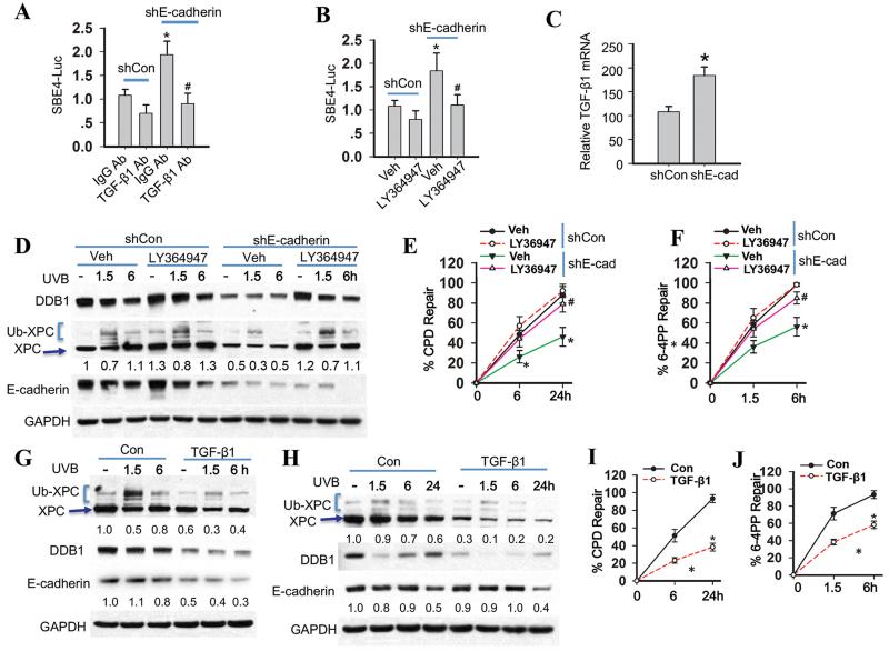
Fig. 4. The TGF-β pathway is required for suppression of nucleotide excision repair by E-cadherin loss.
(A) Luciferase reporter assay of the SBE4 luciferase reporter in HaCaT cells transfected with shCon or shE-cadherin and then treated with normal IgG or anti-TGF-β antibody for 24 h. (mean±S.D. (error bars), n=3; *, p<0.05, compared with the shCon group; #, p<0.05, compared with the shE-cadherin group). (B) Luciferase reporter assay of the SBE4 promoter in HaCaT cells transfected with shCon or shE-cadherin and then treated with vehicle or the TGF-β pathway inhibitor LY364947 (2 μM) for 24 h. (C) Real time RT-PCR analysis of TGF-β1 mRNA levels in HaCaT cells transfected with shCon or shE-cadherin. (D) Immunoblot analysis of E-cadherin, XPC, Ub-XPC, DDB1, and GAPDH in HaCaT cells transfected with shCon or shE-cadherin and then treated with vehicle or LY364947 (2 μM) for 24 h and then collected at 0, 1.5 and 6 h post-UVB (20 mJ/cm2). The results were obtained from three independent experiments. (E-F) Quantification of repair percentage (%) of CPD (E) and 6-4PP (F) in HaCaT cells transfected with shCon or shE-cadherin and then treated with vehicle or TGF-β inhibitor LY364947 (2 μM) for 24 h and collected at 0, 1.5 and 6 h post-UVB (20 mJ/cm2) for 6-4PP and 0, 6 and 24 h post-UVB (20 mJ/cm2) for CPD. *, P < 0.05, compared with shCon group; #, P < 0.05, compared with shE-cadherin/Veh groups. (G) Immunoblot analysis of E-cadherin, XPC, Ub-XPC, DDB1, and GAPDH in HaCaT cells treated with vehicle or TGF-β1 (10 ng/ml) for 48 h and then collected at 0, 1.5 and 6 h post-UVB (20 mJ/cm2). The results were obtained from three independent experiments. (H) Immunoblot analysis of E-cadherin, XPC, Ub-XPC, DDB1, and GAPDH in NHEK cells treated with vehicle or TGF-β1 (10 ng/ml) for 48 h and then collected at 0, 1.5 and 6 h post-UVB (20 mJ/cm2). Protein levels in D, G and H were quantified using ImageJ software (below each band in arbitrary units). (I-J) Quantification of repair percentage (%) of CPD and 6-4PP in NHEK cells treated with vehicle or TGF-β1 (10 ng/ml) for 48 h and then collected at 0, 1.5 and 6 h post-UVB (20 mJ/cm2) for 6-4PP and 0, 6 and 24 h post-UVB (20 mJ/cm2) for CPD. *, P < 0.05, compared with Con groups.

