

Abstract
Background. Only Clostridium botulinum strain IBCA10-7060 produces the recently described novel botulinum neurotoxin type H (BoNT/H). BoNT/H (N-terminal two-thirds most homologous to BoNT/F and C-terminal one-third most homologous to BoNT/A) requires antitoxin to toxin ratios ≥1190:1 for neutralization by existing antitoxins. Hence, more potent and safer antitoxins against BoNT/H are needed.
Methods. We therefore evaluated our existing monoclonal antibodies (mAbs) to BoNT/A and BoNT/F for BoNT/H binding, created yeast-displayed mutants to select for higher-affinity-binding mAbs by using flow cytometry, and evaluated the mAbs' ability to neutralize BoNT/H in the standard mouse bioassay.
Results. Anti-BoNT/A HCC-binding mAbs RAZ1 and CR2 bound BoNT/H with high affinity. However, only 1 of 6 BoNT/F mAbs (4E17.2A) bound BoNT/H but with an affinity >800-fold lower (equilibrium dissociation binding constant [KD] = 7.56 × 10⁻⁸ M) than its BoNT/F affinity (KD = 9.1 × 10⁻¹¹ M), indicating that the N-terminal two-thirds of BoNT/H is immunologically unique. The affinity of 4E17.2A for BoNT/H was increased >500-fold to KD = 1.48 × 10⁻¹⁰ M (mAb 4E17.2D). A combination of mAbs RAZ1, CR2, and 4E17.2D completely protected mice challenged with 280 mouse median lethal doses of BoNT/H at a mAb dose as low as 5 µg of total antibody.
Conclusions. This 3-mAb combination potently neutralized BoNT/H and represents a potential human antitoxin that could be developed for the prevention and treatment of type H botulism.
Keywords: monoclonal antibodies, botulinum toxin, botulism, Clostridium botulinum, botulinum neurotoxin type H, bioterrorism, select agents
Botulinum neurotoxin (BoNT) is the most poisonous substance known [1] and has 3 functional domains [2]: a binding domain (HC), a translocation domain (HN), and a catalytic domain (LC). The HC binds receptors on the presynaptic membrane [3–6], leading to BoNT endocytosis. After endocytosis, the HN forms a channel across the endosomal membrane enabling delivery of the LC into the cytoplasm [7–9]. The LC is a zinc endopeptidase that cleaves SNARE proteins, thereby blocking synaptic vesicle fusion and acetylcholine release [10]. Serotypes are defined immunologically in the standard mouse bioassay and differentiated from each other by the inability of polyclonal immunoglobulin G (IgG) antibodies that neutralize one serotype to neutralize the other serotypes [11–14]. By this definition, BoNT exists as 8 different serotypes, A–H [15–18]. Toxin serotypes A, B, E, and F are further subdivided into subtypes or genetic variants (eg, A1–8 and F1–7), based on immunologic and sequence differences [19].
BoNT causes botulism in humans, is a widely used therapeutic [20], and is also a Tier 1 (mass casualty capable) potential bioweapon [21, 22]. For these reasons, antitoxins neutralizing all BoNT serotypes are needed [23]. A heptavalent (targeting serotypes A–G) equine botulism antitoxin (BAT) produced from immunized horses is licensed in the United States for the treatment of botulism [24]. As a foreign protein, BAT is immunogenic, and hypersensitivity reactions, including serum sickness and asystole, have been reported [24]. Botulism Antitoxin Heptavalent (A,B,C,D,E,F,G) – (Equine) (BAT) is an immunoglobulin F(ab′)2 whose 7 serotype-specific components have short serum half-lives (7.5–34.2 hours), which preclude its effective use for prophylaxis and which may predispose to relapse of botulism after treatment [25]. As an alternative, highly potent human monoclonal antibody (mAb)–based antitoxins composed of 3 mAbs that bind nonoverlapping epitopes [26] are being developed, with the most advanced having completed phase 1 human testing [27].
BoNT/H is produced by bivalent C. botulinum strain IBCA10-7060 that also produces BoNT/B2 [15, 16]. Compared with the other 7 BoNT serotypes, BoNT/H has an N-terminal two-thirds that is most homologous to BoNT/F and a C-terminal one-third that is most homologous to BoNT/A [16]. In its initial description, BoNT/H was not neutralized by existing polyclonal antitoxins, which included a US Army–supplied equine polyclonal heptavalent (targeting serotypes A–G) F(ab′)2 botulinum antitoxin at anti-A and anti-F antitoxin to toxin potency ratios as high as 595:1 [15]. Neutralization only occurred at an anti-A antitoxin ratio of 1190:1 [15]. In more recent work, BoNT/H was neutralized by research antitoxins at ratios ranging from 20:1 to 200:1; for neutralization, using the therapeutic licensed BAT, a ratio of >500:1 was required [18]. We therefore evaluated our existing mAbs to BoNT/A and BoNT/F for BoNT/H binding, evolved one of these mAbs for higher-affinity BoNT/H binding, and thereby created a 3-mAb combination that potently neutralizes BoNT/H.
METHODS
Bacterial Strain and Production of BoNT/H
C. botulinum type Bh strain IBCA10-7060 culture filtrate was prepared, sterilized, and titrated by the mouse bioassay as previously described [15]. Culture filtrates were concentrated approximately 5-fold, using Amicon concentrators (Millipore, Bedford, Massachusetts), and the resultant concentrations (50% mouse lethal doses [MLD50] per milliliter) of BoNT/B2 and BoNT/H were determined (Supplementary Materials).
Mouse Bioassay Neutralization Studies With Equine Polyclonal Antitoxins
Mouse bioassay neutralization studies were performed using sterile culture filtrate and monovalent polyclonal equine antitoxins A, B, and F (Centers for Disease Control and Prevention, Atlanta, Georgia) under institutional animal care and use committee–approved protocols, as previously described [15].
Mouse Bioassay Neutralization Studies With Monoclonal Antibodies
Mixtures of either 2 or 3 monoclonal antibodies in equimolar amounts were prepared to a final protein concentration of 1 mg/mL in phosphate-buffered saline (PBS). mAb mixtures were serially diluted with gel phosphate diluent, and 0.1 ml of these mixtures were combined with 0.5 mL of culture filtrate, incubated at room temperature for 30 minutes, and then injected intraperitoneally, as is done in the standard mouse bioassay with equine monovalent polyclonal antitoxins (Supplementary Materials). In these studies, BoNT/B activity in culture filtrate was neutralized by using either polyclonal anti-B antitoxin or a combination of 3 mAbs known to bind to and potently neutralize BoNT/B2 [28].
mAb Generation, Production, Purification, and Characterization
All immunoglobulin G (IgG) antibodies were produced recombinantly from stable Chinese hamster ovary (CHO) cell lines by cloning the VH and Vk genes of scFv as previously described [26, 29]. All mAbs were expressed with human γ1/k constant domains and purified on protein G (Pharmacia). IgG purity was assessed by sodium dodecyl sulfate–polyacrylamide gel electrophoresis, and the concentration was determined by measuring the absorbance at 280 nm.
Screening of mAbs for Binding to BoNT/H by Enzyme-Linked Immunosorbent Assay (ELISA)
Meso Scale Diagnostic (MSD) 96-well plates were coated with a BoNT-specific IgG (as described below) at a final concentration of 4 µg/mL, 30 µL/well, and incubated overnight at 4°C. After blocking with 2% milk powder in PBS for 30 minutes, 1:1 PBS-diluted culture filtrate samples (pH 7.4) were added to each well. Binding was detected by adding an equimolar mixture of SULFO-TAG-labeled mAbs CR2 and RAZ1. Plates were read in a MSD SECTOR Imager 2400 instrument.
Measurement of BoNT/H and BoNT/B2 Concentrations in Culture Filtrate by ELISA
The concentrations of BoNT/H and BoNT/B2 in culture filtrate were determined from standard curves as previously described [30]. For measurement of the BoNT/B2 concentration, 2B18.3 IgG was used for capture, and SULFO-TAG-labeled 1B10.1 IgG was used for detection. For quantification of the BoNT/H concentration, 6F5.1 IgG was used for capture, and SULFO-TAG-labeled RAZ1 was used for detection. Pure BoNT/A1 (for BoNT/H measurements) and BoNT/B1 (Metabiologics; for BoNT/B2 measurements) were used to construct standard curves. Culture filtrate or BoNT/B1 standard was measured in triplicate, using 3 wells on the same plate. Plates were coated with either 6F5.1 or 2B18.3 IgG at a final concentration of 4 µg/mL, 30 µL/well, and incubated overnight at 4°C. Serially diluted culture filtrate samples or BoNT standards were added to each well, incubated for 1 hour, and washed, and SULFO-TAG detection mAb was added. Plates were read in a SECTOR Imager 2400 instrument.
Measurement of BoNT/H and BoNT/B2 Concentrations by Flow Fluorimetry
The concentrations of BoNT/H and BoNT/B2 in culture filtrate were measured with a KinExA 3200 instrument (Sapidyne) as previously described [29, 31], using mAbs RAZ1 and 1B10.1, respectively. Culture filtrate containing BoNT/H and BoNT/B2 was studied at a concentration estimated to be >10-fold above the value of the equilibrium dissociation binding constant [KD] of the mAb for its target toxin to generate a concentration-controlled curve for greater accuracy in measuring BoNT concentrations. mAb-containing solutions were serially diluted 2-fold 13 times in a constant concentration of culture filtrate, from >10-fold above to <0.01-fold below the estimated BoNT/H or BoNT/B2 concentrations, to capture a complete binding curve. After equilibrium was achieved, samples were passed over a flow cell packed with Sepharose 4 Fast Flow beads (GE Healthcare) covalently coupled with the measuring mAb. An Alexa-647-labeled mAb binding a nonoverlapping BoNT epitope (4E17.2A for BoNT/H, B6.1 for BoNT B2) was then passed over the flow cell, producing a signal proportional to the free BoNT in each sample. An analysis curve yielding values for KD and the binding concentrations of the BoNTs was generated using KinExA Pro software (version 4.0.12) and a 1:1 reverse-binding model.
Measurement of Antibody-Binding Affinity and Kinetics by Flow Fluorimetry
The affinity (measured by the KD) for BoNT/A1, BoNT/B2 (kind gift of Eric Johnson), BoNT/F1, BoNT/F5 (kind gift of Susan Maslanka), and BoNT/H was measured in a KinExA 3200 instrument as described above and previously [29, 32], using 1:2 serial dilutions of 13 samples of mAb in a constant concentration of pure BoNT/A1 (Metabiologics), pure BoNT/B2, or culture filtrate containing BoNT/H at a concentration of ≤10-fold above the estimated KD of the interaction, to ensure a KD-controlled analysis curve. The association rate constant (kon) was measured using either pure BoNT or culture filtrate by passing 0.5-mL volumes over a fresh BoNT-binding bead pack as the mixture came to equilibrium, at intervals of approximately 700 seconds. Free BoNT was detected at each interval with Alexa-647–labeled antibody as described above. The time-dependent exponential decrease in concentration of free BoNT as a function of time was fitted to a standard bimolecular rate equation, using KinExA Pro software to determine kon. koff was calculated from the product of kon × KD.
Measurement of Yeast-Displayed scFv Affinity for BoNT/H
KD was also determined using yeast-displayed scFv and flow cytometry as described previously [29, 33]. Yeasts were incubated with 5 different concentrations of crude BoNT/H in diluted culture filtrate that ranged from 10 times above to 10 times below the KD at 4°C for 30 minutes in florescence-activated cell-sorting buffer with proteinase inhibitor and 20 mM ethylenediaminetetraacetic acid (EDTA). Binding of BoNT/H to yeast-displayed scFv was detected after incubation with 2 µg/mL of RAZ1 IgG, followed by incubation with 1 µg/mL of goat anti-human phycoerythrin and anti-SV5-647. Each KD was determined in triplicate.
RESULTS
Structural Analysis of BoNT/H
BoNT/H has an N-terminal third that is most homologous to the hybrid subtype BoNT/F5, a unique middle third that is most homologous to BoNT/F1, and a C-terminal third that is most homologous to BoNT/A [16]. BoNT/H and BoNT/A1 are only 49.8% identical at the amino acid level but share 92.2% identity in their HCC and 73.1% identity in their HCN domains [16] (Figure 1). BoNT/H is only 52.8% identical to BoNT/F1, with hybrid subtype BoNT/F5 [35] having higher identity (61.8%), especially in its LC (80.1%) [16]. To visualize how these differences influence BoNT/A and BoNT/F antibody binding, BoNT/H amino acid differences were modeled on the structures of BoNT/A1 and BoNT/F1 (Figure 1). BoNT/H and BoNT/A1 share significant surface identity in their HCC domains and to a lesser extent their HCN domains (Figure 1), suggesting that BoNT/A1 antibodies that bind these domains could bind BoNT/H. For the remainder of BoNT/H (its LC and HN), the surface identities with BoNT/A1 and BoNT/F1 are comparable to the low identities observed between the BoNT/A1 and BoNT/F1 serotypes (Figure 1). These low identities made it unlikely that mAbs binding the HN or LC of BoNT/A1 or BoNT/F1 would bind BoNT/H.
Figure 1.

Amino acid and structural identity of botulinum neurotoxin type H (BoNT/H) holotoxin and domains compared to BoNT/A and BoNT/F. The table indicates the amino acid percentage identity between the sequence of BoNT/H holotoxin and its domains [34] and the other BoNTs indicated. The figure shows the surface amino acid differences between BoNT/H and BoNT/A1 (A), BoNT/F1 (B), and BoNT/F5 (C) and the difference between BoNT/F1 and BoNT/A1 (D). Identical amino acids are colored white, and nonidentical amino acids are in increasing shades of red as a function of the relatedness in amino acid side chain. The models were constructed using pyMol on the X-ray crystal structure of BoNT/A1 (3BTA; A and D) or on a model of BoNT/F built from the X-ray crystal structure of the BoNT/F1 light chain (LC; 2A97) and the BoNT/A1 heavy chain (HC; 3BTA; B and C).
Identification of mAbs Likely to Bind BoNT/H
On the basis of structural analyses, >100 published [26, 29, 32, 36, 37] and unpublished human or murine mAbs were surveyed to identify mAbs that might bind BoNT/H, based on the criteria that they bound either the BoNT/A1 HC or the BoNT/F1 HN or LC (Table 1 and Figure 2). Identified mAbs bound the BoNT serotype used for immunization with high affinity (KD = 8.7 × 10⁻⁹–1.5 × 10⁻¹² M). mAb 4E17.2 was cross-reactive with several BoNT serotypes, binding multiple subtypes of BoNT/A, BoNT/B, BoNT/E, and BoNT/F [32]. Three mAbs (3D12, CR1, and 4E17.2) had been mapped to the specific amino acids that were energetically important for binding [37, 38]. These amino acids in BoNT/H were identical for 3D12 and were highly conserved for CR1 and 4E17.2, suggesting a high probability of their binding to BoNT/H.
Table 1.
Characteristics of Monoclonal Antibodies (mAbs) Binding Botulinum Neurotoxin Type A1 (BoNT/A1) and BoNT/F1
| mAb | Species | Immunogen | Epitope | BoNT/A1 KD (×10−9 M) |
BoNT/B2 KD (×10−9 M) |
BoNT/F1 KD (×10−9 M) |
BoNT/F5 KD (×10−9 M) |
|---|---|---|---|---|---|---|---|
| BoNT/A mAbs | |||||||
| 3D12 | Human | Pentavalent toxoid | HCC1 | 0.061 | NB | NB | NM |
| RAZ1 | Human | Pentavalent toxoid | HCC1 | 0.002 | NB | NB | NM |
| S25 | Mouse | BoNT/A1 HC | HCC2 | 1.69 | NB | NB | NM |
| CR1 | Humanized | BoNT/A1 HC | HCN1 | 0.002 | NB | NB | NM |
| CR2 | Humanized | BoNT/A1 HC | HCN1 | 0.01 | NB | NB | NM |
| B4 | Human | BoNT/A1 HC | HCN2 | 0.095 | NB | NB | NM |
| 4E17.2 | Human | Pentavalent toxoid | HN1 | 0.002 | >100 | 0.664 | NM |
| BoNT/F mAbs | |||||||
| 6F3 | Mouse | BoNT/F1 HC/holotoxin | HC | NB | NB | 2.39 | NB |
| 6F4 | Mouse | BoNT/F1 HC/holotoxin | HC | NB | NB | 1.17 | NB |
| 6F6 | Mouse | BoNT/F1 HC/holotoxin | HN2 | NB | NB | 0.049 | NB |
| 6F7 | Mouse | BoNT/F1 HC/holotoxin | LC | NB | NB | 0.57 | NB |
| 6F11 | Mouse | BoNT/F1 HC/holotoxin | HN2 | NB | NB | 0.001 | 11.8 |
| 6F12 | Mouse | BoNT/F1 HC/holotoxin | HN2 | NB | NB | 0.14 | 13.5 |
| 6F13 | Mouse | BoNT/F1 HC/holotoxin | HN3 | NB | NB | 0.33 | 0.13 |
Epitope indicates the domain bound by the mAb and is arbitrarily numbered on the basis of the overlap with the other mAbs.
Abbreviations: NB, no detectable binding; NM, not measured.
Figure 2.
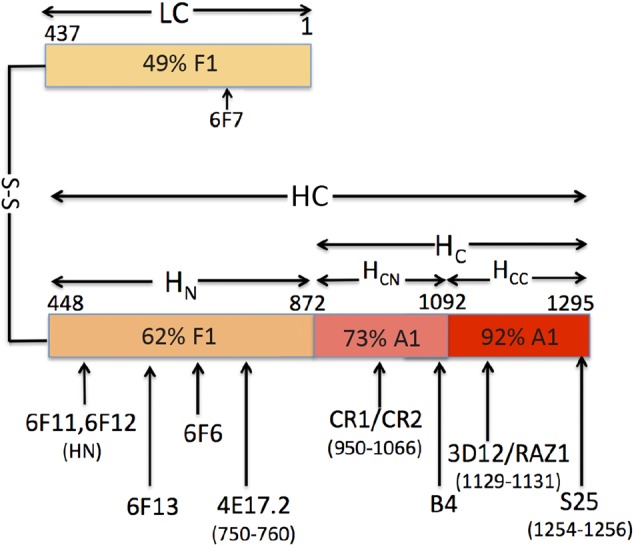
Monoclonal antibodies (mAbs) hypothesized to bind to botulinum neurotoxin type H (BoNT/H), based on its sequence identity to the HC of BoNT/A1 and the LC and HN of BoNT/F1. Where known, the amino acids comprising the epitope are indicated in parentheses. Numbering is based on the numbering of BoNT/A1. The percentage identity of each domain with BoNT/F1 or BoNT/A1 is indicated. The terms “LC” (“light chain”; molecular weight, 50 000 Da) and “HC” (“heavy chain”; molecular weight, 100 000 Da) are historical and refer to the relative molecular weights of the 2 polypeptide chains of BoNT after reduction of the disulfide bond.
Determination of Amounts of BoNT/B2 and BoNT/H in IBCA10-7060 Culture Filtrates
A sandwich ELISA was developed as previously described (Supplementary Figure 1) [30] to measure the concentrations of each BoNT present in culture filtrate. By ELISA, the culture filtrate concentration of BoNT/B2 was 35.3 nM (5.3 µg/mL) and that of BoNT/H was 11.3 nM (1.7 µg/mL). The concentrations of BoNT/B2 (33 nM; 4.96 µg/mL) and BoNT/H (7.53 nM; 1.13 µg/mL) were also measured using flow fluorimetry in a KinExA 3200 instrument (Figure 3). Values for the 2 methods were within 7% of each other for BoNT/B2 and within 34% for BoNT/H. The greater discrepancy for BoNT/H likely resulted from less accurate ELISA measurements due to lack of pure BoNT/H for standard curve construction and the binding of BoNT/B2 to the cross-reactive 4E17.2 mAb. We repeated measurement of BoNT/B2 and BoNT/H concentrations, using the KinExA 3200 instrument, on 4 independent culture filtrates of strain IBCA10-7060. Mean BoNT/B2 concentrations (±SD) were 6.89 ± 3.58 µg/mL, and mean BoNT/H concentrations (±SD) were 1.31 ± 0.93 µg/mL. On one culture filtrate sample, mouse lethality was determined before and after the addition of anti-B antitoxin, enabling measurement of lethality due to BoNT/B2 and to BoNT/H (Supplementary Table 1). The relative lethality of BoNT/B2 to that of BoNT/H in this culture filtrate was 24-fold as previously reported [15], with 4.4-fold due to the higher concentration of BoNT/B2 and 5.47-fold due to the higher specific activity of BoNT/B2.
Figure 3.

Botulinum neurotoxin type B2 (BoNT/B2) and BoNT/H concentrations in culture supernatants, measured by KinExA. Monoclonal antibody (mAb) binding curves and BoNT concentration and percentage residual error for the BoNT concentration calculated from the binding curves for BoNT/B2 (top panel) and BoNT/H (bottom panel).
Evaluation of Existing mAbs for Binding to BoNT/H
Fourteen BoNT/A and BoNT/F mAbs (Table 1) were screened for binding to BoNT/H by ELISA, including 2 control BoNT/F HC mAbs (6F3 and 6F4) not expected to bind to BoNT/H. For the BoNT/A mAbs, 3D12 and its derivative, RAZ1, gave the strongest signals, with binding also observed for CR1, CR2 (the CR1 derivative), S25, and 4E17.2 (Figure 4). For the BoNT/F mAbs, 6F12 gave the strongest signal, with no binding observed for BoNT/F HC mAbs 6F3 and 6F4.
Figure 4.

Enzyme-linked immunosorbent assay (ELISA) of monoclonal antibody (mAb) binding to botulinum neurotoxin type H (BoNT/H) in culture supernatant. ELISA signals denote the binding of the indicated BoNT/A and BoNT/F mAbs to BoNT/H in culture supernatant. The concentration of BoNT/H was estimated to be approximately 1 nM. The indicated mAb was coated on an ELISA plate, with binding detected using an equimolar mixture of mAbs CR2 and RAZ1.
mAb affinity for BoNT/H was measured by flow fluorimetry in a KinExA 3200 instrument [29, 32, 37]. RAZ1 bound to BoNT/H with very high affinity (KD = 4.96 × 10⁻¹² M, similar to its affinity for BoNT/A1; Table 2). CR2 bound to BoNT/H with high affinity (KD = 5.37 × 10⁻⁹ M), approximately 400-fold lower than its affinity for BoNT/A1. This finding is consistent with observed amino acid differences in the CR2 epitope on the BoNT/H HCC. Some of these amino acid differences are identical to those in BoNT/A2 [37], and the CR2 KD for BoNT/H is only 10-fold lower than its KD for BoNT/A2 [37]. mAb S25 bound BoNT/H with moderate affinity (KD = 1.44 × 10⁻⁸ M). We also measured the affinity for BoNT/H of BoNT/A mAbs Aa (KD = 3 × 10⁻¹² M) and Ab (KD = 2.14 × 10⁻⁹ M), 2 of the 3 mAbs in XOMA 3AB [27].
Table 2.
Affinities and Binding Kinetics of Monoclonal Antibodies (mAbs) Binding Botulinum Neurotoxin Type H (BoNT/H)
| mAb | BoNT/H | kon (/Ms) | koff (/s) | BoNT/F1 | kon (/Ms) | koff (/s) | BoNT/B2 |
|---|---|---|---|---|---|---|---|
| KD (×10−9 M) | KD (×10−9 M) | KD (×10−9 M) | |||||
| S25 | 14.43 | NM | NM | NB | … | … | NB |
| RAZ1 | 0.005 | 4.50e + 06 | 2.16e − 05 | NB | … | … | NB |
| CR2 | 5.37 | 2.92e + 06 | 1.57e − 02 | NB | … | … | NB |
| 4E17.2A/6F5.1 | 75.59 | 1.63e + 5 | 1.27e − 02 | 0.091 | 1.42e + 06 | 1.29e − 04 | 35.9 |
| 4E17.2B/6F5.2 | 9.05 | 6.90e + 04 | 6.24e − 04 | 0.003 | 1.10e + 06 | 3.58e − 06 | 93.6 |
| 4E17.2C/6F5.3 | 1.28 | 4.01e + 05 | 5.13e − 04 | 0.162 | 1.05e + 06 | 1.70e − 04 | 117.3 |
| 4E17.2D/6F5.4 | 0.148 | 5.67e + 05 | 8.24e − 05 | 0.015 | 1.63e + 06 | 2.41e − 05 | 512.3 |
KD and kon were measured by flow fluorimetry in a KinExA 3200 instrument, and koff was calculated as KD * kon. The affinity of 3D12 and CR1, lower affinity parental mAbs of RAZ1 and CR2, respectively, was not measured.
Abbreviation: NB, no detectable binding.
Only 1 of 6 BoNT/F mAbs had measurable binding affinity for BoNT/H. mAb 4E17.2A, a higher-affinity derivative of mAb 4E17.2, bound BoNT/H with moderate affinity (KD = 7.56 × 10⁻⁸ M), approximately 800-fold lower than its affinity for BoNT/F1 (KD = 9.1 × 10⁻¹¹ M; Table 2). Despite binding by ELISA, mAbs 6F6, 6F7, 6F10, 6F11, and 6F12 did not show binding in the KinExA assay at BoNT/H concentrations up to 500 nM, indicating that their KD must be lower than 1 µM. Similar instances are known of ELISA-positive binding associated with very poor affinity for antigen when the solution KD is measured [28]. This discordance presumably results from avid solid-phase ELISA binding that does not occur in solution.
Increasing the Affinity of 4E17.2A for BoNT/H
4E17.2A affinity for BoNT/H was increased by using yeast-displayed libraries of 4E17.2A scFv CDRH1, CDRH3, CDRL1, and CDRL3 mutants selected for higher-affinity binding, using flow cytometry after labeling with culture filtrate (Supplementary Figure 2) [29]. It was postulated that selection for higher-affinity BoNT/F1 binding would result in increased affinity for BoNT/H, so additional selections were done using pure BoNT/F1 to also avoid improving the affinity of cross-reactive 4E17.2A for BoNT/B2, rather than BoNT/H. For selections done on BoNT/F1, a higher-affinity mAb (4E17.2B) was identified that had 3 mutations in CDRH1 and a 30-fold and 8.4-fold increase in affinity for BoNT/F1 and BoNT/H, respectively (Table 2 and Supplementary Figure 2). For selections done on BoNT/H, mutations were identified in CDRH3, L1, and L2 that each increased the affinity of yeast-displayed scFv 3–4-fold for BoNT/H. Mutations in these CDRs were combined into a single mAb (4E17.2C), which had a 59-fold higher affinity for BoNT/H. The affinity of 4E17.2C was further increased 8.6-fold by randomly introducing mutations into the 4E17.2C scFv gene and selecting for higher-affinity BoNT/H binding, yielding mAb 4E17.2D (KD = 1.48 × 10⁻¹⁰ M) with a >500-fold affinity for BoNT/H. During the affinity maturation process, the affinity of 4E17-based mAbs for BoNT/B2 decreased 14-fold to 5.12 × 10⁻⁷ M (Table 2).
Neutralization of BoNT/H in Mice by mAb Combinations
All mice receiving as little as 8.25 µg of the mAb pair RAZ1 and CR2 survived challenge with 28 MLD50 of BoNT/H but had mild botulism symptoms (Table 3). Addition of a low-affinity third mAb (4E17.2B) showed equivalent protection. Replacement of 4E17.2B with the higher-affinity 4E17.2C resulted in 5 of 8 mice surviving a 72.5 LD50 challenge. Combining RAZ1 and CR2 with the highest-affinity 4E17.2D protected all mice challenged with 280 LD50 of BoNT/H at a mAb dose as low as 5 µg total antibody (Table 3). No mice survived at a 2.5 µg mAb dose, resulting in an ED50 (the dose at which 50% of mice survive) estimated to be 3.75 µg. A combination of 1 IU of polyclonal anti-BoNT/A, 1 IU of polyclonal anti-BoNT/B, and 1 IU of polyclonal anti-BoNT/F did not protect mice challenged with 280 LD50 of BoNT/H (Table 3).
Table 3.
Protection of Mice From the Lethal Effects of Botulinum Toxin Type H (BoNT/H) by Combinations of Monoclonal Antibodies (mAbs) and Polyclonal Antibodies (pAbs)
| A. Double and triple mAb combination and BoNT/H challenge dosea |
||||
|---|---|---|---|---|
| Antibody Combinations | BoNT/H MLD50/Mouse | Dose of mAbs per Mouse (µg) | Mouse Survival After Intraperitoneal Injection of |
|
| BoNT Culture Filtrate Only | BoNT Culture Filtrate Plus mAb Mixture | |||
| RAZ1, CR2 | 28 | 66 | 0/4 | 4/4a |
| 28 | 33 | 0/4 | 4/4a | |
| 28 | 16.5 | 0/4 | 4/4a | |
| 28 | 8.25 | 0/4 | 4/4a | |
| RAZ1, CR2, 4E17.2B | 28 | 100 | 0/4 | 4/4a |
| 28 | 50 | NDb | 4/4a | |
| 28 | 25 | NDb | 4/4a | |
| 28 | 12.5 | NDb | 4/4a | |
| RAZ1, CR2, 4E17.2C | 1160 | 50 | 0/2 | 0/8 |
| 290 | 50 | 0/2 | 1/8 | |
| 72.5 | 50 | 0/2 | 5/8 | |
| 18.1 | 50 | 0/2 | 8/8 | |
| B. Comparison of mAb combination to monovalent polyclonal antitoxin combinationc |
||||
| Mouse Survival after Intraperitoneal injection of |
||||
| BoNT/H MLD50/Mouse |
Dose of Abs per Mouse |
BoNT Culture Filtrate Only | BoNT Culture Filtrate Plus Antibody Mixture | |
| RAZ1, CR2, 4E17.2D | 280 | 100 µg total mAb | 0/2 | 4/4 |
| Polyclonal anti-A, B, F | 280 | 1.0 IU of each pAb | 0/2 | 0/4 |
| C. Determination of mAb combination neutralizing potency by serial dilutiona |
||||
| BoNT/H MLD50/Mouse | Dose of mAb/ Mouse (µg) | Mouse Survival After Intraperitoneal Injection of |
||
| BoNT Culture Filtrate Only | BoNT Culture Filtrate Plus mAbs |
|||
| RAZ1, CR2, 4E17.2D | 280 | 50 | 0/4 | 4/4 |
| 280 | 5 | 0/4 | 4/4 | |
| 280 | 2.5 | 0/4 | 0/4 | |
| 280 | 1.25 | 0/4 | 0/4 | |
a The BoNT/B in the culture filtrate was neutralized by including in the intraperitoneal injection mixture an excess of a 3-mAb combination that specifically neutralizes BoNT/B.
b The experiment was not done (ND) because the first (control) experiment demonstrated that the 28 LD50s BoNT/H in the CF was lethal for all mice tested.
c BoNT/B was neutralized by an excess of polyclonal anti-BoNT/B or mAb combination; mice were observed for 8 days. Note failure of the polyclonal anti-A immunoglobulin G (IgG) plus polyclonal anti-F IgG combination to neutralize BoNT/H.
DISCUSSION
The results demonstrate the value of having developed well-characterized mAb binding epitopes that are conserved between subtypes of BoNT/A, /B, /C, /D, /E, and /F. Detailed knowledge of the serotype specificity, domain bound, and in some instances, fine epitope structure allowed rapid in silico prediction and experimental confirmation of mAbs that would bind BoNT/H. All 3 BoNT/A mAbs binding the more homologous HCC and 1 of 2 BoNT/A mAbs binding the less homologous HCN bound to BoNT/H with high affinity. However, only a single BoNT/F mAb bound BoNT/H, with only an affinity 800-fold lower than its affinity for BoNT/F. These binding and affinity differences demonstrate that the low sequence and surface identity between the LC and HN of BoNT/H and BoNT/F translate into immunologic uniqueness between these serotypes. mAbs and antitoxins generated by immunization with BoNT/F would be unlikely to bind or neutralize BoNT/H, as shown in this work and as reported by others [15, 18].
These studies provide mAbs useful for the specific diagnosis of type H botulism. Four mAbs binding nonoverlapping epitopes with high affinity were identified that could be used to develop mass spectrometry–based or other assays to detect BoNT/H in environmental samples, blood, or stool [39, 40]. A BoNT/H-specific sandwich ELISA would require development of a BoNT/H-specific mAb.
The humanized and human mAbs reported here may also be developed as BoNT/H therapeutic and prophylactic antitoxins. An equimolar combination of the human mAb RAZ1 and the humanized mAb CR2 neutralized BoNT/H in vivo with a potency of 3400 MLD50/mg (calculated using data from Table 3). While we did not titer the 2-mAb potency in IU, this calculated potency can be approximated into IU by dividing by 10 000 MLD50/IU [26], which yields 0.34 IU/mg. mAbs Aa and Ab, 2 of the 3 component mAbs in XOMA 3AB, also bound BoNT/H with affinities slightly higher than the affinities of RAZ1 and CR2. XOMA 3AB is a 3-mAb-combination pharmaceutical product being developed to treat type A botulism that has completed human phase 1 testing without adverse safety effects [27]. Based on relative affinities, the potency of in vivo neutralization of BoNT/H by XOMA 3AB should be at least equal to that of the RAZ1/CR2 combination. Adding the human mAb 4E17.2D to RAZ1 and CR2 resulted in a 3-mAb combination that potently neutralized BoNT/H (whereas combining polyclonal anti-A plus polyclonal anti-F antitoxins did not neutralize BoNT/H; Table 3). As little as 5 µg of the 3-mAb combination completely neutralized 280 MLD50, indicating a potency of at least 56 000 LD50/mg (5.6 IU/mg). The increase in potency observed by adding a third mAb is consistent with prior studies [26].
The neutralizing potencies of BAT and the 3-mAb anti-BoNT/H combination described here may be compared. A vial of BAT contains approximately 325 mg of antibody and a minimum potency of 4500 IU of anti-BoNT/A [24]. As reported elsewhere, BAT is >500-fold less potent for BoNT/H than for BoNT/A1 [18]. As a result, the potency of a vial of BAT for BoNT/H would be 4500 IU/500, or <9 IU. This translates into 0.028 IU/mg of BAT, >10-fold less potent than that of the mAb pair RAZ1 plus CR2 (0.34 IU/mg) and 200-fold less potent than the triple mAb combination RAZ1, CR2, and 4E17.2D (5.6 IU/mg).
To ensure adequate treatment, BoNT antitoxins such as BAT are given in substantial excess of the amount of systemic BoNT expected to be in the patient. At Cmax a single vial of BAT provides enough anti-BoNT/A antibody to neutralize 26 900 MLD50 of BoNT/A per milliliter of serum [24]. Based on the preceding calculations, the Cmax value equates to a neutralizing capacity of <54 MLD50/mL for BoNT/H. One human foodborne botulism patient had a serum BoNT concentration of 32 MLD50/mL [41], with the highest foodborne botulism serum concentration ever detected being 1800 LD50/mL of BoNT/A [42]. Serum concentrations of BoNT that might result from its bioterrorist use are unknown. Comparison of the estimated BoNT/H neutralizing activity of BAT (<54 MLD50/mL) with reported BoNT serum concentrations (up to 1800 LD50/mL) indicates that, in some patients, a single vial of BAT might be inadequate to neutralize potential BoNT/H serum concentrations. In addition, the observed persistence of BoNT in serum up to 25 days after exposure [42, 43] and the short half-life of anti-BoNT/A in BAT (8.6 hours) [24] heighten the need to anticipate relapse and to consider early BAT readministration where possible [25]. Such considerations also highlight the need to determine the actual potency of BAT against BoNT/H in the physiological primate disease model and to consider developing BoNT/H-specific antitoxins. XOMA 3AB should be studied as an interim BoNT/H therapeutic because, on a per-milligram basis, it should have neutralizing activity against BoNT/H comparable to BAT, was safe in a phase 1 trial [27], and has a significantly longer serum half-life (10.3–24.0 days for its 3-component mAbs) than the anti-BoNT/A activity in BAT (8.6 hours) [24]. The 3 mAbs described here could be further developed into a highly potent 3-mAb combination to BoNT/H. Finally, since BoNT/H has a mosaic structure, it would be prudent to develop mAbs specific to the immunologically unique LC and HN of BoNT/H, as there likely exists in nature the parental BoNT/H whose HC was replaced by a BoNT/A-like HC that would not be neutralized by existing antitoxins.
Supplementary Data
Supplementary materials are available at http://jid.oxfordjournals.org. Consisting of data provided by the author to benefit the reader, the posted materials are not copyedited and are the sole responsibility of the author, so questions or comments should be addressed to the author.
Notes
Acknowledgments. We thank Jessica Payne and Mayuri Panditrao for assistance.
Disclaimer. The opinions, interpretations, and conclusions contained herein are those of the authors and are not necessarily endorsed by the National Institute of Allergy and Infectious Diseases, the National Institutes of Health, or the California Department of Public Health.
Financial support. This work was supported by the National Institute of Allergy and Infectious Diseases (cooperative agreement U01 AI056493 to J. D. M. and grant R01 AI04579 to J. D. M.) and by the California Department of Public Health Infant Botulism Treatment and Prevention Fund.
Potential conflicts of interest. Y. F., J. L., and J. D. M. are inventors of intellectual property in the field of botulinum antitoxins. All other authors report no potential conflicts. All authors have submitted the ICMJE Form for Disclosure of Potential Conflicts of Interest. Conflicts that the editors consider relevant to the content of the manuscript have been disclosed.
References
- 1.Gill DM. Bacterial toxins: a table of lethal amounts. Microbiol Rev 1982; 46:86–94. [DOI] [PMC free article] [PubMed] [Google Scholar]
- 2.Lacy DB, Tepp W, Cohen AC, DasGupta BR, Stevens RC. Crystal structure of botulinum neurotoxin type A and implications for toxicity. Nat Struct Biol 1998; 5:898–902. [DOI] [PubMed] [Google Scholar]
- 3.Dolly JO, Black J, Williams RS, Melling J. Acceptors for botulinum neurotoxin reside on motor nerve terminals and mediate its internalization. Nature 1984; 307:457–60. [DOI] [PubMed] [Google Scholar]
- 4.Dong M, Yeh F, Tepp WH et al. SV2 is the protein receptor for botulinum neurotoxin A. Science 2006; 312:592–6. [DOI] [PubMed] [Google Scholar]
- 5.Montecucco C, Rossetto O, Schiavo G. Presynaptic receptor arrays for clostridial neurotoxins. Trends Microbiol 2004; 12:442–6. [DOI] [PubMed] [Google Scholar]
- 6.Mahrhold S, Rummel A, Bigalke H, Davletov B, Binz T. The synaptic vesicle protein 2C mediates the uptake of botulinum neurotoxin A into phrenic nerves. FEBS Lett 2006; 580:2011–4. [DOI] [PubMed] [Google Scholar]
- 7.Blaustein RO, Germann WJ, Finkelstein A, DasGupta BR. The N-terminal half of the heavy chain of botulinum type A neurotoxin forms channels in planar phospholipid bilayers. FEBS Lett 1987; 226:115–20. [DOI] [PubMed] [Google Scholar]
- 8.Fischer A, Montal M. Single molecule detection of intermediates during botulinum neurotoxin translocation across membranes. Proc Natl Acad Sci U S A 2007; 104:10447–52. [DOI] [PMC free article] [PubMed] [Google Scholar]
- 9.Simpson LL. Kinetic studies on the interaction between botulinum toxin type A and the cholinergic neuromuscular junction. J Pharmacol Exp Ther 1980; 212:16–21. [PubMed] [Google Scholar]
- 10.Schiavo G, Benfenati F, Poulain B et al. Tetanus and botulinum-B neurotoxins block neurotransmitter release by proteolytic cleavage of synaptobrevin. Nature 1992; 359:832–5. [DOI] [PubMed] [Google Scholar]
- 11.Burke GS. Notes on Bacillus botulinus. J Bacteriol 1919; 4:555–70.1. [DOI] [PMC free article] [PubMed] [Google Scholar]
- 12.Burke GS. The occurrence of Bacillus botulinus in nature. J Bacteriol 1919; 4:541–53. [DOI] [PMC free article] [PubMed] [Google Scholar]
- 13.Giménez DF, Ciccarelli AS. Another type of Clostridium botulinum. Zentralbl Bakteriol Orig 1970; 215:221–4. [PubMed] [Google Scholar]
- 14.Moller V, Scheibel I. Preliminary report on the isolation of an apparently new type of CI. botulinum . Acta Pathol Microbiol Scand 1960; 48:80. [DOI] [PubMed] [Google Scholar]
- 15.Barash JR, Arnon SS. A novel strain of Clostridium botulinum that produces type B and type H botulinum toxins. J Infect Dis 2014; 209:183–91. [DOI] [PubMed] [Google Scholar]
- 16.Dover N, Barash JR, Hill KK, Xie G, Arnon SS. Molecular characterization of a novel botulinum neurotoxin type H gene. J Infect Dis 2014; 209:192–202. [DOI] [PubMed] [Google Scholar]
- 17.Lacy DB, Stevens RC. Sequence homology and structural analysis of the clostridial neurotoxins. J Mol Biol 1999; 291:1091–104. [DOI] [PubMed] [Google Scholar]
- 18.Maslanka SE, Lúquez C, Dykes JK et al. A novel botulinum neurotoxin, previously reported as serotype H, has a hybrid-like structure with regions of similarity to the structures of serotypes A and F and is neutralized with serotype A antitoxin. J Infect Dis 2016; 213:379–85. [DOI] [PMC free article] [PubMed] [Google Scholar]
- 19.Hill KK, Smith TJ. Genetic diversity within Clostridium botulinum serotypes, botulinum neurotoxin gene clusters and toxin subtypes. Curr Top Microbiol Immunol 2013; 364:1–20. [DOI] [PubMed] [Google Scholar]
- 20.Chen S. Clinical uses of botulinum neurotoxins: current indications, limitations and future developments. Toxins (Basel) 2012; 4:913–39. [DOI] [PMC free article] [PubMed] [Google Scholar]
- 21.Arnon SS, Schechter R, Inglesby T et al. Botulinum toxin as a biological weapon: medical and public health management. JAMA 2001; 285:1059–70. [DOI] [PubMed] [Google Scholar]
- 22.HHS select agents and toxins. 42 Code of Federal Regulations, Part 73 ed 2012.
- 23.Adalja AA, Toner E, Inglesby TV. Clinical management of potential bioterrorism-related conditions. N Engl J Med 2015; 372:954–62. [DOI] [PubMed] [Google Scholar]
- 24.Full prescribing information: botulism antitoxin heptavalent (A, B, C, D, E, F, G) - (equine). http://www.fda.gov/downloads/BiologicsBloodVaccines/BloodBloodProducts/ApprovedProducts/LicensedProductsBLAs/FractionatedPlasmaProducts/UCM345147.pdf Accessed 22 January 2016.
- 25.Fagan RP, Neil KP, Sasich R et al. Initial recovery and rebound of type F intestinal colonization botulism after administration of investigational heptavalent botulinum antitoxin. Clin Infect Dis 2011; 53:e125–8. [DOI] [PubMed] [Google Scholar]
- 26.Nowakowski A, Wang C, Powers DB et al. Potent neutralization of botulinum neurotoxin by recombinant oligoclonal antibody. Proc Natl Acad Sci U S A 2002; 99:11346–50. [DOI] [PMC free article] [PubMed] [Google Scholar]
- 27.Nayak SU, Griffiss JM, McKenzie R et al. Safety and pharmacokinetics of XOMA 3AB, a novel mixture of three monoclonal antibodies against botulinum toxin A. Antimicrob Agents Chemother 2014; 58:5047–53. [DOI] [PMC free article] [PubMed] [Google Scholar]
- 28.Meng Q, Garcia-Rodriguez C, Manzanarez G et al. Engineered domain based assays to identify individual antibodies in oligoclonal combinations targeting the same protein. Anal Biochem 2012; 430:141–50. [DOI] [PMC free article] [PubMed] [Google Scholar]
- 29.Razai A, Garcia-Rodriguez C, Lou J et al. Molecular evolution of antibody affinity for sensitive detection of botulinum neurotoxin type A. J Mol Biol 2005; 351:158–69. [DOI] [PubMed] [Google Scholar]
- 30.Cheng LW, Stanker LH, Henderson TD, Lou J, Marks JD. Antibody protection against botulinum neurotoxin intoxication in mice. Infect Immun 2009; 77:4305–13. [DOI] [PMC free article] [PubMed] [Google Scholar]
- 31.Blake RC, Pavlov AR, Blake DA. Automated kinetic exclusion assays to quantify protein binding interactions in homogeneous solution. Anal Biochem 1999; 272:123–34. [DOI] [PubMed] [Google Scholar]
- 32.Garcia-Rodriguez C, Geren IN, Lou J et al. Neutralizing human monoclonal antibodies binding multiple serotypes of botulinum neurotoxin. Protein Eng Des Sel 2011; 24:321–31. [DOI] [PMC free article] [PubMed] [Google Scholar]
- 33.Boder ET, Wittrup KD. Yeast surface display for directed evolution of protein expression, affinity, and stability. Methods Enzymol 2000; 328:430–4. [DOI] [PubMed] [Google Scholar]
- 34.Gonzalez-Escalona N, Thirunavukkarasu N, Singh A et al. Draft genome sequence of bivalent Clostridium botulinum strain IBCA10-7060, encoding botulinum neurotoxin B and a new FA mosaic type. Genome Announc 2014; 2:e01275–14. [DOI] [PMC free article] [PubMed] [Google Scholar]
- 35.Raphael BH, Bradshaw M, Kalb SR et al. Clostridium botulinum strains producing BoNT/F4 or BoNT/F5. Appl Environ Microbiol 2014; 80:3250–7. [DOI] [PMC free article] [PubMed] [Google Scholar]
- 36.Amersdorfer P, Wong C, Smith T et al. Genetic and immunological comparison of anti-botulinum type A antibodies from immune and non-immune human phage libraries. Vaccine 2002; 20:1640–8. [DOI] [PubMed] [Google Scholar]
- 37.Garcia-Rodriguez C, Levy R, Arndt JW et al. Molecular evolution of antibody cross-reactivity for two subtypes of type A botulinum neurotoxin. Nat Biotechnol 2007; 25:107–16. [DOI] [PubMed] [Google Scholar]
- 38.Levy R, Forsyth CM, LaPorte SL, Geren IN, Smith LA, Marks JD. Fine and domain-level epitope mapping of botulinum neurotoxin type A neutralizing antibodies by yeast surface display. J Mol Biol 2007; 365:196–210. [DOI] [PMC free article] [PubMed] [Google Scholar]
- 39.Kalb SR, Garcia-Rodriguez C, Lou J et al. Extraction of BoNT/A, /B, /E, and /F with a single, high affinity monoclonal antibody for detection of botulinum neurotoxin by Endopep-MS. PLoS One 2010; 5:e12237. [DOI] [PMC free article] [PubMed] [Google Scholar]
- 40.Kalb SR, Baudys J, Raphael BH et al. Functional characterization of botulinum neurotoxin serotype H as a hybrid of known serotypes F and A (BoNT F/A). Anal Chem 2015; 87:3911–7. [DOI] [PMC free article] [PubMed] [Google Scholar]
- 41.Hatheway CL, Snyder JD, Seals JE, Edell TA, Lewis GE Jr. Antitoxin levels in botulism patients treated with trivalent equine botulism antitoxin to toxin types A, B, and E. J Infect Dis 1984; 150:407–12. [DOI] [PubMed] [Google Scholar]
- 42.Sheth AN, Wiersma P, Atrubin D et al. International outbreak of severe botulism with prolonged toxemia caused by commercial carrot juice. Clin Infect Dis 2008; 47:1245–51. [DOI] [PubMed] [Google Scholar]
- 43.Fagan RP, McLaughlin JB, Middaugh JP. Persistence of botulinum toxin in patients’ serum: Alaska, 1959-2007. J Infect Dis 2009; 199:1029–31. [DOI] [PubMed] [Google Scholar]
Associated Data
This section collects any data citations, data availability statements, or supplementary materials included in this article.