Fig. 1.
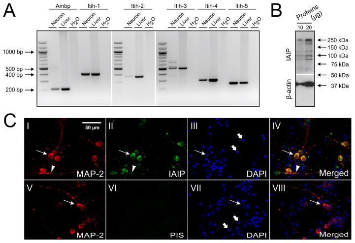
Gene and protein expressions of IAIPs in mouse cultured neurons. (A) RT-PCR shows that mRNAs of Ambp and Itih-1, 2, 3, 4, 5 are expressed in the neurons with expected bp size. Water and mRNA of mouse liver were used as negative and positive controls, respectively. (B) Bands of IaI (250 kDa) and PaI (120 kDa) were detected by Western-immunoblot. (C) Neurons were double-stained with antibodies against MAP-2 (I: red) and IAIPs (II: green). IAIPs are co-localized with MAP-2 and enriched in the cytoplasm (II and IV, arrows) and dendrites (II and IV, arrowheads). PIS and DAPI were used as negative (VI and VIII), and counter-stains, respectively (III and VII). In addition, neuronal nuclei (III and VII, arrows), fragmented cellular debris caused by the preparation procedure, and non-neuronal dividing cells undergoing cell death caused by AraC were stained by DAPI (III and VII, thick arrows).

