Abstract
During evolution the creation of single crossover chimeras between duplicated paralogous genes is a known process for increasing diversity. Comparing the properties of homologously recombined chimeras with one or two crossovers is also an efficient strategy for analyzing relationships between sequence variation and function. However, no well-developed in vitro method has been established to create single-crossover libraries. Here we present an in vitro template-change polymerase change reaction that has been developed to enable the production of such libraries. We applied the method to two closely related toxin genes from B. thuringiensis and created chimeras with differing properties that can help us understand how these toxins are able to differentiate between insect species.
During evolution, organism speciation is accompanied by duplicated gene divergence (subfunctionalization or neofunctionalization), which can confer environmental adaptation advantages for new species. Diversity can subsequently also arise through the creation of single-crossover chimeras between the resulting paralogues. Recent studies in Drosophila have identified a significant number of such chimeric genes that appear to have originated from tandem duplication1. This study proposed that qualitative differences in expression, as well as structural differences between the chimeric and parental genes, could enhance rapid evolution. A number of in vivo methods2,3,4,5 have been developed to create single-crossover chimeric genes, while in vitro homologous recombination methods such as DNA shuffling6,7,8, staggered extension process (StEP)9,10, and other similar methods11,12,13 are best suited to creating multiple-crossover recombination libraries. The three-domain pore-forming Cry toxins of Bacillus thuringiensis show evidence of domain swapping through recombination14 that has resulted in novel specificities. Previous work has also indicated that the specificity of a Cry toxin can be altered through the creation of in vitro chimeras15,16,17,18,19 making this system ideal for developing an in vitro single-crossover method. An advantage of creating these chimeras in vitro is that the cell free systems that are increasingly being used in the discovery of therapeutic proteins20 can be employed to streamline the downstream screening processes. Here we present an in vitro template-change polymerase change reaction developed to enable the production of single-crossover recombination libraries that can mimic those produced in vivo.
Results and Discussion
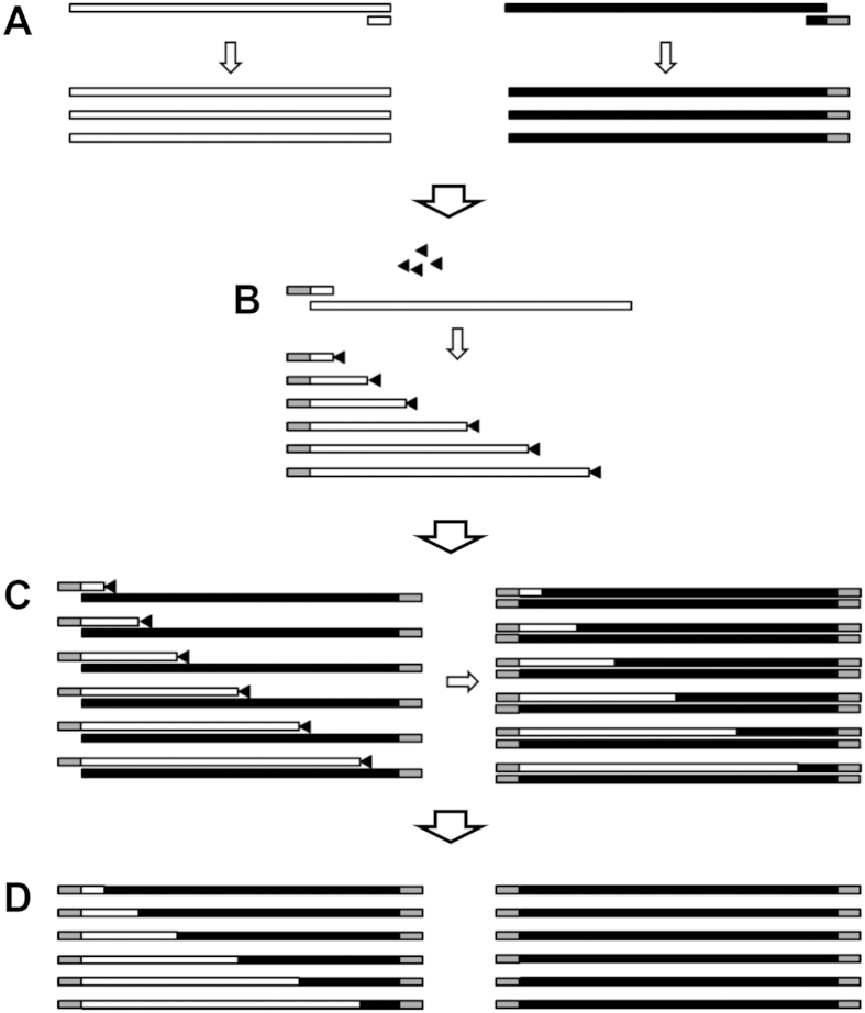
To create single-crossover recombinants incompletely extended chains of one of the parent genes are first produced by asymmetric PCR and then annealed to the full length complementary strand of the second parent gene (also produced by asymmetric PCR). The incompletely extended chains are produced through the inclusion of dideoxyATP in the amplification reaction. The frequency of terminations can be controlled by altering the ddATP/dATP ratio. The hybrid formed between the incomplete chain of parent A and the complete chain of parent B is then extended using KOD DNA polymerase. This polymerase has a number of properties that make it suitable for this procedure: it has a 3′ to 5′ exonuclease proofreading activity that is capable of recognizing and removing the ddATP at the 3′ end of the incomplete strand; it is also capable of extending the incomplete strand even if there are one or two un-matched bases at the 3′ end of the incomplete strand. This latter property means that the two parent genes don’t have to have complete identity at the crossover point to produce a chimera. To create crossovers throughout the length of the gene two recombination reactions were performed one using incomplete cry2Aa chains primed from the N-terminal end of the gene and the other using incomplete cry2Ad chains primed from the C-terminus. The resulting chimeric genes were then amplified using primers that recognized 5′ extensions added to the primers used to generate the templates for the recombination reaction, and which incorporated restriction sites to allow the cloning of the genes into the T7 expression vector pEB for expression in E. coli Rosetta (DE3) cells. The above steps are shown diagrammatically in Fig. 1. In this study, two paralogous Bacillus thuringiensis toxin genes (cry2Aa and cry2Ad) were used to test the efficiency of this method. Both genes are 1,899 base pairs (bp) in length and share 89.57% and 86.10% identity at the DNA and protein levels respectively. The Cry2Aa protein is highly toxic to the insects Ostrinia furnacalis, Plutella xylostella, Chilo suppressalis, and Helicoverpa armigera21, whereas Cry2Ad has little or no toxicity towards these species22. In this study, chimeras were designed with a cry2Aa N-terminus and a cry2Ad C-terminus.
Figure 1. Single-crossover recombination process.
Only the forward recombination process is shown. The small triangles represent ddATP molecules, and the gray regions in primers or DNA fragments represent flanking sequences used for selective amplification. (A) Generation of reverse ssDNA templates from cry2Aa (using primers F and R) and cry2Ad (using primers F’ and LR) by asymmetric PCR. (B) Generation of randomly terminated cry2Aa forward chains using primer LF. (C) Re-annealing and extension of cry2Aa forward chains using the cry2Ad ssDNA template to generate chimeric double-stranded DNA amplicons. (D) Use of the flanking sequence primers (SF and SR) for selective amplification and resolution of the chimeras.
To analyze the recombination efficiency a high-resolution melting (HRM) assay was employed. Two HRM assay primer pairs were designed to amplify the N-terminal (1–157 bp) and C-terminal (1,743–1,899 bp) regions of clones resulting from the recombination reaction. By comparing melting curves, candidates with cry2Aa N-terminal and cry2Ad C-terminal melting-curve signatures were identified as chimeras. Using this method revealed that 89 clones out of 367 tested were chimeric.
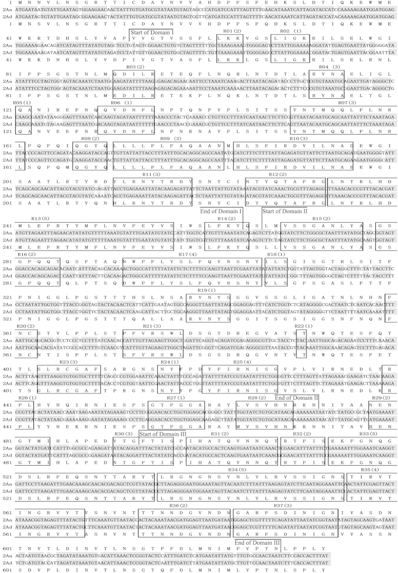
To analyze the recombination site distribution, these 89 chimeras were sequenced and aligned with the cry2Aa and cry2Ad gene sequences. The results showed that 37 different chimeras had been produced with crossover points uniformly distributed between bp 158–1,743 of the coding region (Fig. 2). Using the conditions described each recombination reaction was able to create chimeras within a 1 kb region of the priming site for the incomplete chain synthesis.
Figure 2. Crossover regions of the generated chimeras relative to a cry2Aa-cry2Ad gene alignment.
The crossover occurred in a region containing identical DNA sequences between cry2Aa and cry2Ad, and these are indicated by open rectangles. 2Aa and 2Ad refer to the cry2Aa and cry2Ad genes respectively. R01 to R37 are the crossover regions of chimeras, the numbers in parentheses means the number of sequenced chimeras with crossovers within that region. Regions of sequence identity are indicated by gray shading and the vertical lines represent the boundaries between the three structural domains.
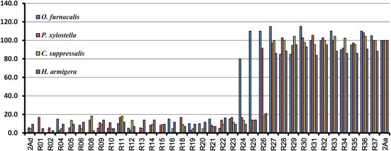
Clones representing each of the 37 chimeras were expressed in E. coli Rosetta (DE3) cells and the toxic properties of those chimeras that produced a protein of the expected size were evaluated in bioassays (Fig. 3, Supplementary Table 2). A general trend observed was that chimeras R01-R23, which contain up to 409 amino acids from Cry2Aa at their N-termini (and a minimum of 224 amino acids from Cry2Ad at their C-terminus) retain the toxicity profile of Cry2Ad. Chimeras R27-R37 containing 170 or fewer amino acids from Cry2Ad at their C-termini possess the toxicity profile of Cry2Aa. Since the junction between domain I and domain II of the Cry2A toxins is at around amino acid 267, and the junction between domains II and III at around amino acid 474, it can be concluded that substitution of domain I from Cry2Aa does not significantly affect the toxicity or specificity of Cry2Ad. Similarly substitution of domain III from Cry2Ad does not significantly affect the toxicity or specificity of Cry2Aa. Interesting data were obtained with the chimeras in which crossovers took place within domain II indicating that the location of the crossover in which toxicity switched from Cry2Aa-like to Cry2Ad-like was insect specific. For O. furnacalis the switch was seen between R23 and R24 whereas for C. suppressalis and H. armigera the switch was between R26 and R27. For P. xylostella the switch was between R25 and R26. There are several reasons why this switch could be insect-specific; it could represent the fact that specific binding of the toxin to the target host cells is mediated by different parts of the toxin in different insects. There is much literature on the involvement of domain II amino acids in the binding of the toxin to host cell receptors14,23,24,25 although there is no clear indication that there is a single binding epitope. An alternative explanation is that insect-specific defense mechanisms can differentially affect the efficacy of the toxin. These might include proteases which cleave the toxin at particular locations26,27 or molecules which sequester the toxin28. Such defense mechanisms could differentiate between the chimeras, that is may be active on one but not another. The differences between hybrids either side of a switch location are relatively small, for example between R23 and R24, which differ in O. furnacalis toxicity, there are only two amino acids changes at positions 410 and 411. Between R25 and R26 there are three changes (amino acids 430, 433 and 439) as there are between R26 and R27 (443, 446 and 447). Various studies have attempted to define regions of Cry2Aa that define specificity to a number of other insects including the mosquito Aedes aegypti29, in agreement with our findings they concluded that different regions of domain II of Cry2Aa were crucial for toxicity to different insects.
Figure 3. Biological activity of the chimeras.
2Aa and 2Ad refer to the Cry2Aa and Cry2Ad toxins respectively, and R01 through R37 represent the individual chimeras. The y-axis indicates the toxin’s relative toxicity following exposure of the test insect to 50 ppm toxin. The relative activity of each toxin was normalized to the activity of Cry2Aa. The original mortality data are shown in Supplementary Table 2.
This example of paralogous gene analysis with cry2Aa and cry2Ad showed that TC-PCR could efficiently create single-crossover chimeras for analyzing relationships between sequence variation and function. By linkage with cell-free technologies such as ribosome display20 or cell line-based technologies, TC-PCR could be broadly used to study structure/function relationships in families of homologous genes. Furthermore, this method can also be applied in directed evolution where one can envision an iterative approach in which successive, low mosaic (ie hybrids with a single crossover), libraries are produced and screened for improved properties.
Methods
Generating ssDNA templates of cry2Aa and cry2Ad
Asymmetric PCR was performed to generate ssDNA copies of cry2Aa and cry2Ad, using plasmid DNA as template. The PCR was performed in 50 μl reaction mixtures containing 10 nM of each dNTP, 50 nM MgSO4, 5 μl 10 × KOD buffer, 0.2 pM forward primer, 20 pM reverse primer and 1.0 unit of KOD DNA polymerase. The reaction conditions were 35 cycles of 94 °C, 1 min; 50 °C (cry2Aa)/54 °C (cry2Ad), 1 min; and 68 °C, 2.5 min followed by an extension at 68 °C for 7 min. After the addition of 40% deionized formamide the amplicons were purified via a PCR Purification Kit (Axygen) and eluted in 30 μl ddH2O. Separate reactions were performed to generate ssDNA copies of both strands for a given gene. All primers used in this study are listed in Supplementary Table 1.
Generation of incomplete extension chains
To generate incomplete extension chains, a 50 μl PCR reaction mixture was prepared consisting of 1 μl ssDNA template, 12 nM ddATP, 0.4 nM each dNTP, 5 μl 10 × Taq buffer, 20 pM primer, and 2.5 units of Taq DNA polymerase. The thermocycling conditions used were as follows: 35 cycles of 94 °C, 1 min; 54 °C, 1 min; and 72 °C, 2.5 min. Following PCR, 40% deionized formamide was added and the products were purified via a PCR Purification Kit (Axygen) and eluted in 25 μl ddH2O.
cry2Aa and cry2Ad gene recombination
A 50 μl reaction mixture consisting of 10 μl of incomplete extension chains, 1 μl of the corresponding ssDNA full length complementary template from the paralogous gene, 5 μl of 10 × KOD buffer, 10 nM of each dNTP, 50 nM MgSO4, 2 μl of dimethyl sulfoxide, and 1.0 unit of KOD DNA polymerase was used to prepare the chimeras. A 5–10 fold excess of incomplete extension chains over template was used. Thermocycling conditions were as follows: 1 cycle of 94 °C, 3 min; 60 °C, 3 min; and 68 °C, 10 min. The recombined products were purified with a PCR Purification Kit (Axygen) and eluted in 30 μl ddH2O.
Selective amplification of cry2Aa-cry2Ad chimeras
A 50 μl reaction mixture consisting of 1 μl recombined products, 1.5 pM of each selection primer, 5 μl of 10 × KOD buffer, 10 nM of each dNTP, 50 nM MgSO4, and 1.0 unit of KOD DNA polymerase was prepared to selectively amplify hybrid genes. PCR was performed using 30 cycles of 94 °C, 1 min; 57 °C, 1 min; and 68 °C, 2.5 min, followed by a final extension step at 68 °C for 7 min. PCR products containing a library of cry2Aa-cry2Ad chimeras were purified using a DNA Gel Extraction Kit (Axygen) and eluted in 30 μl ddH2O.
Library analysis
The library of cry2Aa-cry2Ad chimeras was cloned into the pEB cloning/expression vector (Supplementary Fig. 1) using BamHI and SalI, then introduced into JM109 cells and then plated on agar plates containing X-gal. White colonies were picked and used as templates for colony PCR in a 10 μl reaction mixture consisting of 5 μl of 2 × Taq mix, a colony template, 1 μl of LC green (Idaho, USA), and 1 μl of the appropriate primer pair (P1 and P1′ for head analysis, or P2 and P2′ for tail analysis). Melting curve analysis30,31 was performed on the resulting products to distinguish between cry2Aa and cry2Ad amplicons.
Expression of cry2Aa-cry2Ad chimeras and the parental genes
For E. coli expression studies, plasmids encoding parental and chimeric genes were introduced into E. coli Rosetta (DE3) competent cells. Transformants were grown at 37 °C in LB medium containing ampicillin (100 mg/l) and chloramphenicol (34 mg/l). After culture densities reached 0.5–1.0 absorbance units at 600 nm, isopropyl thiogalactoside was added to cultures at a final concentration of 0.5 mM, and growth continued at 20 °C. Cells were harvested by centrifugation after 10–12 h of incubation, and pellets were resuspended in 50 mM Tris-HCl (pH 8.0) and sonicated. Harvested proteins were analyzed by sodium dodecyl sulfide-polyacrylamide gel electrophoresis and protein concentrations were determined using the Quantity One gel analysis software package (Bio-Rad, USA).
Bioassays
Bioassays were performed with O. furnacalis, C. suppressalis, H. armigera using newly hatched larvae at 28 °C and 70% relative humidity. Larvae were fed an artificial diet containing cell lysates with 50 ppm of each toxin protein. For. P. xylostella second-instar larvae were fed cabbage leaves dipped into lysates containing 50 ppm of each protein and assayed at 26 °C and 70% relative humidity. Twenty larvae were used for each protein tested, and 3 independent experiments were performed in each case. Mortalities were checked after 7 days for O. furnacalis, C. suppressalis and H. armigera or after 2 days for P. xylostella.
Additional Information
How to cite this article: Shu, C. et al. In vitro template-change PCR to create single crossover libraries: a case study with B. thuringiensis Cry2A toxins. Sci. Rep. 6, 23536; doi: 10.1038/srep23536 (2016).
Supplementary Material
Acknowledgments
This study was supported by the National High Technology Research and Development Program of China (863 Program) (No. 2011AA10A203), the National Natural Science Foundation of China (No. 31301731 and 31272115). We would like to thank Dr. Lanzhi Han and Miss Yingping Liang from Institute of Plant Protection, Chinese Academy of Agricultural Sciences for providing the insect larvae and advice on bioassay. We also would like to thank Dr. Bruce Tabashnik (Department of Entomology, University of Arizona) for the valuable suggestions.
Footnotes
Author Contributions C.S. conceived and designed the experiments, C.S., J.Z., K.H. and G.L. performed the experiments, F.S., N.C., D.H. and X.L. helped perform the analysis with constructive discussions, C.S., N.C. and J.Z. performed the data analyses and wrote the manuscript. All authors reviewed the manuscript.
References
- Rogers R. L. & Hartl D. L. Chimeric genes as a source of rapid evolution in Drosophila melanogaster. Mol. Biol. Evol. 29, 517–529 (2012). [DOI] [PMC free article] [PubMed] [Google Scholar]
- Abastado J. P., Darche S., Godeau F., Cami B. & Kourilsky P. Intramolecular recombination between partially homologous sequences in Escherichia coli and Xenopus laevis oocytes. Proc. Natl. Acad. Sci. USA 84, 6496–6500 (1987). [DOI] [PMC free article] [PubMed] [Google Scholar]
- Orr-Weaver T. L., Szostak J. W. & Rothstein R. J. Yeast transformation: a model system for the study of recombination. Proc. Natl. Acad. Sci. USA 78, 6354–6358 (1981). [DOI] [PMC free article] [PubMed] [Google Scholar]
- Cusano A. M., Mekmouche Y., Meglecz E. & Tron T. Plasticity of laccase generated by homeologous recombination in yeast. FEBS J. 276, 5471–5480 (2009). [DOI] [PubMed] [Google Scholar]
- van der Heijden T. et al. Homologous recombination in real time: DNA strand exchange by RecA. Mol. Cell 30, 530–538 (2008). [DOI] [PubMed] [Google Scholar]
- Crameri A., Raillard S. A., Bermudez E. & Stemmer W. P. DNA shuffling of a family of genes from diverse species accelerates directed evolution. Nature 391, 288–291 (1998). [DOI] [PubMed] [Google Scholar]
- Stemmer W. P. Rapid evolution of a protein in vitro by DNA shuffling. Nature 370, 389–391 (1994). [DOI] [PubMed] [Google Scholar]
- Wang J., Zhang Q., Huang Z. & Liu Z. Directed evolution of a family 26 glycoside hydrolase: endo-beta-1, 4-mannanase from Pantoea agglomerans A021. J. Biotechnol 167, 350–356 (2013). [DOI] [PubMed] [Google Scholar]
- Zhao H., Giver L., Shao Z., Affholter J. A. & Arnold F. H. Molecular evolution by staggered extension process (StEP) in vitro recombination. Nat. Biotechnol. 16, 258–261 (1998). [DOI] [PubMed] [Google Scholar]
- Zhao H. & Zha W. In vitro ‘sexual’ evolution through the PCR-based staggered extension process (StEP). Nat. Protoc. 1, 1865–1871 (2006). [DOI] [PubMed] [Google Scholar]
- Shao Z., Zhao H., Giver L. & Arnold F. H. Random-priming in vitro recombination: an effective tool for directed evolution. Nucleic. Acids Res. 26, 681–683 (1998). [DOI] [PMC free article] [PubMed] [Google Scholar]
- Bergquist P. L. & Gibbs M. D. Degenerate oligonucleotide gene shuffling. Methods Mol. Biol. 352, 191–204 (2007). [DOI] [PubMed] [Google Scholar]
- Gibbs M. D., Nevalainen K. M. & Bergquist P. L. Degenerate oligonucleotide gene shuffling (DOGS): a method for enhancing the frequency of recombination with family shuffling. Gene 271, 13–20 (2001). [DOI] [PubMed] [Google Scholar]
- de Maagd R. A., Bravo A. & Crickmore N. How Bacillus thuringiensis has evolved specific toxins to colonize the insect world. Trends Genet. 17, 193–199 (2001). [DOI] [PubMed] [Google Scholar]
- Schnepf E. et al. Bacillus thuringiensis and its pesticidal crystal proteins. Microbiol. Mol. Biol. Rev. 62, 775–806 (1998). [DOI] [PMC free article] [PubMed] [Google Scholar]
- Ge A. Z., Shivarova N. I. & Dean D. H. Location of the Bombyx mori specificity domain on a Bacillus thuringiensis delta-endotoxin protein. Proc. Natl. Acad. Sci. USA 86, 4037–4041 (1989). [DOI] [PMC free article] [PubMed] [Google Scholar]
- Widner W. R. & Whiteley H. R. Location of the dipteran specificity region in a lepidopteran-dipteran crystal protein from Bacillus thuringiensis. J. Bacteriol. 172, 2826–2832 (1990). [DOI] [PMC free article] [PubMed] [Google Scholar]
- de Maagd R. A., Weemen-Hendriks M., Stiekema W. & Bosch D. Bacillus thuringiensis delta-endotoxin Cry1C domain III can function as a specificity determinant for Spodoptera exigua in different, but not all, Cry1-Cry1C hybrids. Appl. Environ. Microbiol. 66, 1559–1563 (2000). [DOI] [PMC free article] [PubMed] [Google Scholar]
- Senthil-Nathan S. A review of biopesticides and their mode of action against insect pests. In Environmental Sustainability (pp. 49–63). Springer: India, (2015). [Google Scholar]
- Murray C. J. & Baliga R. Cell-free translation of peptides and proteins: from high throughput screening to clinical production. Curr. Opin. Chem. Biol. 17, 420–426 (2013). [DOI] [PubMed] [Google Scholar]
- van Frankenhuyzen K. Insecticidal activity of Bacillus thuringiensis crystal proteins. J. Invertebr. Pathol. 101, 1–16 (2009). [DOI] [PubMed] [Google Scholar]
- Zhang J. et al. Cloning, Expression and Insecticidal Activity of cry2Ad Gene from Bacillus thuringiensis. Biotechnology Bulletin 10, 039 (2009). [Google Scholar]
- Howlader M. T. et al. Alanine scanning analyses of the three major loops in domain II of Bacillus thuringiensis mosquitocidal toxin Cry4Aa. Appl. Environ. Microbiol. 76, 860–865 (2010). [DOI] [PMC free article] [PubMed] [Google Scholar]
- Rajamohan F., Cotrill J. A., Gould F. & Dean D. H. Role of domain II, loop 2 residues of Bacillus thuringiensis CryIAb delta-endotoxin in reversible and irreversible binding to Manduca sexta and Heliothis virescens. J. Biol. Chem. 271, 2390–2396 (1996). [DOI] [PubMed] [Google Scholar]
- Rajamohan F. et al. Single amino acid changes in domain II of Bacillus thuringiensis CryIAb delta-endotoxin affect irreversible binding to Manduca sexta midgut membrane vesicles. J. Bacteriol. 177, 2276–2282 (1995). [DOI] [PMC free article] [PubMed] [Google Scholar]
- Fortier M., Vachon V., Frutos R., Schwartz J. L. & Laprade R. Effect of insect larval midgut proteases on the activity of Bacillus thuringiensis Cry toxins. Appl. Environ. Microbiol. 73, 6208–6213 (2007). [DOI] [PMC free article] [PubMed] [Google Scholar]
- Keller M. et al. Digestion of delta-endotoxin by gut proteases may explain reduced sensitivity of advanced instar larvae of Spodoptera littoralis to CryIC. Insect. Biochem. Mol. Biol. 26, 365–373 (1996). [DOI] [PubMed] [Google Scholar]
- Milne R., Wright T., Kaplan H. & Dean D. Spruce budworm elastase precipitates Bacillus thuringiensis delta-endotoxin by specifically recognizing the C-terminal region. Insect. Biochem. Mol. Biol. 28, 1013–1023 (1998). [DOI] [PubMed] [Google Scholar]
- Morse R. J., Yamamoto T. & Stroud R. M. Structure of Cry2Aa suggests an unexpected receptor binding epitope. Structure 9, 409–417 (2001). [DOI] [PubMed] [Google Scholar]
- Shu C. et al. Characterization of cry9Da4, cry9Eb2, and cry9Ee1 genes from Bacillus thuringiensis strain T03B001. Appl. Microbiol. Biotechnol. 97, 9705–9713 (2013). [DOI] [PubMed] [Google Scholar]
- Li H. et al. Detection and identification of vegetative insecticidal proteins vip3 genes of Bacillus thuringiensis strains using polymerase chain reaction-high resolution melt analysis. Curr. Microbiol. 64, 463–468 (2012). [DOI] [PubMed] [Google Scholar]
Associated Data
This section collects any data citations, data availability statements, or supplementary materials included in this article.


