Abstract
Epithelioid multinodular osteoblastoma is a rare variant of osteoblastoma characterized by numerous nodules of epithelioid osteoblasts surrounding bony trabeculae, as well as clusters of epithelioid osteoblasts without osteoid formation. It commonly occurs in the gnathic bones of the face and spine, and has a male predominance. To date, only 26 cases of epithelioid multinodular osteoblastoma have been reported and described in detail in the literature. Lucas et al. (Hum Pathol 25:117–134, 1994) described 43 cases of a variant of osteoblastoma that he termed epithelioid multifocal osteoblastoma. These both likely represent the same entity. Here, we report another case of this rare variant of osteoblastoma. An 18-year-old male patient presents with a periapical radiolucency in the region of vital tooth #30. The surgeon’s differential diagnosis for this radiolucent lesion was ameloblastoma versus cyst. An incisional biopsy of the lesion revealed well-vascularized fibrous connective tissue containing a multinodular tumor composed of collections of epithelioid cells with osteoblastic differentiation surrounding zones of hyalinization and bony trabeculae. Multinucleated giant cells and rare typical mitotic figures were noted. Additionally present within the tumor were clusters of epithelioid osteoblasts without bony trabeculae. Residual immature viable bone trabeculae were noted surrounding the tumor. A diagnosis of epithelioid multinodular osteoblastoma was rendered. In this paper we present a rare case of epithelioid multinodular osteoblastoma of the mandible, provide a general review of the literature, and highlight the unique histological features that help differentiate this tumor from tumors classified as conventional osteoblastoma, aggressive osteoblastoma, pseudoanaplastic osteoblastoma and, most importantly, low-grade or osteoblastoma-like osteosarcoma.
Keywords: Multifocal osteoblastomas, Epithelioid multinodular osteoblastoma, Multinodular osteoblastomas, Epithelioid osteoblastomas
Introduction
Osteoblastoma is an uncommon, benign bone forming neoplasm. It comprises 1 % of all bone tumors and only 3.5 % of all benign bone tumors [1]. It commonly occurs in the second to fourth decade of life and has a male predilection [1]. Osteoblastomas are seen frequently in the vertebral column and in the long bones of the appendicular skeleton [1, 2]. About 10–12 % of osteoblastomas occur within the gnathic bones with a predilection for the mandible [1, 2].
An array of histological variants of osteoblastomas has been identified. Among these variants, Filippi et al. [3] reported 26 cases of epithelioid multinodular osteoblastomas that were radiographically single lesions exhibiting a multinodular growth pattern of epithelioid cells with and without osteoid formation. According to these authors, these tumors have similar histological features as aggressive osteoblastomas, including the presence of epithelioid osteoblasts and blue bone matrix. The epithelioid multinodular osteoblastomas did not, however, present with sheets of epithelioid osteoblasts between bony trabeculae and did not recur as seen with aggressive osteoblastomas. Additionally, these tumors did not permeate bone or have atypical cellular features, which differentiate these lesions from low-grade osteosarcomas.
In this report, we report an additional case of epithelioid multinodular osteoblastoma to those previously described cases and aim to better delineate the histologic features in order to facilitate differentiation from both benign and malignant tumors that have similar microscopic appearances. We will show the histological differences between conventional osteoblastoma, aggressive osteoblastoma, pseudoanaplastic (pseudosarcomatous) osteoblastoma and low-grade or osteoblastoma-like osteosarcoma.
Clinical and Radiographic Features
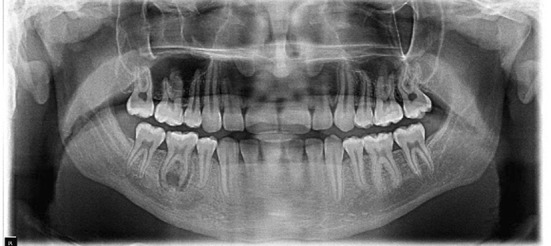
An 18-year-old male patient presented to the Center for Oral and Maxillofacial Surgery as a referral from the patient’s general dentist. The general dentist noted a swelling on the buccal aspect of tooth #30. A panoramic radiograph was taken and a periapical radiolucency in the lower right quadrant of the mandible associated with vital tooth #30 was noted (Fig. 1). The surgeon’s pre-operative differential diagnosis for this lesion was cyst versus ameloblastoma. Incisional biopsy was performed and tissue was submitted for microscopic examination.
Fig. 1.

Panoramic preoperative radiograph reveals an irregular periapical radiolucency in the lower right quadrant of the mandible within the radicular and intraradicular areas of tooth #30. Widening of the PDL space is also noted
Microscopic Features and Diagnosis
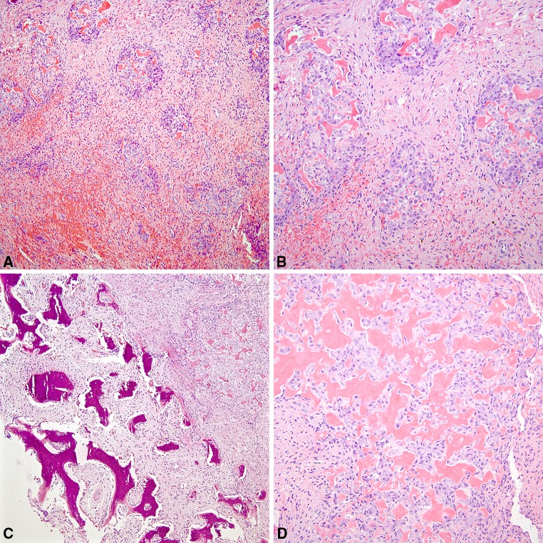
Histological examination of the incisional biopsy revealed a well-vascularized fibrous connective tissue containing a multinodular tumor composed of collections of epithelioid cells with osteoblastic differentiation surrounding zones of hyalinization and bony trabeculae (Figs. 2a–d). Clusters of epithelioid osteoblasts without bone trabeculae are also seen throughout the tumor. The tumor cells are round to ovoid in shape with ample pale cytoplasm and contain vesicular nuclei with prominent nucleoli. Rare typical mitotic figures and scattered multinucleated giant cells were identified. The connective tissue stroma also contained hemorrhagic foci and hemosiderin deposits. Residual immature viable bone trabeculae were present surrounding the tumor. No entrapment or permeation of lamellar bone was noted. Based on these histological features, a diagnosis of epithelioid multinodular osteoblastoma was rendered.
Fig. 2.
Photomicrographs of Epithelioid Multinodular Osteoblastoma. a A well-vascularized fibrous connective tissue stroma containing multiple tumor nodules of varying size and shape composed of epithelioid osteoblasts surrounding zones of hyalinization and bony trabeculae (hematoxylin and eosin, original magnification, 10×). b Clusters of epithelioid osteoblasts are seen associated with and without bony trabeculae in a well-vascularized fibrous connective tissue (hematoxylin and eosin, original magnification, 20×). c Clusters of epithelioid osteoblasts intimately surrounding areas of hyalinization are noted on the right side, and on the left side, residual immature viable bone trabeculae are present adjacent to the tumor nodules. (hematoxylin and eosin, original magnification, 20×). d The epithelioid osteoblasts are round to ovoid in shape with ample, pale cytoplasm and vesicular nuclei surrounding bone matrix (hematoxylin and eosin, original magnification, 40×)
Treatment and Follow-Up
After the histological diagnosis was established, the oral surgeon extracted tooth #30, thoroughly curetted the area of radiolucency, and submitted the specimen for histological analysis. Then a bone graft in the area of extracted tooth #30 was immediately placed. The diagnosis of the excisional biopsy was consistent with the previous diagnosis of epithelioid multinodular osteoblastoma. The patient was asked to return for a follow up in six months and will be followed every six months thereafter.
Discussion
Osteoblastomas are uncommon benign tumors of the bone that present with a broad array of histological features. There are many similarities and differences among the various histological variants of osteoblastomas. These overlapping histological features can pose a challenge in the proper diagnosis of an osteoblastic lesion, especially when the lesion presents with intermediate features of malignancy (Table 1).
Table 1.
Comparison of the histological features of conventional osteoblastoma, epithelioid or aggressive osteoblastoma, epithelioid multinodular osteoblastoma, pseudoanaplastic osteoblastoma, and osteoblastoma-like osteosarcoma [3–6, 10–14]
| Conventional osteoblastoma | Epithelioid or aggressive osteoblastoma | Epithelioid multinodular osteoblastoma | Pseudoanaplastic osteoblastoma | Osteoblastoma-like osteosarcoma | |
|---|---|---|---|---|---|
| Tumor cells | Plumps osteoblasts with round to ovoid basophilic nuclei and abundant eosinophilic cytoplasm Occasional multinucleated giant cells |
Sheets of large epithelioid osteoblasts between trabeculae of bone Epithelioid osteoblast with eccentric round to ovoid nucleus with prominent nucleoli set in abundant eosinophilic cytoplasm with an area of clearing Occasional typical mitotic figures and mild cellular pleomorphism |
Multiple nodules of epithelioid osteoblasts Epithelioid osteoblast with eccentric round to ovoid nucleus with prominent nucleoli set in abundant eosinophilic cytoplasm with clearing Occasional typical mitotic figures Lack of cytological atypia |
Osteoblasts with large, bizarre, smudged nuclei rimming bony trabeculae, or scattered individually or clustered within the stroma Osteoclasts are also seen Rare to absent mitoses No atypical mitoses |
Osteoblast-like cells that have hyperchromatic round, ovoid or spindle shaped nuclei with prominent nucleoli Rare atypical mitotic figures Variable number of multinucleated giant cells |
| Bone matrix | Long interconnecting seams of osteoid with occasional prominent basophilic reversal lines Single row of osteoblasts rimming and forming bridges between trabeculae Prominent vascularity |
Large irregular bone trabeculae Sheets of large epithelioid osteoblasts between bony trabeculae Osteoblastic rimming of bony trabeculae Blue bone matrix |
Nodules of epithelioid osteoblasts surrounding areas of osteoid Lace-like osteoid and blue bone matrix Mature lamellar bone at the periphery of the tumor Cartilaginous differentiation |
Areas of classical osteoblastoma seen with irregular seams of osteoid rimmed by single row of osteoblasts Large irregular osteoid and woven bone surrounded by anaplastic, hyperchromatic osteoblasts and osteoclasts Prominent stromal vascularity |
Unorganized proliferation of osteoid or calcified immature bone Minimal osteoid production between epithelioid osteoblasts “Pseudorosette-like appearance” of osteoid Anaplastic cells within the bony matrix Lack of vascularity |
| Periphery of the tumor | Well-defined No permeation of host bone |
Not well defined Maturation at the periphery of the lesion with woven bone No permeation of host bone |
Maturation at the periphery of the lesion with woven bone No permeation of host bone |
Irregularly defined edges Entrapment of lamellar bone |
Irregularly defined edges Permeation of host bone |
Conventional osteoblastomas (CO) are made up of anastomosing trabeculae of bone set in a loose, well-vascularized fibrous connective tissue stroma [4]. Numerous osteoblasts are seen rimming the bony trabeculae and the bridging of osteoblasts is seen between these trabeculae. Epithelioid osteoblasts are occasionally seen in these tumors [3]. No cytological atypia and rare mitoses are associated with these tumors. CO is well delineated and does not permeate the surrounding bone [1, 2, 4].
Aggressive or epithelioid osteoblastoma (AO) is another histological variant of osteoblastoma. This rare type of osteoblastoma is characterized by numerous large epithelioid osteoblasts which rim the bony trabeculae and form sheets within intertrabecular spaces [5, 6]. Scattered osteoclast-like giant cells and rare mitotic figures are seen. Unlike CO, AO is less vascular, locally aggressive, has a high recurrence rate, and is not well demarcated at the peripheral bone margin [5, 6]. The lack of cellular atypia and permeation into the host bone, however, separates AO from low-grade osteosarcomas.
Epithelioid multinodular osteoblastoma (EMO) is yet another rare variant of osteoblastoma. This entity presents with a unique histological pattern characterized by multiple nodules of tumor composed of epithelioid osteoblasts with or without bone matrix. Jaffe et al. [7] first described this multinodular osteoblastic tumor. Jaffe et al. and many other authors at the time used the terms “multifocal osteoid osteoma” or “multifocal sclerosing osteoblastoma” to describe this entity. Then in 1981, Schajowicz [8] reported several cases of “multifocal osteoblastoma” and described the tumor as containing multiple nodules of epithelioid osteoblasts encased by a substantial amount of sclerotic bone.
Although many authors have previously reported cases with this distinctive multifocal pattern, Lucas et al. [9] were the first authors to describe these tumors as having a unique histological pattern and call them “epithelioid multifocal osteoblastoma”. In their paper, Lucas et al. reported 43 out of 306 osteoblastoma cases (14 %) studied as having a multifocal pattern. They observed that these nodules in the osteoblastic tumors were mostly composed of epithelioid osteoblasts and about 10 % of these lesions occurred in the jaws. These tumors are likely to represent the same entity described by Filippi et al. [3].
Filippi et al. [3] described the clinical, radiographic and histological features of 26 epithelioid multinodular osteoblastomas (Table 2). Clinically, these lesions were seen in the mandible more often than in the maxilla (35 vs. 19 %). Epithelioid multinodular osteoblastomas were more prevalent in males than in females with a median age of 17.8 years. All 26 patients, in their study, presented with nonspecific symptoms, such as pain, swelling, or both. Radiographic features were available for 9 of the 26 tumors reported and these nine tumors presented with varying degrees of bone expansion. According to the authors, five of the nine lesions presented with benign features, two with indeterminate features and two with malignant features. Histologically, tumors were composed of multiple aggregates of epithelioid osteoblasts arranged in a multinodular pattern. These collections of osteoblasts were seen with and without bone matrix formation. The osteoblastic tumors cells contained eccentrically placed nuclei with prominent nucleoli set in a background of abundant eosinophilic cytoplasm. Multinucleated giant cells were occasionally noted within the lesion. Blue bone matrix and lacy osteoid were seen in a large number of the tumors studied. Cartilaginous differentiation within the matrix was also seen in three of twenty-six osteoblastomas. No mitotic activity or foci of necrosis were noted.
Table 2.
Comparison of our case with previously reported cases of epithelioid multinodular osteoblastoma [3]
| Our case (2015) | Filippi et al. [3] (26 cases) |
|
|---|---|---|
| Age | 18 years | 17.8 years |
| Sex | Male | Males > Females (3.6:1) |
| Location | Mandible | Mandible 9/26 Maxilla 5/26 Skull 2/26 Appendicular skeleton 7/26 Other 3/26 |
| Signs and symptoms | Swelling | Swelling, pain or both |
| Radiographic description | Irregular radiolucency with widening of the PDL space | All 26 cases showed bone expansion 2/3 of patients had well-demarcated lesions 2/3 of patients had lesions encircled by reactive sclerotic bone 9 cases with radiographic imaging: – “Cortical destruction/penetration” 2 of 9 cases – “Purely lytic” 1 of 9 cases – “Subtle, thin, whorls of ossification in a gently curvilinear pattern” 3 of 9 cases Summary 5/9 benign features, 2/9 malignant features and 2/9 indeterminate features |
| Treatment | Curettage | Curettage (25/26) and resection (1/26) |
| Follow up | No recurrence noted with 6 months follow up in December 2015 | 6 months to 29 years No recurrences or metastases |
In this study by Filippi et al., the tumors were treated with curettage with one tumor from the clavicle being resected. Follow-up information was obtained for fourteen of the twenty-six patients and the follow-up time ranged from 6 months to 29 years. No recurrences or metastases were associated with these lesions.
In our study, an 18-year-old male patient presented with a painless swelling in the buccal aspect of vital tooth #30 in the right mandible. Upon radiographic examination, an irregular periapical radiolucency within the radicular and intraradicular areas and widening of the PDL space of tooth #30 were noted (Fig. 1). The histological, clinical and radiographic features in our study conform to those cases reported by Filippi et al. These include a multinodular growth pattern of epithelioid osteoblasts with or without osteoid formation, clinical presentation of swelling associated with the lesion, and presence of an ill-defined radiolucency in the area of the lesion (Table 2).
Our case has many overlapping histological features with those cases of epithelioid multinodular osteoblastomas reported by Filippi et al. The presence of intermediate features such as numerous large epithelioid cells, multinodular pattern of the tumor and the presence of mitotic figures, however, could raise concern and cause the clinician to consider a malignant or pseudomalignant bone lesion in their differential diagnosis.
Pseudoanaplastic (pseudosarcomatous) osteoblastoma (PMO) is an extremely rare type of osteoblastoma and is characterized by benign clinical and radiographic features. Of the 4000 cases of bone tumors studied by Bahk and Mirra [11], only 0.35 % of the tumors had features of PMOs. These tumors presented as benign-appearing radiographic lesions that possessed a non-aggressive clinical behavior. According to Bahk and Mirra [11], these lesions did not locally recur or metastasize. Histologically, these lesions are characterized by giant, bizarre-appearing cells with karyorrhectic or smudged nuclei that resemble mitotic figures, and had rare to no typical mitoses without atypical mitoses [11]. Cheung et al. [12] also discuss a case of pseudomalignant osteoblastoma presenting as a typical osteoblastic tumor with atypical osteoblasts and absence of mitoses. They also state that the tumor presents with atypical architectural features such as the increased cellularity and permeative infiltrative pattern at the periphery [12]. These histological features are unique to PMOs and readily distinguish these lesions from other histological variants of osteoblastoma.
Osteoblastoma-like osteosarcomas (OLO) are uncommon low-grade osteosarcomas that look like osteoblastomas and represent about 1.1 % of osteosarcomas [13, 14]. Histologically, these tumors contain large epithelioid osteoblasts like those seen in AO and EMO. However, these lesions also present with spindle cells with hyperchromatic nuclei with prominent nucleoli, scattered atypical mitotic figures, and an irregular lacy bone matrix surrounded and entrapped by anaplastic cells. Additionally, these tumors are seen infiltrating the surrounding host bone.
While epithelioid multinodular osteoblastomas share similar histological features with CO, AO, PMO, and OLO, the presence of bony matrix within the clusters of numerous large epithelioid osteoblasts, the lack of cytological atypia or pleomorphism, and the absence of permeation of surrounding host bone are distinguishing features that separate this entity from CO, AO, PMO, and OLO. Distinguishing these osteoblastic tumors with many common histological features is crucial for the proper diagnosis, treatment, and prognosis of these tumors.
Compliance with Ethical Standards
Disclosure
All authors declare that there are no financial conflicts associated with this study and that the funding source has no role in conceiving and performing the study.
References
- 1.Unni KK, Inwards CY. Dhalin’s bone tumors: general aspects and data on 10,165 cases. 6. Philadelphia: Lippincott Williams & Wilkins; 2010. [Google Scholar]
- 2.Dorfman HD, Czerniak B. Bone tumors. 1. St. Louis: Mosby, Inc; 1998. [Google Scholar]
- 3.Filippi RZ, Swee RG, Unni KK. Epithelioid multinodular osteoblastoma: a clinicopathologic analysis of 26 Cases. Am J Surg Pathol. 2007;31:1265–1268. doi: 10.1097/PAS.0b013e31803402e7. [DOI] [PubMed] [Google Scholar]
- 4.Jones AC, Prihoda TJ, Kacher JE, et al. Osteoblastoma of the maxilla and mandible: a report of 24 cases review of the literature, and discussion of its relationship to osteoid osteoma of the jaws. Oral Surg Oral Med Oral Pathol. 2006;102:639–650. doi: 10.1016/j.tripleo.2005.09.004. [DOI] [PubMed] [Google Scholar]
- 5.Britt JD, Murphey MD, Castle JT. Epithelioid osteoblastoma. Head Neck Pathol. 2012;6:451–454. doi: 10.1007/s12105-012-0356-5. [DOI] [PMC free article] [PubMed] [Google Scholar]
- 6.Harrington C, Accurso B, Kalmar JR, et al. Aggressive osteoblastoma of the maxilla: a case report and review of the literature. Head Neck Pathol. 2011;5:165–170. doi: 10.1007/s12105-010-0234-y. [DOI] [PMC free article] [PubMed] [Google Scholar]
- 7.Jaffe HL, Mayer L. An osteoblastic osteoid tissue-forming tumor of metacarpal bone. Arch Surg. 1932;24:550–564. doi: 10.1001/archsurg.1932.01160160022002. [DOI] [Google Scholar]
- 8.Shajowicz F. Tumors and tumor-like lesions of bones and joints. New York: Springer; 1981. pp. 47–56. [Google Scholar]
- 9.Lucas DR, Unni KK, Mcleod RA, et al. Osteoblastoma: clinicopathologic study of 306 cases. Hum Pathol. 1994;25:117–134. doi: 10.1016/0046-8177(94)90267-4. [DOI] [PubMed] [Google Scholar]
- 10.Bertoni F, Bacchini DD, Donati D, et al. Osteoblastoma-like osteosarcoma. The Rizzoli Institute experience. Mod Pathol. 1993;6:707–716. [PubMed] [Google Scholar]
- 11.Bahk WJ, Mirra JM. Pseudoanaplastic tumors of bone. Skelet Radiol. 2004;33:641–648. doi: 10.1007/s00256-004-0826-2. [DOI] [PubMed] [Google Scholar]
- 12.Cheung FMF, Wu WC, Lam CKL. Diagnostic criteria for pseudomalignant osteoblastoma. Histopathology. 1997;31:196–200. doi: 10.1046/j.1365-2559.1997.5870820.x. [DOI] [PubMed] [Google Scholar]
- 13.Abramovici L, Kenan S, Hytiroglou P, et al. Osteoblastoma-like osteosarcoma of the distal tibia. Skelet Radiol. 2002;31:179–182. doi: 10.1007/s00256-001-0458-8. [DOI] [PubMed] [Google Scholar]
- 14.Dorfman HD, Weiss SW. Borderline Osteoblastic tumors: problems in the differential diagnosis of aggressive osteoblastoma and low-grade osteosarcoma. Semin Diagn Pathol. 1984;1:215–234. [PubMed] [Google Scholar]


