Abstract
A case of clear cell odontogenic carcinoma of the oral cavity is described in this sine qua non radiology-pathology correlation article. CT demonstrated a solid and cystic mass arising from the mandible. Histology demonstrated variably-sized nests of clear to pale eosinophilic cells with occasional central necrosis embedded in a hyalinized to fibrocellular stroma. The specimen was also positive for the characteristic rearrangement of the EWSR1 (22q12) locus in 93.5 % of interphase cells.
Keywords: Oral cavity, Clear cell odontogenic carcinoma, CT, Pathology
History
The patient is a 65-year-old female who initially noticed a mass in her right oral cavity. Biopsy at an outside institution demonstrated an invasive carcinoma with clear cell to squamoid features. The patient decided to proceed with alternative therapy. Unfortunately, the tumor continued to grow and the patient returned 2 years later. Oral cavity exam revealed firm exophytic mass lesion centered on right anterior mandible alveolus, with associated loss of dentition and tenderness to palpation in that area.
Radiographic Features
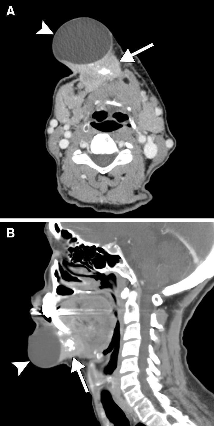
Contrast-enhanced CT demonstrated a large exophytic solid and cystic mass arising from the right mandibular body, where there were irregular lytic changes (Fig. 1). The soft tissue component invaded the overlying subcutaneous tissues and underlying floor of mouth and intrinsic muscles of the tongue. In addition, there was a dominant cystic component within the subcutaneous tissues. The mass also contained several punctate bone fragments displaced away from both sides of the eroded portions of the mandible, which indicated a centrifugal growth pattern centered in the mandible.
Fig. 1.

Axial a and sagittal, b contrast-enhanced CT images show a mass (arrows) arising from the body of the mandible with a large cystic component (arrowheads). There is irregular destruction of the mandible and the soft tissue and cystic components invade the overlying subcutaneous tissues and underlying floor of mouth and intrinsic muscles of the tongue
Diagnosis
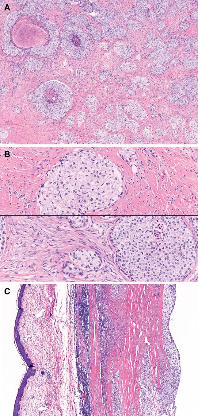
The resected specimen consisted of a 7 cm solid mass with an adjacent 4 cm unilocular cyst. Histology showed an invasive carcinoma with variably-sized nests of clear to pale eosinophilic cells with occasional central necrosis in larger nests (Fig. 2a). The stroma varied from hyalinized to fibrocellular and the cells had abundant clear cytoplasm with well-defined cell borders and nuclei with wrinkled, irregular nuclear membranes and scattered mitoses (Fig. 2b). The subcutaneous unilocular cyst was lined by tumor cells (Fig. 2c). Neither keratinization, intercellular bridges, nor surface mucosal in situ disease were appreciated. Bony, lymphovascular, and perineural invasion were present.
Fig. 2.

Histology showed an invasive carcinoma with variably-sized nests of clear to pale eosinophilic cells with occasional central necrosis in larger nests (a). The stroma varied from hyalinized (top) to more cellular (bottom) (b). The subcutaneous unilocular cyst was lined by tumor cells (c)
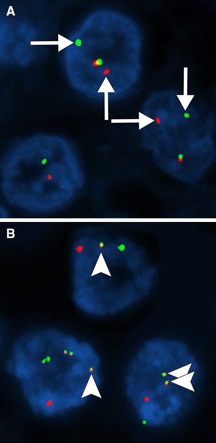
The differential diagnosis included a squamous cell carcinoma with extensive glycogenation, clear cell odontogenic carcinoma, hyalinizing clear cell carcinoma of salivary gland, and metastatic carcinoma (including clear cell renal cell carcinoma and urothelial carcinoma). Immunostains showed that tumor cells were positive for high molecular weight keratin and p40, but negative for S100 and SMA. The immunoprofile could be compatible with both squamous and urothelial carcinoma; however, these were not favored due to the lack of a squamous in situ component and the absence of renal or urinary bladder lesions. The lack of a stromal vascular network and positivity for p40 also mitigated the possibility of renal cell carcinoma. Myoepithelial carcinoma was also considered, but not favored due to absence of S100 and SMA staining. Therefore, clear cell odontogenic carcinoma and hyalinizing clear cell carcinoma of salivary gland were favored, both of which have similar immunoprofiles and molecular genetics. However, the clinical and radiographic findings of a mass centered in the mandible supported clear cell odontogenic carcinoma, and breakapart probe confirmed rearrangement of the EWSR1 (22q12) locus in 93.5 % of interphase cells (Fig. 3a). Dual fusion probe also confirmed EWSR-ATF1 rearrangement in 94.5 % of cells (Fig. 3b). Based on the overall constellation findings, the diagnosis of clear cell odontogenic carcinoma was made.
Fig. 3.

Breakapart probe (a) confirmed rearrangement of the EWSR1 (22q12) locus in 93.5 % of interphase cells (arrows). Dual fusion probe confirmed EWSR1-ATF1 rearrangement (arrowheads) in 94.5 % of cells (b). Courtesy of Julia A Bridge, MD, FACMG
Treatment
The patient was started on carboplatin/cetuximab. After two doses, further treatment was put on hold per the patient’s request because she became very weak and felt that the tumor was still increasing in size. Mandibular resection, partial glossectomy, neck dissection, and chimeric free fibular reconstruction with right thigh skin graft to flap was subsequently performed. There was no evidence of gross tumor recurrence at 6 months follow-up.
Discussion
According to the 2005 WHO classification, there are several types of odontogenic carcinomas, including metastasizing ameloblastoma, ameloblastic carcinoma (primary and secondary), primary intraosseous squamous cell carcinoma (solid type, derived from keratocystic odontogenic tumor, and derived from odontogenic cysts), ghost cell odontogenic carcinoma, and clear cell odontogenic carcinoma [1]. Clear cell odontogenic carcinoma is a rare entity that tends to arise from the body of the mandible during the fifth through seventh decades and has a female preponderance [2].
Clear cell odontogenic carcinoma tends to be an aggressive tumor with a destructive growth pattern. The diagnostic imaging findings tend to reflect this behavior, in which there is osseous destruction associated with a heterogeneously enhancing mass that can invade the adjacent soft tissues. Thus, the imaging differential diagnosis includes squamous cell carcinoma, minor salivary gland carcinomas, and metastatic tumors to the jaws. Alternatively, clear cell odontogenic carcinoma can present as a unilocular cystic lesion that mimics a benign odontogenic lesion, such as ameloblastoma [3]. Clear cell odontogenic carcinoma can potentially metastasize, particularly to the lungs and lymph nodes [4].
On histology, clear cell odontogenic carcinoma should be considered in the differential diagnosis of jaw tumors with prominent clear cell components [5]. The main differential diagnosis for lesions with clear cell features on histology includes ameloblastoma, calcifying epithelial odontogenic tumor, squamous cell carcinoma, and salivary gland tumors, such as mucoepidermoid carcinoma, myoepithelial carcinoma, and hyalinizing clear cell carcinoma, as well as certain metastatic tumors, such as renal, urothelial, and thyroid carcinomas [2]. In particular, clear cell odontogenic carcinomas often demonstrate peripheral palisading nuclei, monotonous nuclear size with occasional raisinoid nuclei, stromal hyalinization, and perineural invasion, but no evidence of dentin or enamel matrix production, and are negative for myoepithelial markers (S100, GFAP, SMA, calponin) [6].
Molecular analysis is helpful for further characterization, as the EWSR1 rearrangement is present in 83 % of clear cell odontogenic carcinomas. EWSR1-ATF1 translocation can occur in both clear cell odontogenic carcinoma and hyalinizing clear cell carcinoma of salivary gland, which also have comparable histologic and immunophenotypic features [7].
Since clear cell odontogenic carcinoma can be an aggressive tumor with a tendency to recur, wide surgical resection with confirmation of tumor-free margins is optimal. Lymph node dissection and adjuvant radiation therapy may be considered in selected cases [8]. BRAFV600E-activating mutation is present in some odontogenic carcinomas and can potentially serve as a biomarker for molecular-targeted therapy [9].
Conflicts of interest
None.
References
- 1.Barnes L, Eveson JW, Reichart P, Sidransky D, editors. World Health organization classification of tumours. Pathology & genetics. head and neck tumours. Lyon: IARC Press; 2005. [Google Scholar]
- 2.Avninder S, Rakheja D, Bhatnagar A. Clear cell odontogenic carcinoma: a diagnostic and therapeutic dilemma. World J Surg Oncol. 2006;12(4):91. doi: 10.1186/1477-7819-4-91. [DOI] [PMC free article] [PubMed] [Google Scholar]
- 3.Kim M, Cho E, Kim JY, Kim HS, Nam W. Clear cell odontogenic carcinoma mimicking a cystic lesion: a case of misdiagnosis. J Korean Assoc Oral Maxillofac Surg. 2014;40:199–203. doi: 10.5125/jkaoms.2014.40.4.199. [DOI] [PMC free article] [PubMed] [Google Scholar]
- 4.Krishnamoorthy R, Ravi Kumar AS, Batstone M. FDG-PET/CT in staging of clear cell odontogenic carcinoma. Int J Oral Maxillofac Surg. 2014;43:1326–1329. doi: 10.1016/j.ijom.2014.06.008. [DOI] [PubMed] [Google Scholar]
- 5.Swain N, Dhariwal R, Ray JG. Clear cell odontogenic carcinoma of maxilla: a case report and mini review. J Oral Maxillofac Pathol. 2013;17:89–94. doi: 10.4103/0973-029X.125226. [DOI] [PMC free article] [PubMed] [Google Scholar]
- 6.Bilodeau EA, Hoschar AP, Barnes EL, Hunt JL, Seethala RR. Clear cell carcinoma and clear cell odontogenic carcinoma: a comparative clinicopathologic and immunohistochemical study. Head Neck Pathol. 2011;5:101–107. doi: 10.1007/s12105-011-0244-4. [DOI] [PMC free article] [PubMed] [Google Scholar]
- 7.Bilodeau EA, Weinreb I, Antonescu CR, Zhang L, Dacic S, Muller S, et al. Clear cell odontogenic carcinomas show EWSR1 rearrangements: a novel finding and a biological link to salivary clear cell carcinomas. Am J Surg Pathol. 2013;37:1001–1005. doi: 10.1097/PAS.0b013e31828a6727. [DOI] [PubMed] [Google Scholar]
- 8.Ebert CS, Jr, Dubin MG, Hart CF, Chalian AA, Shockley WW. Clear cell odontogenic carcinoma: a comprehensive analysis of treatment strategies. Head Neck. 2005;27:536–542. doi: 10.1002/hed.20181. [DOI] [PubMed] [Google Scholar]
- 9.Diniz MG, Gomes CC, Guimarães BV, Castro WH, Lacerda JC, Cardoso SV, et al. Assessment of BRAFV600E and SMOF412E mutations in epithelial odontogenic tumours. Tumour Biol. 2015. (Epub ahead of print). [DOI] [PubMed]


