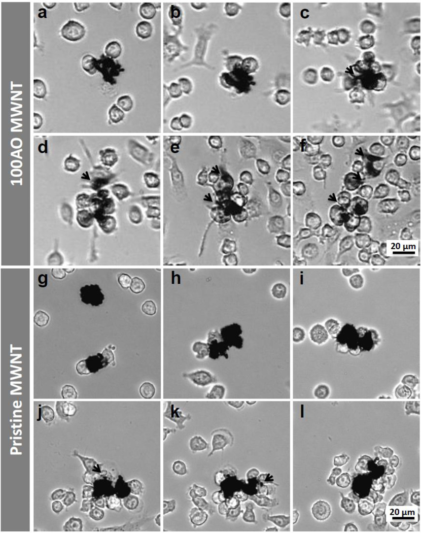
Figure 2.
Selected light micrographs from live cell imaging videos of N9 microglia exposed to (a–f) 100AO and (g–l) pristine MWNTs for 48 hours (see also supplementary video 2). Images here are acquired (a,g) 1 hour (b,h) 3 hours (c,i) 12 hours (d,j) 30 hours (e,k) 40 hours and (f,l) 48 hours after exposure. Irregular opaque particles ~20 µm in diameter are identified as MWNT aggregates. N9 microglia surround 100AO MWNT aggregates about 12 hours after exposure (c). Small aggregates (<10 µm in diameter) were broken up from the main aggregate continually (c-f, marked with black arrows). Microglia are observed to surround pristine MWNT aggregates at ~30 hours (j). The majority of pristine MWNT aggregates remain intact after 48 hours, although some small aggregates (<4 µm in diameter) appear to have been broken away from the main aggregate (black arrows, j,k).

