Abstract
Purpose
Vertebral fragility fractures are often treated by injecting bone cement into the collapsed vertebral bodies (vertebroplasty). The mechanisms by which vertebroplasty induces pain relief are not completely understood yet and recent debates cast doubt over the outcome of the procedure. The controversy is intensified by inconsistent results of randomized clinical trials and biomechanical studies that have investigated the effectiveness or the change in biomechanical response due to the reinforcement. The purpose of this study was to evaluate the effectiveness of vertebroplasty, by varying the relevant treatment parameters and (a) computationally predicting the improvement of the fracture risk depending on the chosen treatment strategy, and (b) identifying the determinants of a successful treatment.
Methods
A Finite Element model with a patient-specific failure criterion and direct simulation of PMMA infiltration in four lumbar vertebrae was used to assess the condition of the bone under compressive load before and after the virtual treatment, simulating in a total of 12000 virtual treatments.
Results
The results showed that vertebroplasty is capable of reducing the fracture risk by magnitudes, but can also have a detrimental effect. Effectiveness was strongly influenced by interactions between local bone quality, cement volume and injection location. However, only a moderate number of the investigated treatment strategies were able to achieve the necessary improvement for preventing a fracture.
Conclusions
We conclude that the effectiveness of vertebroplasty is sensitive to the patient’s condition and the treatment strategy.
Introduction
Osteoporosis is a common disease that manifests itself in fractures occurring at different anatomical sites, e.g. at the spine, the hip, or the wrist, with consequent morbidity and mortality [1–3]. Augmentation through the injection of a reinforcing biomaterial (percutaneous vertebroplasty, PVP), is a popular, minimally invasive intervention for the stabilization of fractured vertebral bodies. The utility and effectiveness of PVP is most often evaluated on the basis of three measures: economic value (i.e. treatment and opportunity costs vs. effect, cost-effectiveness), short- and long-term pain relief and the incidence of new fractures either at the treated, adjacent or remote levels. Cost-effectiveness determines whether, given a cost level that society is willing to spend per quality-adjusted life-year (QALY), PVP is an acceptable treatment strategy or not. Hence, cost-effectiveness depends directly on the other two measures. Both were in the past and more recently addressed by numerous studies, with controversial results and conclusions on the utility and effectiveness of PVP [4]. One potential cause of the discrepancy might be the fact that these studies were driven by different objective targets. For example, immediate pain relief in a short period of time is important for elderly patients while a sustainable treatment outcome is beneficial for younger patients with an acute vertebral fracture. Two studies [5, 6] with a sham control intervention have reported clinical outcomes at one and six months, respectively, in patients with osteoporotic compression fractures up to one year old and cast doubts on the effectiveness of PVP.
In both studies, patients were randomized to undergo either a PVP or a sham procedure. Buchbinder et al. [5] concluded that”similar improvements were seen in both groups with respect to pain at night and at rest, physical functioning, quality of life, and perceived improvement”. The study of Kallmes et al. [6] found similar results and showed that improved disability and pain scores were noted immediately following both procedures however, a trend towards a higher rate of clinically meaningful improvement in pain (30% decrease from the baseline) was observable in the PVP group. These results have been met with some disbelief among physicians treating patients with vertebral compression fractures, and concerns have been raised on the design of both studies (ethical concerns, small sample size, crossover between the groups, i.e. patients were able to guess they were being treated with the sham procedure, and the sham procedure could itself have promoted pain relief, inclusion of patients with subacute and chronic fractures instead of acute fractures) [7–11]. The psychological effect of care and daily attention are believed to account for the decrease of the visual analogue score in conservative treatment groups during the first week of the treatment. Robinson and Olerud [4] concluded that PVP is not better than placebo and PVP cannot be recommended as a standard treatment for vertebral compression fractures. Conflicting with these results are other studies showing the utility and cost-effectiveness [11–13] of PVP, demonstrating both short-term and sustainable pain relief [14–22] and statistically significant differences in life expectancy in favour of PVP [23].
Several biomechanical studies have investigated the consequences of the augmentation on adjacent or remote, non-augmented levels. Most of them [24–32] concluded that new fractures might be the consequence of an adjacent rigid reinforcement. However, new compression fractures after the treatment could also be the consequence of the natural progression of the disease, since initial fractures are also a strong predictor of further vertebral fractures [33]. Inconsistently, clinical studies [17, 20] reported that the incidence of new fractures was not different after PVP, compared with conservative treatment after one year of follow-up. The major limitations of the biomechanical studies that could explain these divergent conclusions were the limited number of specimens and/or experimental trials (a problem generally associated with cadaveric testing) [25, 26, 28–32, 34–36] and Finite Element (FE) models that were limited to the study of a single case [26, 29, 30], constructed on the basis of non-realistic, predefined cement distributions [24, 31, 37–39], cement distributions reconstructed from patient radiographs [40] or peripheral quantitative computed tomography (pQCT) scans of human cadaveric specimens [41] with potentially suboptimal cement placement and/or filling strategy. Furthermore, the complex composite properties of cement-augmented bone have been neglected. A fundamental limitation of such biomechanical studies is the evaluation of physical units (endplate deflection [24, 30], failure load [28], maximum principal strain [30], pressure [26, 29, 30], stresses [29, 37, 39, 41], stiffness [30, 31] or strength [34, 37, 38] that form the basis for the inference as to whether PVP induces adjacent fractures or is efficient from the biomechanical perspective. It has been shown that absolute values alone, e.g. the bone mineral density (BMD), are often suboptimal predictors of osteoporotic fractures [42–44], since the distributions of those values of the different populations can overlap and do not allow sharp discrimination between the populations. The establishment of FE models from computed tomography (CT) data has been successful to investigate patient specific bone strength in-vitro and these perform better in the explanation of failure load variability among different subjects [42, 45]. These models are considered capable of capturing most of the tissue properties which contribute to bone strength and simulate the mechanical consequence of general external boundary conditions. Therefore, an FE model with the material properties derived from a clinical CT scan and an appropriate failure criterion is feasible to reflect and assess the mechanical condition or stability of the individual patient’s vertebrae prior to treatment, i.e. (a) how close a given vertebra is to failure or (b) the amount of mechanical stabilization needed to prevent the failure. Extending the FE model and failure criterion with a constitutive law, which describes accurately the response of the augmented bone cement composite, allows subsequently to locally predict the increase or decrease of fracture risk state induced by PVP and whether the positive changes go far enough to prevent the subsequent fracture of the bone. Furthermore, until now, no evidence has been provided as to which extent cement type, cement volume, cannula placement and other treatment parameters affect the aforementioned loading pattern in the spine and the final outcome of the treatment. This study aims therefore to systematically investigate potential PVP strategies in an in-silico environment that allows for the creation of large set of virtually performed PVP treatment cases. Comparative values are used to measure the change of the local factor of fracture risk and therefore give an indication, (a) if vertebroplasty is an efficient treatment from the biomechanical point of view, and (b) what the important determinants of a positive outcome are.
Methods
Specimens and μCT data
Four human vertebra (Table 1), provided by the International Institute for the Advancement of Medicine (IIAM, 175 May Street, Edison, NJ 08837, http://www.iiam.org/), from two male donors were used in this study. The specimens were scanned using a high resolution computed tomography scanner (XtremeCT, Scanco Medical AG, Brüttisellen, Switzerland) at the Maastricht University Medical Center. The scans, performed in air, were taken at a nominal isotropic resolution of 41 μm and segmented using a global threshold. A mesh morphing technique [46] was applied to map a template finite element (FE) mesh of a vertebra with 51119 elements onto the segmented CT data for each of the specimens. Following, for each element in the mesh the density, trabecular spacing (Tb.Sp) and fabric were calculated for a 4 mm spherical region centered at the element centroid from the original high-resolution images [47].
Table 1. Specimen data.
The volumetric bone mineral density (vBMD) was estimated based on the bone mineral content and volume of the elements of the FE meshes that did not have any nodes on the surface of the models in order not to include any cortical shell in the vBMD calculations.
| Specimen | Level | Donor | Age (years) | Gender | vBMD (g/cm3) |
|---|---|---|---|---|---|
| 1 | L1 | I | 88 | M | 0.187 |
| 2 | T12 | I | 88 | M | 0.360 |
| 3 | L1 | II | 70 | M | 0.113 |
| 4 | T12 | II | 70 | M | 0.187 |
The meshes were then subsequently used to carry out a sequence of mechanical and flow FE simulations as described in detail below.
FE simulations—pre-augmentation fracture risk
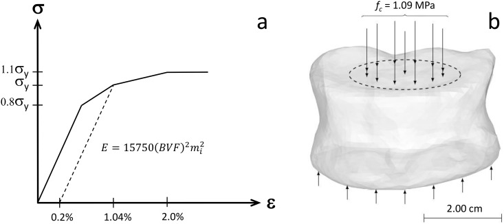
Heterogeneous, orthotropic material constants were mapped to the bone elements of the FE meshes based on approach introduced by Zysset et al. [48]. The principal material directions of the elements were assumed to be aligned with the fabric tensor of the elements. Orthotropic elastic properties then were assigned for each element using a relationship based on density and fabric [49]. The material response was assumed to be non-linear with a yield strain in compression of 1.04%, in agreement with the study of [50] (Fig 1). Tension-compression symmetry in the mechanical properties was assumed. A pressure of 1.09 MPa was applied on the superior end-plate, uniformly distributed over the nucleus pulposus area, occupying 43% of the disc area [51] as shown in Fig 1. The inferior end-plate of the FE models was constrained against translation in all directions. The rationale of this was to mimic a high nucleus pressure during moderate daily activities, e.g. as observed in a healthy male subject when lifting a 20 kg weight and holding it close to the body [52].
Fig 1. Finite Element Analysis setup.
(a) Principal material direction stress-strain response mapped to the elements of the FE meshes. The mi is the normalized eigenvalue of the fabric tensor corresponding to the specific orthotropic plane. The modulus-BVF-fabric relationship was introduced by Garcia et al. [49]. The yield strain, 1.04%, is in accordance with the study of Bayrakhtar et al. [50]. The corresponding yield stress (σy) was determined based on a 0.2% offset rule. Ultimate stress was set to 1.1σy in accordance with the study of Helgason et al. [53]. (b) A compressive load of fc = 1.09 MPa was evenly distributed over the estimated nucleus pulposus contact area of the FE models. The models were constrained against displacement in all directions on the inferior side of the vertebrae.
Failure in the bone elements was assumed to occur if principal strains in tension (εb,t) or compression (εb,c) exceeded 1.5% and -2.0% respectively. The factor of fracture risk (FFR) was computed for each element (i) as the maximum of the ratio between the element principal strains (ε1,i or ε3,i) and the corresponding principal strain threshold (εb,t or εb,c) i.e.:
| (1) |
FE simulations—cement flow
After calculating the pre-augmentation fracture risk, flow simulations were carried out to simulate the distribution of cement within the specimens during vertebroplasty using different cannula placements, cement types and cement volumes. To this end the local heterogeneous hydraulic permeability of the cancellous bone was predicted by the FE bone volume fraction (BVF), the degree of anisotropy (DA) and the trabecular spacing (Tb.Sp) [54]. The flow behavior was governed by a mixed-boundary Darcy formulation [55]. The rheology of the bone cements was experimentally determined in a plate rheometer setup [56]. The deformation rate of a fluid that is infiltrating a porous medium such as the cancellous bone cannot be estimated or predicted at the continuum length scale, however it is an important determinant of the fluid rheology. Therefore, a numerical upscaling scheme was applied to relate the pore-scale viscosity of the cement, which is governed by the fluid deformation rate, to the apparent or Darcy viscosity at the continuum length scale [56]. In the simulations the infiltration of the vertebral body and displacement of the bone marrow by the virtually injected cement, i.e. the multicomponent flow of the bone cement and marrow was simulated. The cement injections were virtually performed at 50 randomly chosen locations within each of the FE models. The assumed locations of the injection device cannula were kept consistent according to the mesh morphing procedure across all four vertebrae specimens. Simulations were carried out for ten different clinically relevant cement volumes (V = 1, 2, 3, 4, 5, 6, 7, 8, 9, 10 mL).
FE simulations—post-augmentation fracture risk
After the flow simulations, the material properties of the augmented elements in the FE models were mapped using the lookup Table approach introduced by Helgason et al. [57]. The rule of mixture in the present study was extended to the rule of mixture in Helgason et al. [57], to take into account that some elements of the FE meshes have their pore space only partially filled after the flow simulations. A reduction factor, ξ = 10%, was introduced to capture the fact that PMMA has lower strength at body temperature than when tested at room temperature [58]. The extended rule of mixture used in the present study was thus:
| (2) |
Where:
i: refers to the element number
j: refers to the cement type (see Table 2).
Table 2. Properties related to the mechanical response of the PMMA cements used in present study.
E: Modulus of elasticity; Φ: rule of mixture constant. The cements were selected from a range of commercial products (Simplex P; Stryker Howmedica Osteonics, Mahwah, NJ. Vertecem; Synthes GmbH, Solothurn, Switzerland. Active OS; Kyphon, Sunnyvale, CA), where the liquid phase was modified in our lab, in some cases, for the purpose of getting a wide range of stiffness for the cements used in the study; A: modified Simplex P. B: modified Simplex P. C: Modified Vertecem. D: Vertecem. E: Simplex P. F: Active OS.
| Cement | E (MPa) | εcem (%) | Φ (-) |
|---|---|---|---|
| A | 510 | 7.00 | 0.40 |
| B | 1434 | 7.10 | 0.40 |
| C | 2003 | 5.44 | 0.40 |
| D | 2697 | 5.10 | 0.29 |
| E | 2869 | 5.00 | 0.29 |
| F | 5193 | 3.93 | 0.15 |
BVFi: bone volume fraction
CVFi: cement volume fraction
ηi = anisotropy ratio
Φj = cement specific rule of mixture constant (see Table 2).
CVFi∙(1-ξ)∙σc,j + BVFi∙σb: is an approximation for the temperature adjusted upper limit response (σUL) for an augmented element where σc,j and σb are the uniaxial stress response of a given cement (j) and the tissue level properties of bone respectively.
c = CVFi/(1-BVFi) is a constant that adjusts the uniaxial stress response of the augmented elements to take incomplete augmentation of an element after flow simulations into account. If the pore space is completely filled after the flow simulations CVF is equal to the pore space = 1-BVF and thus c = 1.
The moduli of elasticity of the evaluated cements encompassed a range of E = 0.5 − 5.2 GPa, as defined in Table 2. The properties of the augmented elements were assumed to be orthotropic with the same principal material axes as the native bone only elements. The specimens were loaded and supported in the same manner as in the pre-augmentation simulation case and the post-augmentation risk of fracture for the augmented elements was calculated using:
| (3) |
Where εcomp,t and εcomp,c are the strain thresholds assumed to apply for each augmented element. These thresholds were defined with the following rule of mixture:
| (4) |
| (5) |
The FFRpost for bone bone elements was calculated using Eq 1.
Post-processing of data
To quantify the effect of a given treatment strategy an organ fracture risk improvement factor (OFRI) was calculated according to:
| (6) |
Where Ω refers to all elements of the FE model and FFRpre and FFRpost refer to the pre- and post-augmentation factor of fracture risk indicators, respectively. The OFRI is a measure of change in peak factor of fracture risk due to the treatment. A value for OFRI below unity indicates that a given treatment strategy increases the calculated risk of fracture compared to the FFRpre risk but a value of OFRI above unity indicates a reduced calculated risk of fracture after the treatment.
For each specimen an N-way ANOVA (MATLAB R2012b, The Mathworks, Natick, USA), with the study parameters as the analysis factors, was performed to identify the effect of the treatments. p-values for the linear, two- and three-factor interactions were computed. p < 0.05 was considered as statistically significant.
Results
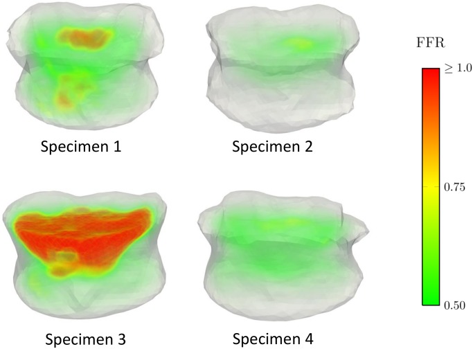
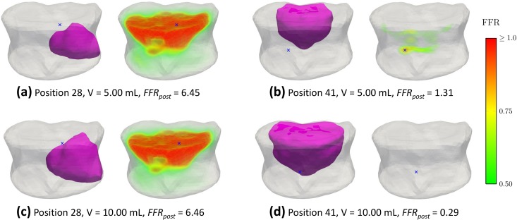
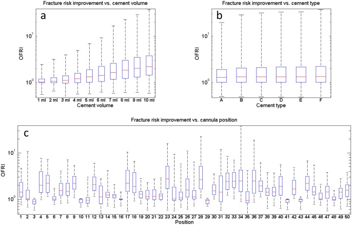
The local distribution of FFRpre for all the specimens is illustrated in Fig 2. All of the specimens have a maximum predicted FFRpre higher than one (Table 3) indicating that fracture is predicted for all the specimens prior to the augmentation. Sample simulated cement spreading patterns and local FFRpost -distributions for two cannula placement trials and two injection volumes are shown in Fig 3. The results for predicted values of FFR for pre and post-augmentation risk of fracture, as well as fracture risk improvement indicator (OFRI), are listed in Table 3. The organ fracture risk improvement indicator (OFRI) ranged from 0.47 to 37.23 depending on specimen, position, injected volume and cement type (Fig 4). Out of all treatments across all specimens, 76.6% resulted in an improved predicted fracture risk (OFRI > 1), 1.5% in no-effect (OFRI = 1) and 21.8% in a negative effect (OFRI < 1). Approximately 40% of the treatment strategies resulted in FFRpost being lower than unity but this number varied considerably between specimens, being 21, 74, 3, and 68% for specimens 1 to 4, respectively.
Fig 2. Pre-treatment initial factor of fracture risk (FFRpre).
Distribution according to FE simulations for all of the specimens.
Table 3. Pre- and post-augmentation risk for fracture (FFRpre and FFRpost) and fracture risk improvement indicator (OFRI).
| Specimen | FFRpre | FFRpost | OFRI |
|---|---|---|---|
| 1 | 1.95 | 0.29–3.43 | 0.57–6.76 |
| 2 | 1.08 | 0.03–2.28 | 0.47–37.23 |
| 3 | 6.41 | 0.29–11.60 | 0.55–22.44 |
| 4 | 1.06 | 0.17–1.79 | 0.59–6.21 |
Fig 3. Comparison of pre- and post-augmentation fracture risk.
The local change of the fracture risk following two treatment strategies and cannula placement trials with cement type F. The blue markers indicate the location of maximum fracture risk occurrence. (a) The asymmetric cement placement leads to a detrimental effect with a cement volume of V = 5.00 mL and even a large cement volume (V = 10.0 mL) is incapable of preventing a fracture (c). (b) A more centrally placed cement volume of V = 5.00 mL is nearly adequate to prevent the organ from overloading. (d) Injecting a volume of V = 10.0 mL prevents the organ from overloading.
Fig 4. Post-augmentation fracture risk.
The organ fracture risk improvement as a function of the injection volume (a), the cement type (b) and cannula position (c). The cannula position explains 47.2% of the variation in fracture risk reduction after treatment, and also highlights that a poorly planned intervention can even have a detrimental effect. The treatment outcome is also strongly related to cement volume (22.8% of the variability). The cement type only has a marginal influence on the overall effect (0.2% of the variability).
The N-way ANOVA indicated that 9.9% of the variance in OFRI is explained by specimen variability, 22.8% of the variance in OFRI is explained by cement volume, 0.2% of the variance in OFRI explained by cement type, 47.2% of the variance in OFRI explained by cannula position and 19.9% of the variance is unexplained. All the variability was found to be significant (p < 0.05).
Discussion
The aim of this study was to systematically investigate potential percutaneous vertebroplasty strategies in an in-silico environment to establish whether the treatment is efficient from a biomechanical point of view, and what the important determinants of a positive outcome are.
The findings of this study demonstrate that the outcome of vertebroplasty from a biomechanical perspective is complex and dependent on multiple factors. We found that PVP considerably improved the strength and reduced the calculated fracture risk in 76.8% of the treatment strategies simulated, but that the largest contributor to variation in outcome is associated with the cannula placement. However, a treatment can increase the predicted fracture risk and highlights that PVP can even have a detrimental effect. This has also been reported by Higgins et al. [36]. Generally, a good treatment outcome is associated with a cannula placed close to the central aspect of the vertebral body. This finding is consistent with the conclusions of other studies, where asymmetric placement of cement has been shown to reduce the biomechanical stability of osteoporotic vertebra [24, 31].
Biomechanical studies have reported that small amounts of cement resulted in a statistically significant increase in terms of strength [25, 36, 41] or stiffness [31, 35], compared to untreated vertebrae. Our data support this hypothesis only partially. For all cement volumes, we found treatment strategies that affected fracture risk both positively and negatively; however, with a clear trend towards higher cement volumes resulting in lower predicted fracture risk. Although our data suggests that large injection volumes are beneficial, it has been proposed that subsequent vertebral compression fractures of adjacent or remote levels may be caused by the increased stiffness of the treated vertebrae as a result of the amount of cement injected [28–30, 59] and are biomechanically not optimal [31]. The in-silico models in the present study however, are not capable of predicting the fracture risk in adjacent or remote levels. Therefore, this risk is not included in our models, but although indicated, large cement volumes do not have to be implicitly beneficial. Additionally, other biomechanical [60] and clinical studies [17, 61] found no statistically significant correlation between cement volume and pain relief. Therefore, large cement volumes should not be injected on a routine basis. Furthermore, the potential biomechanical benefits of increased cement volumes should be considered against the risks of cement or marrow extravasation.
We found that only 40% of the treatment strategies across all specimens resulted in a post treatment fracture risk (FFRpost) below unity. However, the absolute values of the FFR factor have to be interpreted with care because the current in-silico prediction of the fracture risk is based on a single simplified load case. In addition, it is unknown to which extent bone can be loaded without causing pain, therefore we defined relatively conservative strain thresholds for defining failure from a biomechanical point of view. The specimen variability in the predicted fracture risk after treatment is of interest. Only 3% of the treatment strategies resulted in a FFRpost factor below unity for specimen 3 but 67% for specimen 4 from the same donor. This could, in our opinion, further support the conclusion that the treatment of osteoporotic vertebrae needs to be carefully planned in order to capture all the weaknesses when augmenting the cancellous compartment. These results thus only conditionally support the statement that small cement volumes on the order of 30% (4–8 mL, [60]) are sufficient to achieve good outcomes [17, 25, 31, 35] because by studying our data in more detail we found e.g. that no cannula positions would produce a FFRpost below unity with a cement volume smaller than 6 ml for specimen 3.
It has been reported that highly osteoporotic patients may receive the least amount of improvement in vertebral mechanical strength after vertebroplasty [62] and PVP is unnecessary for a subgroup of patients with osteoporotic fractures [9]. Moreover, the data of this study highlight the sensitivity of the mechanical response on the treatment parameters [31, 61]; critics would say that pain relief by vertebroplasty can only be achieved by chance, whereas optimists argue that the uncertainty can be eliminated by optimization techniques. The computational model presented in this study is a first attempt to solve this optimization problem by considering the individual patient’s local bone condition prior to treatment and investigating different treatment strategies with the novelty of a rapid, patient-specific prediction of the ultimate bone strength and cement spreading pattern. The advantage and importance of a patient-specific procedure to maximize the mechanical benefits while minimizing the risk of complications have been pointed out by Higgins et al. [36]. Furthermore, the cement spreading pattern is said to be influential on the stiffening and strengthening of the vertebral body [24, 31]. The FE model incorporates a simplified but patient-specific mechanical model of the vertebra, with only compressive loading and prophylactic vertebroplasty simulated. This has the advantage that multiple treatment scenarios can be rapidly computed and compared, which is considered valid and well-accepted for the accurate prediction of compressive strength and fracture risk [39, 63]. It is recommended, however, that a conclusive assessment of treatment effectiveness be verified through the simulation of functional spinal units (FSUs) including fractured vertebrae in order to assess fracture risk at adjacent or remote levels, incorporating spectra of multi-axial loading and age-related daily activities.
There are several limitations in our study that need to be addressed. First, we are using intact specimens in our simulations, whereas PVP is generally performed to treat fractured vertebrae in the clinic. The FE models would thus only directly simulate the prophylactic augmentation of vertebrae at risk, which is a potential future development of clinical procedures. Nevertheless, we believe that carrying this study out on fractured specimens would only further strengthen our main conclusion from this work, which is that treatment needs to be carefully planned in order to have a biomechanical effect, as fractured vertebrae demonstrate a high degree of heterogeneity in the local bone properties. We believe that the added complexity of fracture patterns in the specimens would most likely contribute to increase the number of unsuccessful treatment outcomes. Second, we are only simulating unipedicular injection of cement but bi-pedicular injection procedures are also used in clinical practice. However, the goal of the present study was not to find out which treatment strategy to use, which could be subject specific anyway, but rather to determine which treatment parameters the outcome is most sensitive to.
In summary our results suggest that: (a) the outcome and effectiveness of vertebroplasty is strongly determined the patient’s bone condition prior to treatment, which defines the baseline and the amount of mechanical stabilization required for the prevention of subsequent fractures; (b) not all treatment strategies are suitable for accomplishing this mechanical stabilization; and (c) the risk of treatment complications (subsequent fractures in adjacent or remote levels, cement leakage) might be a limiting factor for a treatment success. Whilst the latter has been recognized and thoroughly discussed in literature, until now the first two aspects were not given enough attention and might at least partially explain the controversial conclusions drawn from the randomized controlled trials that support or question the effectiveness of vertebroplasty.
Supporting Information
This Matlab file contains the predicted risk of fracture before and after augmentation for all of the 12000 simulations cases of the study.
(MAT)
Data Availability
All relevant data are within the paper and its Supporting Information files.
Funding Statement
This work was funded by the European Research Council project FP7-ICT-223865-VPHOP.
References
- 1.Kanis JA, Johnell O. Requirements for DXA for the management of osteoporosis in Europe. Osteoporos Int 2005;16: 229–38. [DOI] [PubMed] [Google Scholar]
- 2.Haleem S, Lutchman L, Mayahi R, Grice JE, Parker MJ. Mortality following hip fracture: Trends and geographical variations over the last 40 years. Injury-International Journal of the Care of the Injured 2008;39: 1157–1163. [DOI] [PubMed] [Google Scholar]
- 3.Lau E, Ong K, Kurtz S, Schmier J, Edidin A. Mortality following the diagnosis of a vertebral compression fracture in the Medicare population. Journal of Bone and Joint Surgery-American Volume 2008;90A: 1479–1486. [DOI] [PubMed] [Google Scholar]
- 4.Robinson Y, Olerud C. Vertebroplasty and kyphoplasty-A systematic review of cement augmentation techniques for osteoporotic vertebral compression fractures compared to standard medical therapy. Maturitas 2012;72: 42–49. 10.1016/j.maturitas.2012.02.010 [DOI] [PubMed] [Google Scholar]
- 5.Buchbinder R, Osborne RH, Ebeling PR, Wark JD, Mitchell P, Wriedt C, et al. A Randomized Trial of Vertebroplasty for Painful Osteoporotic Vertebral Fractures. New England Journal of Medicine 2009;361: 557–568. 10.1056/NEJMoa0900429 [DOI] [PubMed] [Google Scholar]
- 6.Kallmes DF, Comstock BA, Heagerty PJ, Turner JA, Wilson DJ, Diamond TH, et al. A Randomized Trial of Vertebroplasty for Osteoporotic Spinal Fractures. New England Journal of Medicine 2009;361: 569–579. 10.1056/NEJMoa0900563 [DOI] [PMC free article] [PubMed] [Google Scholar]
- 7.Clark W, Lyon S, Burnes J. Trials of Vertebroplasty for Vertebral Fractures. New England Journal of Medicine 2009;361: 2097–2098. 10.1056/NEJMc096289 [DOI] [PubMed] [Google Scholar]
- 8.Baerlocher MO, Munk PL, Liu DM, Tomlinson G, Badii M, Kee ST, et al. Clinical Utility of Vertebroplasty: Need for Better Evidence. Radiology 2010;255: 669–674. 10.1148/radiol.10092107 [DOI] [PubMed] [Google Scholar]
- 9.Andersson GB. Surgery: vertebroplasty: one solution does not fit all. Nat Rev Rheumatol 2009;5: 662–3. 10.1038/nrrheum.2009.233 [DOI] [PubMed] [Google Scholar]
- 10.Sambrook P. Between a rock and a hard place: What is the evidence for vertebroplasty and kyphoplasty? IBMS BoneKEy 2009;5: 385–388. [Google Scholar]
- 11.Klazen CA, Lohle PN, de Vries J, Jansen FH, Tielbeek AV, Blonk MC, et al. Vertebroplasty versus conservative treatment in acute osteoporotic vertebral compression fractures (Vertos II): an open-label randomised trial. Lancet 2010;376: 1085–92. 10.1016/S0140-6736(10)60954-3 [DOI] [PubMed] [Google Scholar]
- 12.Edidin AA, Ong KL, Lau E, Schmier JK, Kemner JE, Kurtz SM. Cost-effectiveness analysis of treatments for vertebral compression fractures. Appl Health Econ Health Policy 2012;10: 273–84. 10.2165/11633220-000000000-00000 [DOI] [PubMed] [Google Scholar]
- 13.Svedbom A, Alvares L, Cooper C, Marsh D, Strom O. Balloon kyphoplasty compared to vertebroplasty and nonsurgical management in patients hospitalised with acute osteoporotic vertebral compression fracture: a UK cost-effectiveness analysis. Osteoporos Int 2013;24: 355–67. 10.1007/s00198-012-2102-y [DOI] [PMC free article] [PubMed] [Google Scholar]
- 14.Truumees E, Hilibrand A, Vaccaro AR. Percutaneous vertebral augmentation. Spine J 2004;4: 218–29. [DOI] [PubMed] [Google Scholar]
- 15.Heini PF. Vertebroplastik bei hochgradiger osteoporose. Orthopäde 2004: 22–29. [DOI] [PubMed] [Google Scholar]
- 16.Hulme PA, Krebs J, Ferguson SJ, Berlemann U. Vertebroplasty and kyphoplasty: A systematic review of 69 clinical studies. Spine (Phila Pa 1976) 2006;31: 1983–2001. [DOI] [PubMed] [Google Scholar]
- 17.Al-Ali F, Barrow T, Luke K. Vertebroplasty: what is important and what is not. AJNR Am J Neuroradiol 2009;30: 1835–9. 10.3174/ajnr.A1732 [DOI] [PMC free article] [PubMed] [Google Scholar]
- 18.Masala S, Mastrangeli R, Petrella MC, Massari F, Ursone A, Simonetti G. Percutaneous vertebroplasty in 1,253 levels: results and long-term effectiveness in a single centre. Eur Radiol 2009;19: 165–71. 10.1007/s00330-008-1133-4 [DOI] [PubMed] [Google Scholar]
- 19.Masala S, Mammucari M, Angelopoulos G, Fiori R, Massari F, Faria S, et al. Percutaneous vertebroplasty in the management of vertebral osteoporotic fractures. Short-term, mid-term and long-term follow-up of 285 patients. Skeletal Radiol 2009;38: 863–9. 10.1007/s00256-009-0712-z [DOI] [PubMed] [Google Scholar]
- 20.Klazen CA, Venmans A, de Vries J, van Rooij WJ, Jansen FH, Blonk MC, et al. Percutaneous vertebroplasty is not a risk factor for new osteoporotic compression fractures: results from VERTOS II. AJNR Am J Neuroradiol 2010;31: 1447–50. 10.3174/ajnr.A2148 [DOI] [PMC free article] [PubMed] [Google Scholar]
- 21.Blasco J, Martinez-Ferrer A, Macho J, San Roman L, Pomes J, Carrasco J, et al. Effect of vertebroplasty on pain relief, quality of life, and the incidence of new vertebral fractures: a 12-month randomized follow-up, controlled trial. J Bone Miner Res 2012;27: 1159–66. 10.1002/jbmr.1564 [DOI] [PubMed] [Google Scholar]
- 22.Mazzantini M, Carpeggiani P, d'Ascanio A, Bombardieri S, Di Munno O. Long-term prospective study of osteoporotic patients treated with percutaneous vertebroplasty after fragility fractures. Osteoporos Int 2011;22: 1599–607. 10.1007/s00198-010-1341-z [DOI] [PubMed] [Google Scholar]
- 23.Edidin AA, Ong KL, Lau E, Kurtz SM. Life expectancy following diagnosis of a vertebral compression fracture. Osteoporos Int 2013;24: 451–8. 10.1007/s00198-012-1965-2 [DOI] [PubMed] [Google Scholar]
- 24.Tschirhart CE, Roth SE, Whyne CM. Biomechanical assessment of stability in the metastatic spine following percutaneous vertebroplasty: effects of cement distribution patterns and volume. J Biomech 2005;38: 1582–90. [DOI] [PubMed] [Google Scholar]
- 25.Belkoff SM, Mathis JM, Jasper LE, Deramond H. The biomechanics of vertebroplasty. The effect of cement volume on mechanical behavior. Spine (Phila Pa 1976) 2001;26: 1537–41. [DOI] [PubMed] [Google Scholar]
- 26.Baroud G, Nemes J, Heini P, Steffen T. Load shift of the intervertebral disc after a vertebroplasty: a finite-element study. European Spine Journal 2003;12: 421–426. [DOI] [PMC free article] [PubMed] [Google Scholar]
- 27.Baroud G, Heini P, Nemes J, Bohner M, Ferguson S, Steffen T. Biomechanical explanation of adjacent fractures following vertebroplasty. Radiology 2003;229: 606–7; author reply 607–8. [DOI] [PubMed] [Google Scholar]
- 28.Berlemann U, Ferguson SJ, Nolte LP, Hein PF. Adjacent vertebral failure after vertebroplasty—A biomechanical investigation. Journal of Bone and Joint Surgery-British Volume 2002;84B: 748–752. [DOI] [PubMed] [Google Scholar]
- 29.Polikeit A, Nolte LP, Ferguson SJ. The effect of cement augmentation on the load transfer in an osteoporotic functional spinal unit—Finite-element analysis. Spine (Phila Pa 1976) 2003;28: 991–996. [DOI] [PubMed] [Google Scholar]
- 30.Wilcox RK. The biomechanical effect of vertebroplasty on the adjacent vertebral body: a finite element study. Proceedings of the Institution of Mechanical Engineers Part H-Journal of Engineering in Medicine 2006;220: 565–572. [DOI] [PubMed] [Google Scholar]
- 31.Liebschner MA, Rosenberg WS, Keaveny TM. Effects of bone cement volume and distribution on vertebral stiffness after vertebroplasty. Spine (Phila Pa 1976) 2001;26: 1547–54. [DOI] [PubMed] [Google Scholar]
- 32.Baroud G, Bohner M. Biomechanical impact of vertebroplasty. Postoperative biomechanics of vertebroplasty. Joint Bone Spine 2006;73: 144–50. [DOI] [PubMed] [Google Scholar]
- 33.Wasnich RD. Vertebral fracture epidemiology. Bone 1996;18: 179S–183S. [DOI] [PubMed] [Google Scholar]
- 34.Tohmeh AG, Mathis JM, Fenton DC, Levine AM, Belkoff SM. Biomechanical efficacy of unipedicular versus bipedicular vertebroplasty for the management of osteoporotic compression fractures. Spine (Phila Pa 1976) 1999;24: 1772–6. [DOI] [PubMed] [Google Scholar]
- 35.Luo J, Daines L, Charalambous A, Adams MA, Annesley-Williams DJ, Dolan P. Vertebroplasty: only small cement volumes are required to normalize stress distributions on the vertebral bodies. Spine (Phila Pa 1976) 2009;34: 2865–73. [DOI] [PubMed] [Google Scholar]
- 36.Higgins KB, Harten RD, Langrana NA, Reiter MF. Biomechanical effects of unipedicular vertebroplasty on intact vertebrae. Spine (Phila Pa 1976) 2003;28: 1540–7; discussion 1548. [PubMed] [Google Scholar]
- 37.Sun K, Liebschner MA. Biomechanics of prophylactic vertebral reinforcement. Spine (Phila Pa 1976) 2004;29: 1428–35; discusssion 1435. [DOI] [PubMed] [Google Scholar]
- 38.Sun K, Liebschner MA. Evolution of vertebroplasty: a biomechanical perspective. Ann Biomed Eng 2004;32: 77–91. [DOI] [PubMed] [Google Scholar]
- 39.Dickey BT, Tyndyk MA, Doman DA, Boyd D. In silico evaluation of stress distribution after vertebral body augmentation with conventional acrylics, composites and glass polyalkenoate cements. J Mech Behav Biomed Mater 2012;5: 283–90. 10.1016/j.jmbbm.2011.08.007 [DOI] [PubMed] [Google Scholar]
- 40.Rohlmann A, Boustani HN, Bergmann G, Zander T. A probabilistic finite element analysis of the stresses in the augmented vertebral body after vertebroplasty. Eur Spine J 2010;19: 1585–95. 10.1007/s00586-010-1386-x [DOI] [PMC free article] [PubMed] [Google Scholar]
- 41.Chevalier Y, Pahr D, Charlebois M, Heini P, Schneider E, Zysset P. Cement distribution, volume, and compliance in vertebroplasty—Some answers from an anatomy-based nonlinear finite element study. Spine (Phila Pa 1976) 2008;33: 1722–1730. [DOI] [PubMed] [Google Scholar]
- 42.Dall'Ara E, Pahr D, Varga P, Kainberger F, Zysset P. QCT-based finite element models predict human vertebral strength in vitro significantly better than simulated DEXA. Osteoporosis International 2012;23: 563–572. 10.1007/s00198-011-1568-3 [DOI] [PubMed] [Google Scholar]
- 43.Orwoll ES, Marshall LM, Nielson CM, Cummings SR, Lapidus J, Cauley JA, Ensrud K, Lane N, Hoffmann PR, Kopperdahl DL, Keaveny TM, Osteoporotic Fractures in Men Study G. Finite element analysis of the proximal femur and hip fracture risk in older men. J Bone Miner Res 2009;24: 475–83. 10.1359/jbmr.081201 [DOI] [PMC free article] [PubMed] [Google Scholar]
- 44.Kopperdahl DL, Aspelund T, Hoffmann PF, Sigurdsson S, Siggeirsdottir K, Harris TB, Gudnason V, Keaveny TM. Assessment of incident spine and hip fractures in women and men using finite element analysis of CT scans. J Bone Miner Res 2014;29: 570–80. 10.1002/jbmr.2069 [DOI] [PMC free article] [PubMed] [Google Scholar]
- 45.Dragomir-Daescu D, Op Den Buijs J, McEligot S, Dai Y, Entwistle RC, Salas C, Melton LJ 3rd, Bennet KE, Khosla S, Amin S. Robust QCT/FEA models of proximal femur stiffness and fracture load during a sideways fall on the hip. Ann Biomed Eng 2011;39: 742–55. 10.1007/s10439-010-0196-y [DOI] [PMC free article] [PubMed] [Google Scholar]
- 46.Grassi L, Hraiech N, Schileo E, Ansaloni M, Rochette M, Viceconti M. Evaluation of the generality and accuracy of a new mesh morphing procedure for the human femur. Med Eng Phys 2011;33: 112–20. 10.1016/j.medengphy.2010.09.014 [DOI] [PubMed] [Google Scholar]
- 47.Hazrati Marangalou J, Ito K, Cataldi M, Taddei F, van Rietbergen B. A novel approach to estimate trabecular bone anisotropy using a database approach. J Biomech 2013;46: 2356–62. 10.1016/j.jbiomech.2013.07.042 [DOI] [PubMed] [Google Scholar]
- 48.Zysset PK, Goulet RW, Hollister SJ. A global relationship between trabecular bone morphology and homogenized elastic properties. J Biomech Eng 1998;120: 640–6. [DOI] [PubMed] [Google Scholar]
- 49.Garcia D, Zysset PK, Charlebois M, Curnier A. A three-dimensional elastic plastic damage constitutive law for bone tissue. Biomech Model Mechanobiol 2009;8: 149–65. 10.1007/s10237-008-0125-2 [DOI] [PubMed] [Google Scholar]
- 50.Bayraktar HH, Morgan EF, Niebur GL, Morris GE, Wong EK, Keaveny TM. Comparison of the elastic and yield properties of human femoral trabecular and cortical bone tissue. J Biomech 2004;37: 27–35. [DOI] [PubMed] [Google Scholar]
- 51.Meijer GJ, Homminga J, Hekman EE, Veldhuizen AG, Verkerke GJ. The effect of three-dimensional geometrical changes during adolescent growth on the biomechanics of a spinal motion segment. J Biomech 2010;43: 1590–7. 10.1016/j.jbiomech.2010.01.028 [DOI] [PubMed] [Google Scholar]
- 52.Wilke HJ, Neef P, Caimi M, Hoogland T, Claes LE. New in vivo measurements of pressures in the intervertebral disc in daily life. Spine (Phila Pa 1976) 1999;24: 755–62. [DOI] [PubMed] [Google Scholar]
- 53.Helgason B, Viceconti M, Runarsson TP, Brynjolfsson S. On the mechanical stability of porous coated press fit titanium implants: a finite element study of a pushout test. J Biomech 2008;41: 1675–81. 10.1016/j.jbiomech.2008.03.007 [DOI] [PubMed] [Google Scholar]
- 54.Widmer RP, Ferguson SJ. On the interrelationship of permeability and structural parameters of vertebral trabecular bone: a parametric computational study. Comput Methods Biomech Biomed Engin 2012. [DOI] [PubMed] [Google Scholar]
- 55.Widmer RP, Ferguson SJ. A Mixed Boundary Representation to Simulate the Displacement of a Biofluid by a Biomaterial in Porous Media. Journal of Biomechanical Engineering-Transactions of the Asme 2011;133. [DOI] [PubMed] [Google Scholar]
- 56.Widmer Soyka RP, Lopez A, Persson C, Cristofolini L, Ferguson SJ. Numerical description and experimental validation of a rheology model for non-Newtonian fluid flow in cancellous bone. J Mech Behav Biomed Mater 2013;27: 43–53. 10.1016/j.jmbbm.2013.06.007 [DOI] [PubMed] [Google Scholar]
- 57.Helgason B, Stirnimann P, Widmer R, Boger A, Ferguson SJ. Influence of cement stiffness and bone morphology on the compressive properties of bone-cement composites in simulated vertebroplasty. Journal of Biomaterials Research—Part B 2013;101: 364–374. [DOI] [PubMed] [Google Scholar]
- 58.Race A, Mann KA, Edidin AA. Mechanics of bone/PMMA composite structures: an in vitro study of human vertebrae. J Biomech 2007;40: 1002–10. [DOI] [PubMed] [Google Scholar]
- 59.Baroud G, Wu JZ, Bohner M, Sponagel S, Steffen T. How to determine the permeability for cement infiltration of osteoporotic cancellous bone. Med Eng Phys 2003;25: 283–8. [DOI] [PubMed] [Google Scholar]
- 60.Molloy S, Mathis JM, Belkoff SM. The effect of vertebral body percentage fill on mechanical behavior during percutaneous vertebroplasty. Spine (Phila Pa 1976) 2003;28: 1549–54. [PubMed] [Google Scholar]
- 61.Kaufmann TJ, Trout AT, Kallmes DF. The effects of cement volume on clinical outcomes of percutaneous vertebroplasty. AJNR Am J Neuroradiol 2006;27: 1933–7. [PMC free article] [PubMed] [Google Scholar]
- 62.Graham J, Ahn C, Hai N, Buch BD. Effect of bone density on vertebral strength and stiffness after percutaneous vertebroplasty. Spine (Phila Pa 1976) 2007;32: E505–11. [DOI] [PubMed] [Google Scholar]
- 63.Jones AC, Wilcox RK. Finite element analysis of the spine: Towards a framework of verification, validation and sensitivity analysis. Medical Engineering & Physics 2008;30: 1287–1304. [DOI] [PubMed] [Google Scholar]
Associated Data
This section collects any data citations, data availability statements, or supplementary materials included in this article.
Supplementary Materials
This Matlab file contains the predicted risk of fracture before and after augmentation for all of the 12000 simulations cases of the study.
(MAT)
Data Availability Statement
All relevant data are within the paper and its Supporting Information files.


