Abstract
M-cells (microfold cells) are thought to be a primary conduit of intestinal antigen trafficking. Using an established neutralizing anti-RANKL (Receptor Activator of NF-κB Ligand) antibody treatment to transiently deplete M-cells in vivo, we sought to determine whether intestinal M-cells were required for the effective induction of protective immunity following oral vaccination with ΔiglB (a defined live attenuated Francisella novicida mutant). M-cell depleted, ΔiglB-vaccinated mice exhibited increased (but not significant) morbidity and mortality following a subsequent homotypic or heterotypic pulmonary F. tularensis challenge. No significant differences in splenic IFN-γ, IL-2, or IL-17 or serum antibody (IgG1, IgG2a, IgA) production were observed compared to non-depleted, ΔiglB-vaccinated animals suggesting complementary mechanisms for ΔiglB entry. Thus, we examined other possible routes of gastrointestinal antigen sampling following oral vaccination and found that ΔiglB co-localized to villus goblet cells and enterocytes. These results provide insight into the role of M-cells and complementary pathways in intestinal antigen trafficking that may be involved in the generation of optimal immunity following oral vaccination.
Introduction
Oral vaccination serves as an efficacious mechanism to induce potent systemic and mucosal immunity. This route targets the largest immune organ in the body, the gut and its associated lymphoid tissue, which contains 80% of the body's activated B cells [1] and up to 70% of the body’s immunocytes [2]. Oral vaccines, besides being more easily administered, may more successfully stimulate the mucosal immune system as this route allows for direct interaction of the vaccine with mucosal tissues and subsequent induction of antigen-specific mucosal immunity required for clearance of many pathogens, including F. tularensis [3]. The clinical efficacy of oral vaccines has been demonstrated against a variety of pathogens, including poliovirus (Sabin vaccine), rotavirus, Salmonella Typhi, and Vibrio cholerae [4], and this route also has been deemed more cost-effective and amenable to mass administration as minimal training is required for oral vaccination [5].
Our laboratory [3, 6, 7] and others [8–10] have demonstrated success using oral vaccines against pulmonary F. tularensis challenge in both mice [3, 6, 8–10] and rats [7], with LVS [3, 9, 10] and other live attenuated F. tularensis vaccines including U112ΔiglB [6] (referred to as ΔiglB in this paper) and Schu S4 mutants ΔclpB, ΔiglC, and the double mutant Δ0918ΔcapB [8] at varying doses (103–108 CFU). Our studies have demonstrated protection in mice against Schu S4 challenge with low doses (1000 CFU) of LVS [3] or ΔiglB [6] oral vaccination; the protective immunity was accompanied by potent cellular and humoral immune responses, as illustrated by IFN-γ production from antigen-specific T cells and antibody production both locally (intestinal IgA) and systemically (IgG1, IgG2a, and IgA in sera).
The success of oral vaccines has been attributed to the induction of the common mucosal immune system [11, 12] and efficient antigen-sampling involving intestinal M-cells (microfold cells) [2, 13]. M-cells are predominantly found in the follicle-associated epithelium (FAE) of intestinal Peyer’s patches (PP), and have distinctive morphological features, including a unique basolateral invagination which allows for docking and interaction with immune cells from the lamina propria, thus serving as a conduit for antigens trafficked from the lumen to be presented to APCs within the lamina propria [14]. Targeting vaccines to M-cells has been suggested as a potential mechanism for increased induction of immunity [15, 16] and has been attempted in mice, primates, and humans [17, 18]. However, the mechanism(s) by which M-cells may facilitate the induction of protective immunity has yet to be elucidated.
To this end, anti-RANKL neutralizing antibody (αRANKL) treatment has been demonstrated as an effective method to transiently deplete intestinal M-cells [19], and we utilized this treatment regimen in this study to reduce M-cells at the time of oral vaccination with the defined live attenuated mutant ΔiglB [6, 7]. Subsequently, we tested whether depletion of intestinal M-cells at the time of priming altered the immune response to oral vaccination. Additionally, we explored other intestinal cell types as complementary mechanisms in uptake and trafficking of the ΔiglB oral vaccine.
Materials and Methods
Animals
Four to six week old female BALB/c mice were obtained from the National Cancer Institute (Bethesda, MD). Mice were housed at the University of Texas at San Antonio AAALAC accredited facility, in ventilated cages and received food and water ad libitum for all experiments. The only exception to these conditions was for specified imaging experiments, in which mice were moved to wire-bottomed cages the night before the experiment, received water containing 5% sucrose, and were fasted overnight for no more than 16 hrs. All work was done in accordance with the University of Texas at San Antonio Institutional Biosafety Committee (IBC) and Institutional Animal Care and Use Committee (IACUC), who specifically approved this study.
Bacterial (Francisella) challenge may cause pain and distress to animals; however, potential effects on unalleviated pain are naturally occurring and should be considered part of the total immune response. Intervention with analgesics could induce variables to our studies and make the interpretation of data difficult. Thus, animals challenged with F. tularensis were provided nutrient gel cups in the cages so that all animals had direct source of fluids. Mice were monitored and weighed daily. When animals became symptomatic (such as inactivity, sunken eyes, hunched posture piloerection/matted fur), they were monitored twice daily not more than 14 hours apart. Any animal that was clearly terminal as indicated by lack of activity, difficulty in breathing, ruffled fur persisting for 24 hours and dramatic loss of body weight greater than 20% were euthanized in a closed chamber with CO2 (no response to vigorous rear toe pinch) followed by cervical dislocation.
Bacteria
Francisella tularensis live vaccine strain (LVS, lot # 703-0303-016) was obtained from Dr. Rick Lyons at Colorado State University, and Francisella novicida strain U112 was obtained from Dr. Francis Nano at the University of Victoria, Canada. All strains were grown on tryptic soy agar (TSA) or in tryptic soy broth (TSB, both obtained from BD Biosciences) supplemented with 0.1% (w/v) L-cysteine (Fisher Scientific). The vaccine strain F. novicida ΔiglB was generated in our previous report [20] and the cloning strategy for generating mCherry LVS [3] was applied to obtain the mCherry-expressing ΔiglB strain (KKF431) in this study. Dilution plating was used to enumerate titers of stocks.
Intestinal imaging
For intestinal imaging following overnight fasting, mice were anesthetized and orally administered a single 100 L dose of mCherry ΔiglB, which had been grown overnight to OD600 = 1.0 (approximately 109 CFU/mL), using a 22-gauge, 25-mm-long, 1.25-mm-round tip feeding needle via a previously established oral vaccination procedure [3]. No evidence was shown to have LVS delivered to the lungs by this oral gavage inoculation. Mice were sacrificed 90 minutes or 3 hrs post-vaccination to collect the entire intestinal tract for immunohistochemistry (IHC) or cytometry imaging analyses. For IHC, the intestinal tract was embedded into paraffin in sequential segments, then sectioned using a Microm rotary microtome and stained with H&E or PAS. Other sectioned tissues were subjected to confocal imaging analyses with the following antibodies: rhodamine- or FITC-labeled anti-UEA-1 (Vector Labs) for M-cells, anti-cytokeratin-18 and anti-MUC2 (both from Abcam) for GCs, Alexa 647 goat anti-mouse IgG1 (Life Technologies), and FITC goat anti-rabbit Ig secondary antibodies (Jackson Immunoresearch) plus DAPI nuclear stain (Fisher Scientific). Briefly, slides were heat fixed at 65°C for 20–30 minutes and rehydrated through a series of 3 minute long xylene and ethanol baths. Following rehydration, tissues were permeabilized for 10 minutes at room temperature, then blocked with serum for 30 minutes at room temperature. After blocking, slides were stained with primary antibodies overnight at 4°C, rinsed, and stained with secondary antibodies with DAPI for 2 hrs at room temperature. Slides were washed and mounted prior to imaging on the Zeiss 510 Meta laser scanning confocal microscope. For cytometry imaging analysis, excised PP or 5-cm small intestinal segments were subjected to single cell preparation [21] and cell surface labelling with either Alexa Fluor 488 conjugated anti-GP2 mouse mAb (MBL Inc.) or FITC-anti-MUC2 for detection of M-cells in PP or GC in the intestine, respectively. The labeled cells were then visualized and frequency analyzed using the Imagestream MKII (Amnis, EMD-Millipore).
M-cell depletion treatment and Peyer's patch (PP) whole mount staining
Mice were treated i.p. following a previously described protocol [19] with 250 μg of either anti-RANKL antibody (clone IK22-5[19]), or rat Ig (BioXcell) as a mock treatment, on days 0, 2, 4, and 6, for a total of 4 doses. Animals were then sacrificed at defined time points following treatment and PP removed for whole mount staining as previously described [19]. Whole PP were imaged at low magnification with combined low light phase contrast/fluorescence microscopy and with fluorescence microscopy (Zeiss Axioskop) and ImageJ software to quantify the number of M-cells in each PP.
Vaccination, immune response assessment, and F. tularensis challenge
Three groups of mice (n = 3 per group) were treated as described with either αRANKL antibody or rat Ig, or remained untreated, and were orally vaccinated with ΔiglB (103 or 105 CFU in separate experiments) 2 days after final Ab treatment. The fourth group of naïve mice received PBS orally as the non-vaccination control. Animals were rested for 3 weeks and vaccination induced immunity was assessed. For cellular responses, splenocytes (106 cells/well, in triplicate) from individual animals were stimulated with antigens for 24 hrs and the frequency of IFN-γ, IL-2 and IL-17 producing cells was determined by ELISpot as described previously [22]. Antigens included Concanavalin A (1 μg/well) and anti-CD3 (1 μg/mL) as positive controls, hen egg lysozyme (HEL, 1 μg/well) and medium as negative controls, and UV-inactivated ΔiglB (consisting of 1 μg proteins/well) for Ag-specific responses. For humoral responses, mice were bled, and serum Ab titers against ΔiglB were assayed by ELISA for total Ig, IgG1, and IgG2a (all from Southern Biotech) using previously described protocols [3, 6]. For challenge experiments, similarly Ab-treated and vaccinated groups (4) of mice (n = 6–10 per group) were challenged intranasally with LVS or U112 at 30 days after vaccination as previously described [3, 6]. Mice were monitored daily for morbidity and mortality for 30 days after challenge.
Statistical analysis
Statistical analysis was performed using GraphPad Prism 5 software. One way ANOVA with Bonferroni and Tukey multiple comparisons were used to compare naïve, vaccinated, rat Ig vaccinated, and αRANKL vaccinated group immune responses (both cellular and humoral responses). One way ANOVA also was used to compare M-cell counts among these groups. Kaplan-Meyer analysis with the Gehan-Breslow-Wilcoxon test was used for challenge studies.
Results
Putative Francisella tularensis vaccine strain ΔiglB co-localizes to M-cells after oral vaccination
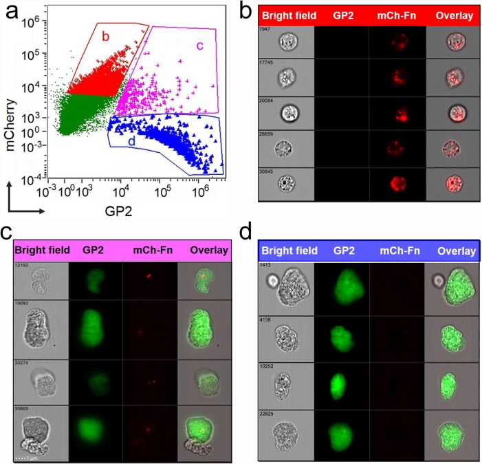
Our laboratory previously demonstrated that orally-administered F. tularensis live vaccine strain (LVS) co-localized to M-cells within 90 minutes [3]. To verify the translocation of ΔiglB via intestinal M-cells, we inoculated mice with mCherry-expressing ΔiglB by oral gavage and collected small intestines 90 minutes later. Single cells were made from excised PPs, labeled with AF-488 conjugated anti-GP2 (a PP M-cell specific antigen [23]) antibody, and analyzed by imaging flow cytometry. As shown in Fig 1, approximately 3% of the examined PP cells are GP2+ M-cells (Fig 1A, population 1C and 1D) and 25% of the M-cells contain mCherry-ΔiglB (Fig 1A, population 1C). mCherry-ΔiglB also was detected in GP2- cells (Fig 1A population 1B, ~9% of total PP cells) which may include antigen presenting cells (dendritic cells and macrophages) closely interacting with M-cells. The intracellular localization of mCherry-ΔiglB in Fig 1A population b and c was estimated to be 98% and 97% using the Internalization Index analysis (IDEAS®), and the representative cell images are displayed in Fig 1B and 1C, respectively. Additionally, mCherry-ΔiglB can be found at the proximity of M-cells in PP by immunohistochemistry visualization at 90 minutes after injection into closed murine ileal loops (data not shown). Collectively, these results suggest M-cells serve as a conduit for antigen-trafficking of orally administered F. tularensis in the intestine.
Fig 1. Uptake of ΔiglB vaccine strain by intestinal M-cells following oral inoculation.
BALB/c mice (4–6 wks) were orally vaccinated with mCherry-expressing ΔiglB (108 CFU/100 μL). Small intestines were collected at 90 min. post-vaccination and Peyer’s patches were excised to generate single cell suspensions. Cells were labeled with AF-488 conjugated α-GP2 antibody and subjected to cytometry imaging analysis. (a) Dot-plot depicts GP2 and mCherry intensity of each examined cell and three gated cell populations: (b) mCherryhiGP2low, (c) mCherryhiGP2hi, and (d) mCherrylowGP2hi. The representative cell images of these three gated populations are shown in (b) ΔiglB residing in non-M-cells, (c) ΔiglB residing in M-cells, and (d) M-cells with no ΔiglB, respectively. Representative images from 2 independent experiments are shown.
Administration of αRANKL antibody transiently depletes M-cells
Impaired M-cell development in mice has been demonstrated in a variety of knockout animals, including LT (lymphotoxin)-α, LT-β, and IL-7R knockouts; these animals, however, suffer severe immune defects as the gut and immune system do not mature properly with these genetic alterations [24]. In order to assess immune function in the absence of M-cells, we instead utilized a transient depletion strategy to temporarily knock-down M-cells in mice, via a neutralizing αRANKL antibody treatment regimen previously demonstrated to successfully reduce intestinal M-cells [19].
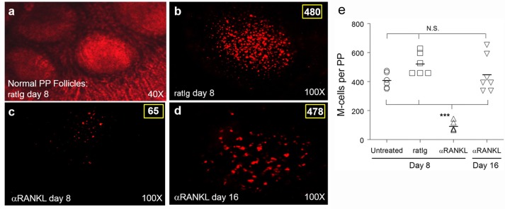
Whole mount imaging was employed to visualize and subsequently quantify M-cells in the PP of untreated, M-cell depleted (αRANKL treated), or rat Ig (ratIg; control Ig to αRANKL) treated animals at the time of vaccination (day 8). As shown in Fig 2, αRANKL treatment significantly depleted M-cells (labeled by α-UEA-1 antibody) within the intestine (p < 0.001 compared to no treatment or mock treatment), from 480 countable M-cells in a mock-treated PP (Fig 2B) to 65 in an αRANKL treated mouse (Fig 2C) with an approximate 90% reduction of M-cells in this case. This depletion was temporary, with M-cells beginning to emerge in follicles by day 12 (6 days after last treatment, data not shown) and completely restored by day 16 (10 days after treatment cessation, Fig 2D) with M-cell counts comparable to an untreated PP or a day 8 mock-treated PP (quantitative data for multiple animals shown in Fig 2E). Similar αRANKL-mediated M-cell depletion was observed when whole mounted PP were analyzed with the more M-cell specific α-GP2 antibody (S1 Fig). These findings confirm the effective, transient depletion of M-cells with αRANKL treatment in BALB/c mice. Additionally, this antibody treatment did not produce detectable physiological or immunological adverse effects in mice (S2 Fig).
Fig 2. M-cells are depleted with αRANKL antibody treatment.
BALB/c mice (n = 3 per group) were treated i.p. with 250 μg of either rat Ig (a-b) or αRANKL antibody IK22-5 (c-d) on days 0, 2, 4, and 6. On day 8 (a-c) or day 16 (d), animals were sacrificed and Peyer's patches (PP) collected and stained with rhodamine-labeled UEA-1 for whole mount imaging. Representative images showing normal morphology of intact (ratIg-treated) PP were taken using combined phase contrast and fluorescence at 40x (a), or labeled M cells (480 in rat Ig-treated PP on day 8, 65 M-cells in αRANKL-treated PP on day 8, or 476 in αRANKL-treated PP on day 16) all with fluorescence alone at 100x (b-d). M-cells in PP whole mounts were quantified for naive, rat Ig-treated, and αRANKL-treated groups (e), with αRANKL treatment inducing a significant decrease in M-cells (***p < 0.001) compared to naive or rat Ig-treated animals at day 8 or αRANKL-treated animals upon PP repopulation (day 16). Representative images from 3 experiments are shown.
M-cell depletion at the time of vaccination does not significantly increase morbidity or mortality following challenge
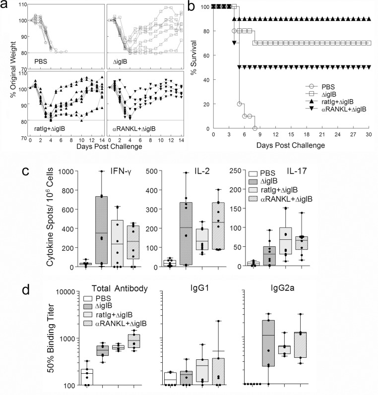
Having shown transient depletion of M-cells, we compared the susceptibility of untreated or mock-treated vaccinated controls with M-cell depleted, vaccinated animals to pulmonary F. tularensis challenge. M-cell depleted/ ΔiglB-vaccinated animals exhibited increased, but not significant, morbidity (prolonged weight loss, hunched posture, ruffled fur, inactivity) and mortality compared to challenged M-cell intact (mock and rat Ig treatment) ΔiglB-vaccinated groups (Fig 3A and 3B). While M-cell depletion deceased survival, there was no significant difference in survival between the three ΔiglB-vaccinated groups (50% for M-cell depleted, 70% for untreated, and 90% for rat Ig treated, Fig 3B) after a 45,000 CFU (~10 LD50) pulmonary challenge with the murine-virulent strain LVS. As expected, all mock (PBS)-vaccinated animals succumbed to challenge at this lethal dose (p < 0.01 compared to vaccinated groups). As this vaccine is so efficacious at the 1000 CFU vaccination dose, we reasoned that the lack of a significant difference between the M-cell intact and M-cell depleted groups was because the vaccine was so well-tolerated and immunogenic. We sought to test this by pushing the boundaries of the protection generated by the vaccine by increasing the challenge dose for the heterotypic LVS challenge and by adding a homotypic U112 challenge, perhaps leading to a significant difference between M-cell depleted and M-cell intact groups. However, we found no significant differences in survival of vaccinated M-cell depleted animals compared to M-cell sufficient control groups (receiving rat Ig or no treatment) with an increased challenge dose (85,000 CFU, ~20 LD50) of LVS (37.5% vs 50%) or with a homotypic (1000 CFU U112; ~100 LD50, 66.7% vs 100%, S3 Fig) pulmonary challenge. These results demonstrate that depletion of M-cells at the time of priming did not significantly abrogate protection against a subsequent pulmonary F. tularensis challenge.
Fig 3. Depletion of M-cells does not affect ability to survive subsequent pulmonary challenge or mount an immune response.
BALB/c mice (n = 10 per group for a & b, n = 3 per group for c & d) were treated i.p. with rat Ig or αRANKL antibody (250 μg) on days 0, 2, 4, and 6, and then, along with an untreated group of mice (indicated as ΔiglB), were vaccinated orally with ΔiglB (1000 CFU for a & b, 105 CFU for c & d) on day 8. Naïve mice receiving PBS orally were used as the non-vaccinated control. (a-b) After 30 days, all animals were intranasally challenged with 45,000 CFU of LVS (~10 LD50) and were monitored daily for weight changes (a) and survival (b). Representative results from 3 experiments are shown. (c-d) After a resting period of 21 days to allow for clearance of the vaccine, mice were bled to obtain sera, and then sacrificed for spleen collection. (c) ELISpots for IFN-γ, IL-2, and IL-17 were performed using single-cell splenocytes stimulated with 1 μg of UV-killed ΔiglB. Spot numbers of 3 individual spleens with triplicate, 9 for each group, are shown with mean ± standard error. Representative of 2 independent experiments. (d) ELISAs were conducted to determine serum antibody responses (total antibody, IgG1, and IgG2a) to UV-killed ΔiglB. Representative results from 3 experiments are shown. * p <0.05, *** p < 0.001.
Depletion of M-cells does not abrogate the vaccination induced cellular or humoral immune responses
Although survival was not significantly altered, we assessed whether cellular or humoral immune responses were decreased following depletion of M-cells. Antigen-specific IFN-γ, IL-2 and IL-17 production by T cells were examined by ELISpot, as these cytokines have been demonstrated to be important for clearance of pulmonary F. tularensis [3, 25, 26]. As shown in Fig 3C, stimulation of splenocytes from ΔiglB-vaccinated animals (whether M-cell depleted or not) with UV-killed ΔiglB, resulted in a higher frequency of IFN-γ, IL-2 and IL-17 production compared to PBS vaccinated mice (p < 0.05). However, there were no significant differences among the 3 vaccinated groups indicating no significant difference between M-cell depleted or non-depleted ΔiglB-vaccinated groups in production of any of the analyzed cytokines following recall with UV-killed ΔiglB.
Humoral responses (Fig 3D) showed a similar pattern to the cellular responses (Fig 3C), with the only significant differences seen between the vaccinated groups and those receiving a PBS mock vaccination for total antibody and no differences with M-cell depletion. Specifically, PBS animals had an average 50% binding titer of 178 in comparison to 551, 630, and 889 for ΔiglB, ratIg+ΔiglB, and αRANKL+ΔiglB groups, respectively. Further isotyping analyses revealed that all three ΔiglB-vaccinated groups of mice produced comparable levels of IgG2a, consistent with a high frequency of IFN-γ producing T cells, suggesting that Th1 type immunity was generated in the vaccinated animals regardless of M-cell depletion. Serum IgA production in all four groups was minimal, again, with no significant differences between the three vaccinated groups (data not shown). Additionally, no significant differences were seen in IgM or IgA production in fecal supernatants (data not shown). Overall, this data suggests that αRANKL antibody treatment and the resulting depletion of M-cells does not abrogate the ability of mice to mount potent cellular or humoral immune responses which provide protection against a pulmonary F. tularensis challenge.
Potential complementary mechanisms for oral ΔiglB vaccine entry in the small intestine
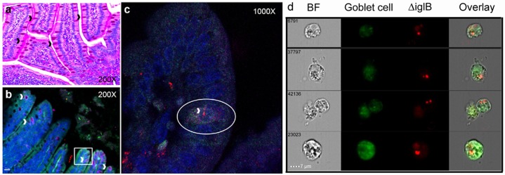
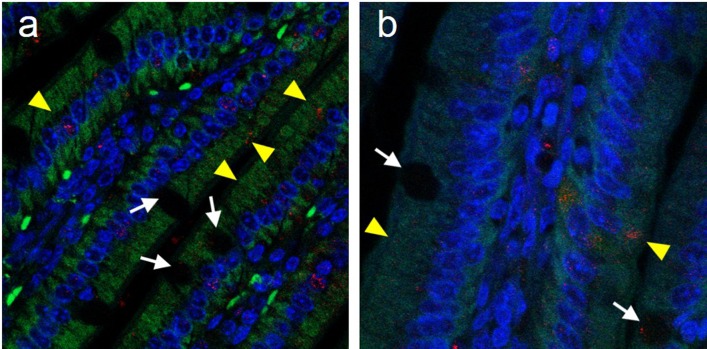
As depletion of M-cells at vaccination did not significantly alter immune responses or affect survival following pulmonary challenge, complementary mechanisms of antigen trafficking and delivery may be involved in generating protection following oral ΔiglB immunization. Several mechanisms of antigen trafficking beside M-cell transcytosis have been reported [27], ranging from extensions of dendritic cell processes through the intestinal epithelial monolayer [28, 29], transepithelial passage [30, 31], translocation through enterocytes [32] and recently, a study by McDole et al.[33] demonstrating that goblet cells (GCs) can take up soluble antigen and interact with CD103+ dendritic cells within intestinal villi. This observation prompted us to investigate whether intestinal GCs interact with orally delivered ΔiglB. Small intestines were collected at 90 min after mCherry-ΔiglB inoculation and analyzed for bacterial uptake by IHC or cytometry imaging. As shown in Fig 4, GCs were readily visible in the periodic acid Schiff (PAS) stained intestinal tissue sections (black arrows, Fig 4A), and by co-staining of the GC surface marker cytokeratin-18 and mucin MUC-2, we observed the presence of mCherry-ΔiglB within GCs by confocal microscopy (white arrowheads, Fig 4B and 4C). Similar observations were made at 3 hrs post-vaccination and within ileal loop sections 90 min after injection (data not shown). Additional flow cytometry imaging analysis (Fig 4D) confirmed the internalization of mCherry-ΔiglB by GCs (which accounted for ~20% of ΔiglB-bearing cells in the preparation) 90 minutes post-vaccination. These results demonstrate that GCs may serve as a novel host cell for F. tularensis and a potential mechanism for soluble and particulate antigens to enter from the intestinal lumen. Additionally, we also detected mCherry-ΔiglB moving through villi other than by GCs in enterocytes following oral vaccination as shown in Fig 5. Kujala et.al, demonstrated that prions can be taken up by FAE enterocytes and released to macrophages in the sub-epithelial dome by exocytosis within Gpa33+ exosomes [32]. Thus, transepithelial passage also could play a role in ΔiglB transcytosis and serve as an additional complementary mechanism for antigen uptake and processing.
Fig 4. Goblet cells can take up Francisella tularensis by 90 minutes after oral vaccination.
BALB/c mice (n = 3) were orally vaccinated with mCherry-ΔiglB (approximately 108 CFU) and rested for 90 minutes prior to sacrifice for collection of whole intestines, which were paraffin embedded, sectioned, and stained for (a) periodic acid Schiff to visualize GCs (black arrowheads) and (b-c) confocal analysis with nuclear stain DAPI (blue), mucin marker anti-MUC-2 (green), and GC surface marker anti-cytokeratin-18 (pink), with GCs shown by white arrowheads. (c) High magnification of a goblet cell (circled), stained with both MUC-2 and cytokeratin-18, which has taken up ΔiglB (red). (d) Single cells were prepared from intestines of similarly vaccinated mice, labeled with FITC-anti-MUC-2 (green), and subjected to cytometry imaging to visualize the uptake of mCherry-ΔiglB (red) by goblet cells. BF, bright field. Representative images from 2 separate experiments are shown.
Fig 5. Uptake of mCherry-ΔiglB by enterocytes.
BALB/c mice (n = 3) were orally vaccinated with mCherry-ΔiglB (approximately 108 CFU) and rested for 90 minutes prior to sacrifice for collection of whole intestines, which were paraffin embedded, sectioned, and stained for confocal analysis with nuclear stain DAPI (blue), and with mCherry-ΔiglB in GCs shown by white arrows and mCherry-ΔiglB in enterocytes shown by yellow arrowheads. (a) 630x and (b) 1000x.
Discussion
Antigen trafficking within the intestine has been primarily attributed to M-cells [34–36], and previous studies have shown that in the absence of M-cells, infections with Yersinia enterocolitica [37], prions [38], and retrovirus [39] were abrogated, and antigen-specific T-cell responses to oral infection with Salmonella typhimurium was impaired [23, 40]. We demonstrate here that a significant reduction (approximately 90%) of M-cells at the time of oral priming did not significantly reduce antigen-specific cellular or humoral responses, or the ability of M-cell depleted animals to survive a subsequent pulmonary challenge with either a homotypic (U112) or heterotypic (LVS) strain of F. tularensis. As M-cells were not completely depleted, we must acknowledge that the remaining M-cells present following depletion treatment may still be functional and allow for trafficking of lumenal antigen. Similar transient M-cell depletion by αRANKL treatment has effectively blocked prion uptake and prevented disease progression [38]. In contrast, our results suggest that M-cells may not serve as the principal mechanism of antigen trafficking, at least for F. tularensis, a bacterium which does not seem to preferentially exploit M-cells for entry as occurs with Salmonella and Shigella. Such redundancy of function speaks directly to the importance of antigen trafficking in the intestine, while at the same time raises questions about the primary role of M-cells in this process, i.e., does antigen need to be trafficked through the M-cell to induce immunity? The role of M-cells in antigen processing of the orally delivered vaccines remains elusive. Although we can not rule out the possible vaccine transcytosis by repopulated M-cells upon cessation of treatment, antigen trafficking via goblet cells and enterocytes may explain the observed lack of significant decreases in immune responses and survival with M-cell reduction at priming.
The mCherry-labeled ΔiglB vaccine strain co-localized to UEA-1 positive regions (presumably M-cells stained with the lectin marker) above Peyer’s patches after oral vaccination (S4B and S4C Fig with a Peyer’s patch shown by H&E staining in S4A Fig). In contrast, M cell depletion by αRANKL treatment did not alter the ability of the ΔiglB vaccine strain to enter regions above and all the way through Peyer’s patches (S4D Fig). Additionally, M-cell depletion did not prevent entry through villi above Peyer’s patches (S4E Fig), suggesting that while M-cells traffic the live attenuated strain out of the intestinal lumen, routes other than M-cells also may serve as conduits for antigen-trafficking of orally administered F. tularensis in the intestine.
As M-cell depletion at the time of priming did not significantly reduce immune responses and survival, we examined complementary antigen trafficking mechanisms and surprisingly discovered that goblet cells and enterocytes were able to take up particulate antigens, including F. tularensis following oral vaccination. We initially hypothesized that trafficking via complementary mechanisms such as GCs may serve to compensate for the depletion of M-cells at the time of priming. However, we did not see an increase in the overall numbers of GCs in M-cell depleted animals (data not shown). At this time, the consequences of losing one or more of these complementary antigen trafficking mechanisms (either via genetic knockout or temporary depletion) are unknown.
As GCs are found throughout the intestinal epithelium but cannot be easily isolated from this area in primary culture, we differentiated a human colon epithelial cell line (HT29) to obtain the GC phenotype [41, 42] for the assessment of antigen uptake in vitro. Our HT29 cells were more viscous in cell culture in galactose and were both cytokeratin 18 and MUC-2 positive, two characteristics used in the seminal study of McDole et al.[33] to distinguish goblet cells in vivo. The GC-like HT29 cells took up both U112 and ΔiglB (S5A Fig, 3 hrs), with significant replication of U112 intracellularly shown at 24 and 48 hrs. In contrast to the parental strain, ΔiglB was deficient for replication in the HT29 cells, which was not unexpected as it cannot multiply in murine or rat macrophages [6, 7]. Nevertheless, this strain was taken into GCs (S5B Fig, Figs 4 and 5), demonstrating that F. tularensis can exploit GCs as a potential host cell, and further suggesting that GCs may serve as an entry point for the vaccine following oral immunization. Our results suggested that GCs can take up vaccine strain ΔiglB, but can GCs facilitate delivery of particulate antigens to an APC, i.e., a dendritic cell (DC)? We infected the differentiated HT29 cell line with ΔiglB. Exosomes isolated from the ΔiglB infected GC culture were able to activate human DCs to express the co-stimulatory marker CD80 (S5C Fig) and induce inflammatory cytokines IL-1β and IL-8 (S5D Fig). These results demonstrate a plausible mechanism by which GCs deliver uptake antigens to APCs via exosomes similarly to enterocytes [32]. The entrance mechanism of bacteria into GC is not known; however, we speculate that F. tularensis may be entering GCs via E-cadherin interactions and at tips of villi in regions of extruded enterocytes, as recently demonstrated for Listeria monocytogenes [43, 44]. Moreover, F. tularensis encodes a putative protein with a homologous binding site to L. monocytogenes InlA, which lends credence to this hypothesis (unpublished observations). These findings extend knowledge within the field of goblet cell biology and may have broader implications for pathogens in both the gastrointestinal and respiratory tracts where these cells are found (i.e., they may suggest alternative mechanisms for pathogen entry into the mucosa to cause illness).
In summary, this study has demonstrated the importance of redundant mechanisms of antigen trafficking in the intestine by M-cells and goblet cells for induction of protective immunity following vaccination. These studies also suggest complementary mechanisms by which immunity can be generated from an oral vaccine, inducing protection against a subsequent challenge in the respiratory compartment. Implications from this study extend beyond F. tularensis into bacterial pathogenesis and mucosal vaccine development. Listeria monocytogenes has already been demonstrated to exploit GCs to exit the intestinal lumen [43, 44]; it is likely, but unknown whether other enteric pathogens utilize GCs for this process, or if GCs are exploited by bacteria for mucosal entry in the respiratory compartment. Additionally, GCs may serve as a useful target for oral vaccines against these and other pathogens, inducing enhanced immunity against subsequent mucosal (gastrointestinal or pulmonary) infection.
Supporting Information
(PDF)
(PDF)
(PDF)
(PDF)
(PDF)
Acknowledgments
The authors would like to acknowledge Dr. Colleen Witt and Robert Cook of the UTSA Research Centers in Minority Institutions (RCMI) Biophotonics Core for help with confocal and two-photon imaging. We also thank Drs. Weidang Li, Rishein Gupta, and Annette Rodriguez for assistance with flow cytometry experiments.
Data Availability
All relevant data are within the paper and its Supporting Information files.
Funding Statement
This work was supported by the UTHSCSA Department of Microbiology and Immunology Microbial Pathogenesis training grant (NIH #T32AI007271), the UTSA Center for Excellence in Infection Genomics training grant (DOD #W911NF-11-1-0136 for BPA), and a UTSA RCMI (Regional Centers in Minority Institutions) grant (#G12MD007591) from the National Institute on Minority Health and Health Disparities. All funders had no role in study design, data collection and analysis, decision to publish, or preparation of the manuscript.
References
- 1.Brandtzaeg P, Baekkevold ES, Morton HC. From B to A the mucosal way. Nat Immunol. 2001;2(12):1093–4. Epub 2001/11/29. 10.1038/ni1201-1093 . [DOI] [PubMed] [Google Scholar]
- 2.Corr SC, Gahan CC, Hill C. M-cells: origin, morphology and role in mucosal immunity and microbial pathogenesis. FEMS Immunol Med Microbiol. 2008;52(1):2–12. Epub 2007/12/18. 10.1111/j.1574-695X.2007.00359.x . [DOI] [PubMed] [Google Scholar]
- 3.Ray HJ, Cong Y, Murthy AK, Selby DM, Klose KE, Barker JR, et al. Oral live vaccine strain-induced protective immunity against pulmonary Francisella tularensis challenge is mediated by CD4+ T cells and antibodies, including immunoglobulin A. Clin Vaccine Immunol. 2009;16(4):444–52. Epub 2009/02/13. 10.1128/CVI.00405-08 [DOI] [PMC free article] [PubMed] [Google Scholar]
- 4.Dietrich G, Griot-Wenk M, Metcalfe IC, Lang AB, Viret JF. Experience with registered mucosal vaccines. Vaccine. 2003;21(7–8):678–83. Epub 2003/01/18. doi: S0264410X02005790 [pii]. . [DOI] [PubMed] [Google Scholar]
- 5.Levine MM. Can needle-free administration of vaccines become the norm in global immunization? Nat Med. 2003;9(1):99–103. Epub 2003/01/07. 10.1038/nm0103-99 . [DOI] [PubMed] [Google Scholar]
- 6.Cong Y, Yu JJ, Guentzel MN, Berton MT, Seshu J, Klose KE, et al. Vaccination with a defined Francisella tularensis subsp. novicida pathogenicity island mutant (DiglB) induces protective immunity against homotypic and heterotypic challenge. Vaccine. 2009;27(41):5554–61. Epub 2009/08/05. 10.1016/j.vaccine.2009.07.034 [DOI] [PMC free article] [PubMed] [Google Scholar]
- 7.Signarovitz AL, Ray HJ, Yu JJ, Guentzel MN, Chambers JP, Klose KE, et al. Mucosal immunization with live attenuated Francisella novicida U112DiglB protects against pulmonary F. tularensis SCHU S4 in the Fischer 344 rat model. PLoS ONE. 2012;7(10):e47639 Epub 2012/11/03. 10.1371/journal.pone.0047639 [DOI] [PMC free article] [PubMed] [Google Scholar]
- 8.Conlan JW, Shen H, Golovliov I, Zingmark C, Oyston PC, Chen W, et al. Differential ability of novel attenuated targeted deletion mutants of Francisella tularensis subspecies tularensis strain SCHU S4 to protect mice against aerosol challenge with virulent bacteria: effects of host background and route of immunization. Vaccine. 2010;28(7):1824–31. Epub 2009/12/19. 10.1016/j.vaccine.2009.12.001 [DOI] [PMC free article] [PubMed] [Google Scholar]
- 9.KuoLee R, Harris G, Conlan JW, Chen W. Oral immunization of mice with the live vaccine strain (LVS) of Francisella tularensis protects mice against respiratory challenge with virulent type A F. tularensis. Vaccine. 2007;25(19):3781–91. Epub 2007/03/10. 10.1016/j.vaccine.2007.02.014 [DOI] [PMC free article] [PubMed] [Google Scholar]
- 10.Skyberg JA, Rollins MF, Samuel JW, Sutherland MD, Belisle JT, Pascual DW. Interleukin-17 protects against the Francisella tularensis live vaccine strain but not against a virulent F. tularensis type A strain. Infect Immun. 2013;81(9):3099–105. Epub 2013/06/19. 10.1128/IAI.00203-13 [DOI] [PMC free article] [PubMed] [Google Scholar]
- 11.Brandtzaeg P. Induction of secretory immunity and memory at mucosal surfaces. Vaccine. 2007;25(30):5467–84. Epub 2007/01/18. 10.1016/j.vaccine.2006.12.001 . [DOI] [PubMed] [Google Scholar]
- 12.Brandtzaeg P. Mucosal immunity: induction, dissemination, and effector functions. Scand J Immunol. 2009;70(6):505–15. Epub 2009/11/13. 10.1111/j.1365-3083.2009.02319.x . [DOI] [PubMed] [Google Scholar]
- 13.Jang MH, Kweon MN, Iwatani K, Yamamoto M, Terahara K, Sasakawa C, et al. Intestinal villous M cells: an antigen entry site in the mucosal epithelium. Proc Natl Acad Sci U S A. 2004;101(16):6110–5. Epub 2004/04/09. 10.1073/pnas.0400969101 [DOI] [PMC free article] [PubMed] [Google Scholar]
- 14.Neutra MR, Mantis NJ, Kraehenbuhl JP. Collaboration of epithelial cells with organized mucosal lymphoid tissues. Nat Immunol. 2001;2(11):1004–9. Epub 2001/10/31. 10.1038/ni1101-1004 . [DOI] [PubMed] [Google Scholar]
- 15.Frey A, Neutra MR. Targeting of mucosal vaccines to Peyer's patch M cells. Behring Inst Mitt. 1997;(98):376–89. Epub 1997/02/01. . [PubMed] [Google Scholar]
- 16.Kim SH, Seo KW, Kim J, Lee KY, Jang YS. The M cell-targeting ligand promotes antigen delivery and induces antigen-specific immune responses in mucosal vaccination. J Immunol. 2010;185(10):5787–95. Epub 2010/10/19. 10.4049/jimmunol.0903184 . [DOI] [PubMed] [Google Scholar]
- 17.Misumi S, Masuyama M, Takamune N, Nakayama D, Mitsumata R, Matsumoto H, et al. Targeted delivery of immunogen to primate M-cells with tetragalloyl lysine dendrimer. J Immunol. 2009;182(10):6061–70. Epub 2009/05/06. 10.4049/jimmunol.0802928 . [DOI] [PubMed] [Google Scholar]
- 18.Roth-Walter F, Bohle B, Scholl I, Untersmayr E, Scheiner O, Boltz-Nitulescu G, et al. Targeting antigens to murine and human M-cells with Aleuria aurantia lectin-functionalized microparticles. Immunol Lett. 2005;100(2):182–8. Epub 2005/05/26. 10.1016/j.imlet.2005.03.020 . [DOI] [PubMed] [Google Scholar]
- 19.Knoop KA, Kumar N, Butler BR, Sakthivel SK, Taylor RT, Nochi T, et al. RANKL is necessary and sufficient to initiate development of antigen-sampling M cells in the intestinal epithelium. J Immunol. 2009;183(9):5738–47. Epub 2009/10/16. 10.4049/jimmunol.0901563 . [DOI] [PMC free article] [PubMed] [Google Scholar]
- 20.Liu J, Zogaj X, Barker JR, Klose KE. Construction of targeted insertion mutations in Francisella tularensis subsp. novicida. Biotechniques. 2007;43(4):487–90, 92. Epub 2007/11/21. doi: 000112574 [pii]. . [DOI] [PubMed] [Google Scholar]
- 21.Geem D, Medina-Contreras O, Kim W, Huang CS, Denning TL. Isolation and characterization of dendritic cells and macrophages from the mouse intestine. J Vis Exp. 2012;(63):e4040 Epub 2012/05/31. 10.3791/4040 [DOI] [PMC free article] [PubMed] [Google Scholar]
- 22.Li W, Murthy AK, Lanka GK, Chetty SL, Yu JJ, Chambers JP, et al. A T cell epitope-based vaccine protects against chlamydial infection in HLA-DR4 transgenic mice. Vaccine. 2013;31(48):5722–8. Epub 2013/10/08. 10.1016/j.vaccine.2013.09.036 [DOI] [PMC free article] [PubMed] [Google Scholar]
- 23.Hase K, Kawano K, Nochi T, Pontes GS, Fukuda S, Ebisawa M, et al. Uptake through glycoprotein 2 of FimH+ bacteria by M cells initiates mucosal immune response. Nature. 2009;462(7270):226–30. Epub 2009/11/13. 10.1038/nature08529 . [DOI] [PubMed] [Google Scholar]
- 24.Adachi S, Yoshida H, Honda K, Maki K, Saijo K, Ikuta K, et al. Essential role of IL-7 receptor alpha in the formation of Peyer's patch anlage. Int Immunol. 1998;10(1):1–6. Epub 1998/03/06. . [DOI] [PubMed] [Google Scholar]
- 25.Surcel HM, Syrjala H, Karttunen R, Tapaninaho S, Herva E. Development of Francisella tularensis antigen responses measured as T-lymphocyte proliferation and cytokine production (tumor necrosis factor alpha, gamma interferon, and interleukin-2 and -4) during human tularemia. Infect Immun. 1991;59(6):1948–53. Epub 1991/06/01. [DOI] [PMC free article] [PubMed] [Google Scholar]
- 26.Cowley SC, Meierovics AI, Frelinger JA, Iwakura Y, Elkins KL. Lung CD4-CD8- double-negative T cells are prominent producers of IL-17A and IFN-gamma during primary respiratory murine infection with Francisella tularensis live vaccine strain. J Immunol. 2010;184(10):5791–801. Epub 2010/04/16. 10.4049/jimmunol.1000362 . [DOI] [PubMed] [Google Scholar]
- 27.Mabbott NA, Donaldson DS, Ohno H, Williams IR, Mahajan A. Microfold (M) cells: important immunosurveillance posts in the intestinal epithelium. Mucosal Immunol. 2013. Epub 2013/05/23. 10.1038/mi.2013.30 . [DOI] [PMC free article] [PubMed] [Google Scholar]
- 28.Rescigno M, Urbano M, Valzasina B, Francolini M, Rotta G, Bonasio R, et al. Dendritic cells express tight junction proteins and penetrate gut epithelial monolayers to sample bacteria. Nat Immunol. 2001;2(4):361–7. Epub 2001/03/29. 10.1038/86373 . [DOI] [PubMed] [Google Scholar]
- 29.Farache J, Koren I, Milo I, Gurevich I, Kim KW, Zigmond E, et al. Luminal bacteria recruit CD103+ dendritic cells into the intestinal epithelium to sample bacterial antigens for presentation. Immunity. 2013;38(3):581–95. Epub 2013/02/12. 10.1016/j.immuni.2013.01.009 . [DOI] [PMC free article] [PubMed] [Google Scholar]
- 30.Amerongen HM, Weltzin R, Farnet CM, Michetti P, Haseltine WA, Neutra MR. Transepithelial transport of HIV-1 by intestinal M cells: a mechanism for transmission of AIDS. J Acquir Immune Defic Syndr. 1991;4(8):760–5. Epub 1991/01/01. . [PubMed] [Google Scholar]
- 31.Fotopoulos G, Harari A, Michetti P, Trono D, Pantaleo G, Kraehenbuhl JP. Transepithelial transport of HIV-1 by M cells is receptor-mediated. Proc Natl Acad Sci U S A. 2002;99(14):9410–4. Epub 2002/07/03. 10.1073/pnas.142586899 [DOI] [PMC free article] [PubMed] [Google Scholar]
- 32.Kujala P, Raymond CR, Romeijn M, Godsave SF, van Kasteren SI, Wille H, et al. Prion uptake in the gut: identification of the first uptake and replication sites. PLoS Pathog. 2011;7(12):e1002449 Epub 2012/01/05. 10.1371/journal.ppat.1002449 [DOI] [PMC free article] [PubMed] [Google Scholar]
- 33.McDole JR, Wheeler LW, McDonald KG, Wang B, Konjufca V, Knoop KA, et al. Goblet cells deliver luminal antigen to CD103+ dendritic cells in the small intestine. Nature. 2012;483(7389):345–9. Epub 2012/03/17. 10.1038/nature10863 [DOI] [PMC free article] [PubMed] [Google Scholar]
- 34.Kraehenbuhl JP, Neutra MR. Molecular and cellular basis of immune protection of mucosal surfaces. Physiol Rev. 1992;72(4):853–79. Epub 1992/10/01. . [DOI] [PubMed] [Google Scholar]
- 35.Neutra MR, Frey A, Kraehenbuhl JP. Epithelial M cells: gateways for mucosal infection and immunization. Cell. 1996;86(3):345–8. Epub 1996/08/09. . [DOI] [PubMed] [Google Scholar]
- 36.Neutra MR, Pringault E, Kraehenbuhl JP. Antigen sampling across epithelial barriers and induction of mucosal immune responses. Annu Rev Immunol. 1996;14:275–300. Epub 1996/01/01. 10.1146/annurev.immunol.14.1.275 . [DOI] [PubMed] [Google Scholar]
- 37.Westphal S, Lugering A, von Wedel J, von Eiff C, Maaser C, Spahn T, et al. Resistance of chemokine receptor 6-deficient mice to Yersinia enterocolitica infection: evidence of defective M-cell formation in vivo. Am J Pathol. 2008;172(3):671–80. Epub 2008/02/09. 10.2353/ajpath.2008.070393 [DOI] [PMC free article] [PubMed] [Google Scholar]
- 38.Donaldson DS, Kobayashi A, Ohno H, Yagita H, Williams IR, Mabbott NA. M cell-depletion blocks oral prion disease pathogenesis. Mucosal Immunol. 2012;5(2):216–25. Epub 2012/02/02. 10.1038/mi.2011.68 [DOI] [PMC free article] [PubMed] [Google Scholar]
- 39.Golovkina TV, Shlomchik M, Hannum L, Chervonsky A. Organogenic role of B lymphocytes in mucosal immunity. Science. 1999;286(5446):1965–8. Epub 1999/12/03. . [DOI] [PubMed] [Google Scholar]
- 40.Kanaya T, Hase K, Takahashi D, Fukuda S, Hoshino K, Sasaki I, et al. The Ets transcription factor Spi-B is essential for the differentiation of intestinal microfold cells. Nat Immunol. 2012;13(8):729–36. Epub 2012/06/19. 10.1038/ni.2352 . [DOI] [PMC free article] [PubMed] [Google Scholar]
- 41.Huet C, Sahuquillo-Merino C, Coudrier E, Louvard D. Absorptive and mucus-secreting subclones isolated from a multipotent intestinal cell line (HT-29) provide new models for cell polarity and terminal differentiation. J Cell Biol. 1987;105(1):345–57. Epub 1987/07/01. [DOI] [PMC free article] [PubMed] [Google Scholar]
- 42.Godefroy O, Huet C, Blair LA, Sahuquillo-Merino C, Louvard D. Differentiation of a clone isolated from the HT29 cell line: polarized distribution of histocompatibility antigens (HLA) and of transferrin receptors. Biol Cell. 1988;63(1):41–55. Epub 1988/01/01. . [PubMed] [Google Scholar]
- 43.Nikitas G, Deschamps C, Disson O, Niault T, Cossart P, Lecuit M. Transcytosis of Listeria monocytogenes across the intestinal barrier upon specific targeting of goblet cell accessible E-cadherin. J Exp Med. 2011;208(11):2263–77. Epub 2011/10/05. 10.1084/jem.20110560 [DOI] [PMC free article] [PubMed] [Google Scholar]
- 44.Tsai YH, Disson O, Bierne H, Lecuit M. Murinization of internalin extends its receptor repertoire, altering Listeria monocytogenes cell tropism and host responses. PLoS Pathog. 2013;9(5):e1003381 Epub 2013/06/06. 10.1371/journal.ppat.1003381 [DOI] [PMC free article] [PubMed] [Google Scholar]
Associated Data
This section collects any data citations, data availability statements, or supplementary materials included in this article.
Supplementary Materials
(PDF)
(PDF)
(PDF)
(PDF)
(PDF)
Data Availability Statement
All relevant data are within the paper and its Supporting Information files.


