Abstract
Directed evolution in Saccharomyces cerevisiae offers many attractive advantages when designing enzymes for biotechnological applications, a process that involves the construction, cloning and expression of mutant libraries, coupled to high frequency homologous DNA recombination in vivo. Here, we present a protocol to create and screen mutant libraries in yeast based on the example of a fungal aryl-alcohol oxidase (AAO) to enhance its total activity. Two protein segments were subjected to focused-directed evolution by random mutagenesis and in vivo DNA recombination. Overhangs of ~50 bp flanking each segment allowed the correct reassembly of the AAO-fusion gene in a linearized vector giving rise to a full autonomously replicating plasmid. Mutant libraries enriched with functional AAO variants were screened in S. cerevisiae supernatants with a sensitive high-throughput assay based on the Fenton reaction. The general process of library construction in S. cerevisiae described here can be readily applied to evolve many other eukaryotic genes, avoiding extra PCR reactions, in vitro DNA recombination and ligation steps.
Keywords: Molecular Biology, Issue 110, Directed evolution, Saccharomyces cerevisiae, in vivo DNA recombination, focused-random mutagenesis, high-throughput screening
Introduction
Directed molecular evolution is a robust, fast and reliable method to design enzymes1, 2. Through iterative rounds of random mutation, recombination and screening, improved versions of enzymes can be generated that act on new substrates, in novel reactions, in non-natural environments, or even to assist the cell to achieve new metabolic goals3-5. Among the hosts used in directed evolution, the brewer's yeast Saccharomyces cerevisiae offers a repertoire of solutions for the functional expression of complex eukaryotic proteins that are not otherwise available in prokaryotic counterparts6,7.
Used exhaustively in cell biology studies, this small eukaryotic model has many advantages in terms of post-translational modifications, ease of manipulation and transformation efficiency, all of which are important traits to engineer enzymes by directed evolution8. Moreover, the high frequency of homologous DNA recombination in S. cerevisiae coupled to its efficient proof-reading apparatus opens a wide array of possibilities for library creation and gene assembly in vivo, fostering the evolution of different systems from single enzymes to complex artificial pathways9-12. Our laboratory has spent the past decade designing tools and strategies for the molecular evolution of different ligninases in yeast (oxidoreductases involved in the degradation of lignin during natural wood decay)13-14. In this communication, we present a detailed protocol to prepare and screen mutant libraries in S. cerevisiae for a model flavooxidase, -aryl-alcohol oxidase (AAO15)-, that can be easily translated to many other enzymes. The protocol involves a focused-directed evolution method (MORPHING: Mutagenic Organized Recombination Process by Homologous in vivo Grouping) assisted by the yeast cell apparatus16, and a very sensitive screening assay based on the Fenton reaction in order to detect AAO activity secreted into the culture broth17.
Protocol
1. Mutant Library Construction
- Choose the regions to be subjected to MORPHING with the help of computational algorithms based on the available crystal structure or homology models18.
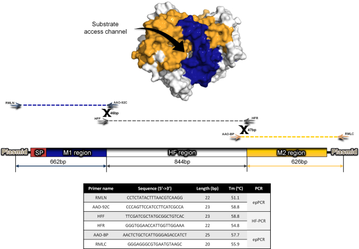
- Here, target two regions of AAO from Pleurotus eryngii for random mutagenesis and recombination (Met[α1]-Val109, Phe392-Gln566), while amplifying the remainder of the gene (844 bp) by high-fidelity PCR (Figure 1). Note: Several segments can be studied by MORPHING in an independent or combined manner16.
- Amplify the targeted areas by mutagenic PCR. Create overlapping areas between segments (~50 bp each) by superimposing PCR reactions of the defined regions.
- Prepare mutagenic PCR of targeted segments in a final volume of 50 µl containing DNA template (0.92 ng/µl), 90 nM oligo sense (RMLN for segment M-I and AAO-BP for segment M-II), 90 nM antisense primer (AAO-92C for segment M-I and RMLC for segment M-II) , 0.3 mM dNTPs (0.075 mM each), 3% (v/v) dimethylsulfoxide (DMSO), 1.5 mM MgCl2, 0.05 mM MnCl2 and 0.05 U/µl Taq DNA polymerase. Primers sequences are detailed in Figure 1.
- Use the following PCR program: 95 °C for 2 min (1 cycle); 95 °C for 45 sec, 50 °C for 45 sec, 74 °C for 45 sec (28 cycles); and 74 °C for 10 min (1 cycle).
- Amplify the non-mutagenic regions with ultra-high fidelity polymerase and include the corresponding areas overlapping the mutagenic segments and/or linearized vector overhangs.
- Prepare reaction mixtures in a final volume of 50 µl containing: DNA template (0.2 ng/µl), 250 nM oligo sense HFF, 250 nM oligo antisense HFR, 0.8 mM dNTPs (0.2 mM each), 3% (v/v) dimethylsulfoxide (DMSO) and 0.02 U/µl iproof DNA polymerase. Primers sequences are detailed in Figure 1.
- Use the following PCR program: 98 °C for 30 sec (1 cycle); 98 °C for 10 sec, 55 °C for 25 sec, 72 °C for 45 sec (28 cycles); and 72 °C for 10 min (1 cycle). Note: With conditions described in 1.2 and 1.3 overlaps of 43 bp (plasmid-M1 region); 46 bp (M1 region-HF region); 47 bp (HF region-M2 region) and 61 bp (M2 region- plasmid) are designed (Figure 1) to favor in vivo splicing in yeast.
- Purify all the PCR fragments (mutagenic and non-mutagenic) with a commercial gel extraction kit according to manufacturer's protocol.
- Linearize the vector such that flanking regions of approximately 50 bp are created that are homologous to the 5´- and 3´-ends of the target gene.
- Prepare a linearization reaction mixture containing 2 µg DNA, 7.5 U BamHI, 7.5 U XhoI, 20 µg BSA and 2 µl of Buffer BamHI 10x in a final volume of 20 µl.
- Incubate the reaction mixture at 37 °C for 2 hr and 40 min. Afterwards, proceed with inactivation at 80 °C for 20 min.
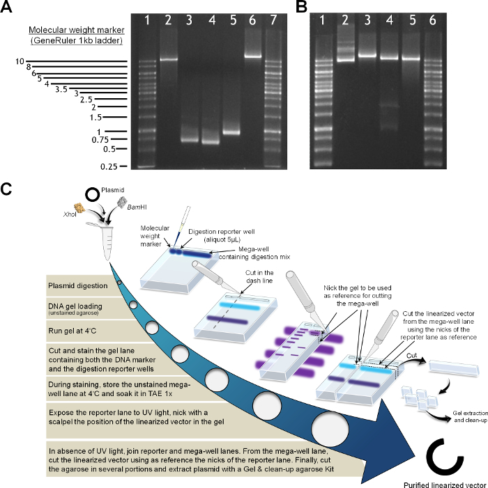
- Purify the linearized vector by agarose gel extraction to avoid contamination with the residual circular plasmid (Figure 2).
- Load the digestion reaction mix into the mega-well of a semi-preparative low melting point agarose gel (0.75%, w:v) as well as an aliquot (5 µl) of the reaction mix in the adjacent well as a reporter.
- Run DNA electrophoresis (5 V/cm between electrodes, 4 ᵒC) and separate the agarose gel corresponding to the mega-well and store it at 4 ᵒC in 1x TAE.
- Stain the lane with the molecular weight ladder and the reporter. Visualize the bands under UV light. Nick the position where the linearized vector places. Note: As the quality of the purified linearized vector is a critical factor for successful recombination and assembly in yeast, avoid gel staining for semi-preparative DNA electrophoresis. The use of dyes and UV exposure for gel extraction may affect the stability of the DNA vector, compromising the in vivo recombination efficiency. As alternative to toxic EtBr dyes, Gel Red and SYBR dyes are commonly used for gel staining.
- In the absence of UV light, identify the linearized vector in the mega-well fragment using the guidance of the nicks in the stained reporter lane so that it can be isolated.
- Extract the linearized vector from agarose and purify it with a commercial gel extraction kit according to manufacturer's protocol. Note: Use high-copy episomal shuttle vectors with antibiotic and auxotrophy markers: In this example we employed the uracil independent and ampicillin resistance pJRoC30 vector, under the control of the yeast GAL1 promoter.
- Prepare an equimolar mixture of the PCR fragments and mix it with the linearized vector at a 2:1 ratio, with no less than 100 ng of linearized plasmid (test different ratios of equimolar library/open vector to achieve good transformation yields).
- Measure the absorbance of the PCR fragments and linearized vector at 260 nm and 280 nm to determine their concentration and purity.
- Transform yeast competent cells with the DNA mixture using a commercial yeast transformation kit (see Table for supplies) according to manufacturer's instructions.
- Here, use a protease deficient and URA3- dependent S. cerevisiae strain, BJ5465. Transform the cells with the parental circularized vector as an internal standard during screening (see below). Additionally, check the background by transforming the linearized vector in the absence of PCR fragments. Note: In case of detecting initial low secretion levels, use S. cerevisiae protease deficient strains like BJ5465 to foster the accumulation of active protein in culture supernatants. If the target enzyme undergoes hyperglycosylation, the use of glycosylation-deficient strains (e.g., Δkre2 that is only capable of attaching smaller mannose oligomers) could be a suitable option.
Plate the transformed cells on SC drop-out plates and incubate them at 30 °C for three days. Plate (on SC drop-out plates supplemented with uracil) URA3-S. cerevisiae cells lacking the plasmid as a negative control for screening (see below).
2. High-Throughput Screening Assay (Figure 3)
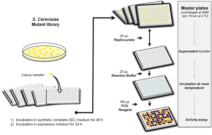
Fill an appropriate number of sterile 96-well plates (23 plates to analyze a library of 2,000 clones) with 50 µl minimal medium per well with the help of a pipetting robot.
- Pick individual colonies from the SC-drop out plates and transfer them to the 96-well plates.
- In each plate, inoculate column number 6 with the parental type as an internal standard and well H1 with URA3-S. cerevisiae cells (in SC medium supplemented with uracil) with no plasmid as a negative control. Note: Well H1 is filled specifically with drop-out media supplemented with uracil. A blank well containing media without cells can be also prepared as an additional sterility control.
Cover the plates with their lids and wrap them in Parafilm. Incubate plates for 48 hr at 30 °C, 225 rpm and 80% relative humidity in a humid shaker.
Remove the Parafilm, add 160 µl of expression medium to each well with the help of the pipetting robot, reseal the plates and incubate them for a further 24 hr. Note: Minimal medium and expression medium are prepared as reported elsewhere19. Secretion levels may vary depending on the gene under study and accordingly, the incubation times must be optimized in each case to synchronize the cell growth in all the wells.
Centrifuge the plates (master plates) at 2,800 x g for 10 min at 4 °C.
Transfer 20 µl of the supernatant from the wells in the master plate to the replica plate using a liquid handling robotic multistation. Note: To favor enzyme secretion it is advisable to replace the native signal peptide of the target protein by signal peptides commonly used for heterologous expression in yeast (e.g., the α factor prepro-leader, the leader of the K1 Killer toxin from S. cerevisiae, or even chimeric versions of both peptides13). Alternatively, the native signal peptide can be exclusively evolved for secretion in yeast.
Add 20 µl of 2 mM p-methoxybenzylalcohol in 100 mM sodium phosphate buffer pH 6.0 with the help of the pipetting robot. Stir the plates briefly with a 96-well plate mixer and incubate them for 30 min at RT.
- With the pipetting robot, add 160 µl of the FOX reagent to each replica plate and stir briefly with the mixer (final concentration of FOX mixture in the well: 100 µM xylenol orange, 250 µM Fe(NH4)2(SO4)2 and 25 mM H2SO4).
- Add several additives to the reagent to enhance sensitivity, such as organic co-solvents (DMSO, ethanol, methanol) or sorbitol17. Here, amplify the response by adding sorbitol to a final concentration of 100 mM (Figure 4).
Read the plates (end-point mode, t0) at 560 nm on a plate reader.
- Incubate the plates at RT until the color develops and measure the absorption again (t1).
- Calculate the relative activity from the difference between the Abs value after incubation and that of the initial measurement normalized to the parental type for each plate (Δt1 - t0).
Subject the best mutant hits to two consecutive re-screenings to rule out false positives. Note: Typically, re-screenings include plasmid isolation from yeast, amplification and purification in Escherichia coli, followed by transformation of fresh yeast cells with the plasmid19. Each selected clone is re-screened in pentaplicate.
Representative Results
AAO from P. eryngii is an extracellular flavooxidase that supplies fungal peroxidases with H2O2 to start attacking lignin. Two segments of AAO were subjected to focused-directed evolution by MORPHING in order to enhance its activity and its expression in S. cerevisiae 19. Irrespective of the foreign enzymes harbored by S. cerevisiae, the most critical issue when constructing mutant libraries in yeast concerns the engineering of specific overlapping regions to favor the splicing between fragments and their cloning into the linearized vector. In the current example, for each PCR reaction, all the fragments had overhangs of approximately 50 bp to promote in vivo splicing in yeast. The number of recombination events is dependent on the number of segments to be assembled and cloned with the linearized vector (i.e., two crossover events took place between the three PCR segments -the two mutagenic segments flanking the non-mutagenized segment- plus two additional crossovers with the linearized vector; Figure 1). According to our experience, overlapping sequences longer than 50 bp decrease the likelihood of internal recombination while they do not improve transformation efficiency.
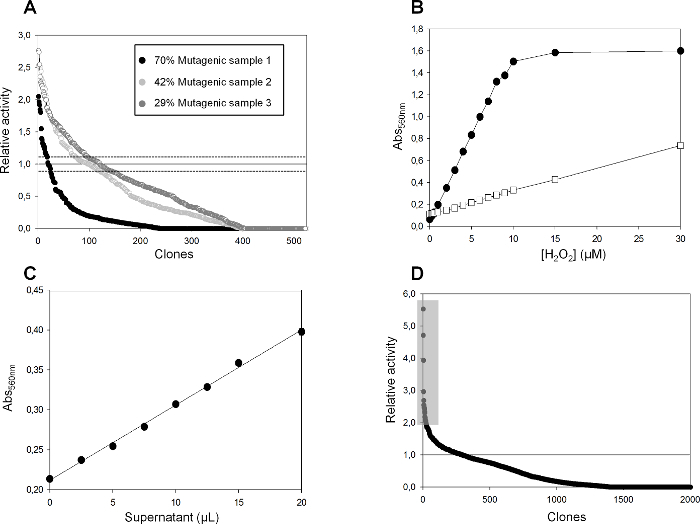
Mutational loads were adjusted by sampling mutant libraries with different landscapes, calculating the number of clones with <10% of the parental enzyme activity, and further checking them by sequencing a random sample of active and non-active variants (Figure 5A). For the determination of the coefficient of variance S. cerevisiae cells were transformed with the parental AAO and plated on SC-drop out plates. Individual colonies were picked and inoculated in a 96 well-plate and the activity of the clones was evaluated from fresh preparations. Mutagenic sample 2 (Taq/MnCl2 0.05 mM) was chosen as the departure point for library construction and screening.
As the biological activity of AAO increases the H2O2 concentration in the reaction medium, we searched for a sensitive and accurate assay to quantify minor changes in H2O2. FOX is a chemical method based on the Fenton reaction20, whereby oxidation by H2O2 drives the reaction of Fe3+ with xylenol orange to form a blue-purple complex (o-cresolsulfone-phthalein 3',3''-bis(methylimino)diacetate ε560 = 1.5 x 10⁴ M-1cm-1). The ferrous oxidation step was amplified by adding sorbitol to enhance the sensitivity of the assay, increasing the propagation of radicals with an apparent ε560 = 2.25 x 10⁵ M-1cm-1 (Figure 4).
The detection limit of this assay (in the µM range) was calculated by the Blank determination method in a 96-well plate with standards in triplicate (0, 0.5, 1, 1.5, 2, 2.5, 3 and 4 µM H2O2) and using several supernatants from S. cerevisiae lacking the URA3- plasmid (Figure 5B). The assay was linear in the presence of sorbitol (up to 8 µM of H2O2), and although linearity was more persistent in the absence of this sugar (at least up to 30 µM of H2O2) the response was weaker (e.g., at 6 µM of H2O2, a 4-fold enhancement was obtained in the presence of sorbitol -deep purple- from an absorbance of 0.24 in its absence -dark orange- (Figure 5B)). The relationship between Abs and the AAO concentration was evaluated with increasing amounts of enzyme (from yeast supernatants) and a linear response was observed; R2 = 0.997 (Figure 5C).
It is notable that the FOX signal was stable for several hours without any apparent interference by the different elements in the culture broth. The estimated sensitivity of FOX was ~0.4 µM of H2O2 produced by the AAO in the supernatantin the presence of sorbitol, and ~2 µM in its absence.
A mutant library of 2,000 clones was constructed and screened with this assay. Several AAO mutants were identified with notably improved secretion and activity against p-methoxybenzyl alcohol (Figure 5D)19.
 Figure 1. MORPHING Protocol for AAO Evolution. Two different regions of AAO were targeted for random mutagenesis and recombination: M1 (blue, 590 bp) that includes the signal peptide (SP); M2 (yellow, 528 bp). The HF region (grey, 844 bp) was amplified with high fidelity polymerases. Mutagenic regions were mapped in the crystal structure of AAO (PDB ID: 3FIM). Please click here to view a larger version of this figure.
 Figure 2. Preparation of PCR Products and the Linearized Vector. (A) Analytical agarose gel (1% w:v) containing a molecular weight marker (1 kb ladder) in lanes 1 and 7; the BamHI and XhoI linearized vector, lane 2; PCR segment M1, lane 3; PCR segment M2, lane 4; PCR segment HF, lane 5; the in vivo reassembled vector linearized with NheI (containing the full AAO gene with regions M-I, HF and M-II), lane 6. (B) Vector linearization, lanes 1 and 6 molecular standards, 1 Kb ladder; plasmid miniprep, lane 2; plasmid linearized with NheI, lane 3; plasmid linearized with BamHI and XhoI, lane 4; linearized plasmid obtained by gel extraction and clean-up after digestion, lane 5. (C) Protocol for plasmid purification. Please click here to view a larger version of this figure.
 Figure 3. High-throughput Screening Protocol. Overview of the process. Please click here to view a larger version of this figure.
 Figure 4. The FOX Method. White-rot fungi attack the cell wall of wood through a Fenton reaction that produces hydroxyl radical OH•. The FOX method couples this reaction to xylenol orange (XO), and the absorbance of the XO-Fe3+ complex is measured at 560 nm. Ferrous oxidation is amplified by the addition of sorbitol to the reagent mixture. Please click here to view a larger version of this figure.
 Figure 5. Mutagenic Landscapes for MORPHING Libraries Using Different Error Prone PCR Conditions and Validation of the Screening Assay. (A) MORPHING landscapes. Solid horizontal line shows the activity of the parental type in the assay while the dashed lines indicate the coefficient of variance of the assay. The percentages indicate the number of clones with less than 10% of the parental enzyme activity. Activities are plotted in descending order. (B) The FOX detection limit was evaluated with increasing concentrations of H2O2 in the presence (black circles) and absence (white squares) of sorbitol. (C) Linear correlation between AAO concentration (transformant supernatants) and Abs560nm. Each point corresponds to the average of 8 experiments and includes the standard deviation. (D) Mutant library landscape. The selected variants (shadowed square) were rescreened as reported elsewhere19. Solid line shows the activity of AAO parental type. Please click here to view a larger version of this figure.
Discussion
In this article, we have summarized most of the tips and tricks employed in our laboratory to engineer enzymes by directed evolution in S. cerevisiae (using AAO as an example) so that they can be adapted for use with many other eukaryotic enzyme systems by simply following the common approach described here.
In terms of library creation, MORPHING is a fast one-pot method to introduce and recombine random mutations in small protein stretches while leaving the remaining regions of the protein unaltered16. Libraries with several mutational loads can be readily prepared and recombined in vivo, along with the linearized plasmid, to generate a full autonomously replicating vector. It is critical that overlapping sequences flank each stretch to allow the fragments of the full gene to be reassembled through in vivo recombination, avoiding extra PCR reactions and in vitro ligation steps. In this protocol, the frequency of crossover events between PCR fragments can be increased by reducing the size of the overlapping regions, although this may compromise the transformation efficiency. Regardless of the DNA polymerases used for mutagenic PCR, the mutational loads can be adjusted by previously constructing and analyzing small mutant library landscapes (Figure 5A). If the GeneMorph II Kit is used, it is still advisable to follow this approach since in vivo DNA recombination can notably modify the mutational loads estimated by the manufacturer. In general terms, mutant landscapes in which 35 - 50% of the total clones screened have less than 10% of the parental activity are suitable for directed evolution campaigns, although this number varies in function of the target protein and its activity. Typically, the analysis of mutant libraries landscapes are further verified by DNA sequencing of a random sample of mutants. In the current example, the Taq DNA polymerase was used due to its high error rate, which is linked to the lack of 3´→5´proof-reading exonuclease activity. The mutational loads in Taq libraries were modified by the addition of different concentrations of MnCl2, but the use of unbalanced dNTPs and/or the reduction in gene template concentrations are also suitable options. Inherent limitations of MORPHING come from the number of segments to be recombined. According to our experience, up to four protein blocks (five crossover events counting the recombination areas with the linearized vector) can be spliced with good transformation yields (~105 clones per transformation reaction). This method can be easily modified to performed multiple site-saturation mutagenesis (e.g., using NDT degenerated primers or creating degeneracy for 22 unique codons) to explore several positions simultaneously while reducing significantly the screening efforts21,22.
The direct "blind" screening protocol for AAO is extremely sensitive and reliable (based on the direct detection of H2O2 regardless of the substrate used by the enzyme), representing a complementary assay to other well established indirect protocols to detect peroxides (mostly coupling peroxidases with colorimetric substrates). Indeed, the FOX assay has been routinely employed to measure H2O2 in biological fluids, and it can now be easily translated into protocols to evolve AAO and any other H2O2 producing enzymes (e.g., glucose oxidases, cellobiose dehydrogenases, glyoxal oxidases, methanol oxidases), particularly for activity on non-natural substrates where responses are otherwise hard to detect.
S. cerevisiae is the most adequate host for directed evolution of eukaryotic genes since it offers high transformation efficiencies (up to 1 x 10⁶ transformants/µg DNA), it performs complex post-translational processing and modifications (including N- and C-terminal processing, and glycosylation) and it exports foreign proteins into the culture broth via a secretory pathway. In addition, well-established molecular biology tools are available to work with this yeast, including uni- or bi-directional episomal (non-integrative) shuttle vectors under the control of promoters of different strengths. Last but not least, its high frequency of homologous DNA recombination has allowed a range of methods to be developed to obtain DNA diversity that are currently being used to evolve single proteins, as well as more complex enzyme pathways8, 12, 13, 23. The in vivo gap repair and the proof-reading device of this yeast can be also employed to create chimeras when recombining different genes (with approx. 60% of DNA sequence identity), as well as to shuffle best offspring/mutations from a directed evolution campaign, or to bring together in vitro and in vivo recombination methods in one round of evolution, thereby enriching mutant libraries in terms of foldability and function.
Disclosures
The authors have nothing to disclose.
Acknowledgments
This work was supported by the European Commission project Indox-FP7-KBBE-2013-7-613549; a Cost-Action CM1303-Systems Biocatalysis; and the National Projects Dewry [BIO201343407-R] and Cambios [RTC-2014-1777-3].
References
- Jäckel C, Hilvert D. Biocatalysts by evolution. Curr. Opin. Biotechnol. 2010;21(6):753–759. doi: 10.1016/j.copbio.2010.08.008. [DOI] [PubMed] [Google Scholar]
- Bornscheuer UT. Engineering the third wave of biocatalysis. Nature. 2012;485(7397):185–194. doi: 10.1038/nature11117. [DOI] [PubMed] [Google Scholar]
- Renata H, Wang ZW, Arnold FH. Expanding the enzyme universe: accessing non-natural reactions by mechanism-guided directed evolution. Angew. Chem. Int. Ed. 2015;54(11):3351–3367. doi: 10.1002/anie.201409470. [DOI] [PMC free article] [PubMed] [Google Scholar]
- Cobb RE, Chao R, Zhao H. Directed evolution: past, present and future. AIChE J. 2013;59(5):1432–1440. doi: 10.1002/aic.13995. [DOI] [PMC free article] [PubMed] [Google Scholar]
- Abatemarco J, Hill A, Alper HS. Expanding the metabolic engineering toolbox with directed evolution. Biotechnol. J. 2013;8(12):1397–1410. doi: 10.1002/biot.201300021. [DOI] [PubMed] [Google Scholar]
- Pourmir A, Johannes TW. Directed evolution: selection of the host organism. Comput Struct Biotechnol J. 2012;2(3):e201209012. doi: 10.5936/csbj.201209012. [DOI] [PMC free article] [PubMed] [Google Scholar]
- Krivoruchko A, Siewers V, Nielsen J. Opportunities for yeast metabolic engineering: lessons from synthetic biology. Biotechnol J. 2011;6(3):262–276. doi: 10.1002/biot.201000308. [DOI] [PubMed] [Google Scholar]
- Gonzalez-Perez D, Garcia-Ruiz E, Alcalde M. Saccharomyces cerevisiae in directed evolution: an efficient tool to improve enzymes. Bioeng Bugs. 2012;3:172–177. doi: 10.4161/bbug.19544. [DOI] [PMC free article] [PubMed] [Google Scholar]
- Alcalde M. Mutagenesis protocols in Saccharomyces cerevisiae by In Vivo Overlap Extension. Methods Mol. Biol. 2010;634:3–14. doi: 10.1007/978-1-60761-652-8_1. [DOI] [PubMed] [Google Scholar]
- Bulter T, Alcalde M. Preparing libraries in Saccharomyces cerevisiae. Methods. Mol. Biol. 2003;231:17–22. doi: 10.1385/1-59259-395-X:17. [DOI] [PubMed] [Google Scholar]
- Ostrov N, Wingler LM, Cornish W. Gene assembly and combinatorial libraries in S. cerevisiae via reiterative recombination. Methods. Mol. Biol. 2013;978:187–203. doi: 10.1007/978-1-62703-293-3_14. [DOI] [PubMed] [Google Scholar]
- Shao Z, Zhao H, Zhao H. DNA assembler, an in vivo genetic method for rapid construction of biochemical pathways. Nucleic Acids Res. 2009;37(2):e16. doi: 10.1093/nar/gkn991. [DOI] [PMC free article] [PubMed] [Google Scholar]
- Alcalde M. Engineering the ligninolytic enzyme consortium. Trends Biotechnol. 2015;33(3):155–162. doi: 10.1016/j.tibtech.2014.12.007. [DOI] [PubMed] [Google Scholar]
- Garcia-Ruiz E. Cascade Biocatalysis: integrating stereoselective and environmentally friendly reactions. Wiley-VCH; 2014. Directed evolution of ligninolytic oxidoreductases: from functional expression to stabilization and beyond; pp. 1–22. [Google Scholar]
- Hernandez-Ortega A, Ferreira P, Martinez AT. Fungal aryl-alcohol oxidase: a peroxide-producing flavoenzyme involved in lignin degradation. Appl. Microbiol. Biotechnol. 2012;93(4):1395–1410. doi: 10.1007/s00253-011-3836-8. [DOI] [PubMed] [Google Scholar]
- Gonzalez-Perez D, Molina-Espeja P, Garcia-Ruiz E, Alcalde M. Mutagenic organized recombination process by homologous in vivo grouping (MORPHING) for directed enzyme evolution. PLoS One. 2014;9:e90919. doi: 10.1371/journal.pone.0090919. [DOI] [PMC free article] [PubMed] [Google Scholar]
- Rhee SG, Chang T, Jeong W, Kang D. Methods for Detection and Measurement of Hydrogen Peroxide Inside and Outside of Cells. Mol. Cells. 2010;29(6):539–549. doi: 10.1007/s10059-010-0082-3. [DOI] [PubMed] [Google Scholar]
- Sebestova E, Bendl J, Brezovsky J, Damborsky J. Computational tools for designing smart libraries. Methods. Mol. Biol. 2014;1179:291–314. doi: 10.1007/978-1-4939-1053-3_20. [DOI] [PubMed] [Google Scholar]
- Viña-Gonzalez J, Gonzalez-Perez D, Ferreira P, Martinez AT, Alcalde M. Focused directed evolution of aryl-alcohol oxidase in yeast using chimeric signal peptides. Appl. Environ. Microbiol. 2015. [DOI] [PMC free article] [PubMed]
- Gay C, Collins J, Gebicki JM. Hydroperoxide Assay with the Ferric-Xylenol orange Complex. Anal. Biochem. 1999;273(2):149–155. doi: 10.1006/abio.1999.4208. [DOI] [PubMed] [Google Scholar]
- Reetz MT. Biocatalysis in organic chemistry and biotechnology: Past, present, and future. J. Am. Chem. Soc. 2013;135(34):12480–12496. doi: 10.1021/ja405051f. [DOI] [PubMed] [Google Scholar]
- Mate DM, Gonzalez-Perez D, Mateljak I, Gomez de Santos P, Vicente AI, Alcalde M. Biotechnology of Microbial Enzymes: Production, Biocatalysis and Industrial Applications. Elsevier; The pocket manual of directed evolution: Tips and tricks. Forthcoming. [Google Scholar]
- Chao R, Yuan Y, Zhao H. Recent advances in DNA assembly technologies. FEMS Yeast Res. 2015;15:1–9. doi: 10.1111/1567-1364.12171. [DOI] [PMC free article] [PubMed] [Google Scholar]


