

Abstract
We report a 63-year-old woman who presented with 1 month of non-productive cough and non-bloody diarrhea. She was on maintenance therapy for a 15-year history of Crohn's disease. Treatment with systemic corticosteroids resulted in rapid improvement of both her diarrhea and respiratory symptoms. Our patient is unique in that she presented with tracheobronchitis during an acute flare of her Crohn's without obvious lung pathology on chest imaging. Tracheobronchitis is a rare manifestation of inflammatory bowel disease that should be considered in Crohn's disease patients presenting with persistent non-infectious cough.
Introduction
Crohn's disease (CD) is an inflammatory bowel disease (IBD) that presents with extraintestinal manifestations more frequently than ulcerative colitis. Extraintestinal manifestations of CD include erythema nodosum, pyoderma gangrenosum, arthritis, uveitis, episcleritis, mouth ulcers, renal stones, thromboembolic disease, and primary sclerosing cholangitis. Though once considered to be rare manifestations of IBD, studies have shown that subclinical pulmonary abnormalities occur in 50–60% of the IBD population.1 These abnormalities are typically independent of smoking status.2 It has been hypothesized that the pathogenesis of these pulmonary manifestations involves the common embryonic origin of the intestine and the lungs from the primitive foregut, the coexistence of mucosa-associated lymphoid tissue in both organs, and the bacterial translocation from the colon to the lungs. The most common lung manifestations of CD include bronchitis, bronchiectasis, bronchiolitis, and subglottic stenosis, but involvement of the trachea is rare.3
Case Report
A 63-year-old, non-smoking woman with a 15-year history of CD presented with 1 month of worsening cough and diarrhea. Her CD had been limited to the colon, and she had been taking mesalamine since diagnosis. Her cough was dry with intermittent post-tussive vomiting. Additionally, she was having up to 10 loose bowel movements daily that were green and non-bloody. The diarrhea was associated with intermittent abdominal pain with no relief on defecation. She denied fevers, chills, rhinorrhea, dysuria, headache, itchy eyes, any sick contacts, new medications, antibiotics, or recent travel. She endorsed mild dyspnea on exertion, but denied any history of pre-existing lung disease or asthma. She denied any changes in appetite and was still tolerating oral intake well.
On admission, she was febrile to 100.4° F and tachycardic to 119 beats per minute. Labs were significant for an elevated white blood cell count of 13.3 cells/mcL, hemoglobin of 8.6 g/dL, and erythrocyte sedimentation rate of 105 mm/hour. Empiric antibiotics were started until a full infectious work-up returned negative, so antibiotics were discontinued after 24 hours without improvement. Chest and abdominal computed tomography (CT) revealed wall thickening of the colon from the proximal sigmoid to the cecum. No abnormalities were noted of the lung or large airways. Guaifenesin with codeine and ipratropium bromide/albuterol sulfate inhalation with diphenhydramine provided mild cough relief. Colonoscopy with random biopsies confirmed moderately active colitis in her terminal ileum and her entire colon, consistent with CD (Figure 1). Bronchoscopy revealed ulcerations throughout her trachea with associated abnormal mucosa described as nodular, erythematous, and edematous, having an almost “cobblestoned” appearance (Figure 2). Pathology showed a squamous papilloma. She was treated with intravenous methylprednisolone with significant improvement of her symptoms over 2 days, and was discharged on a prolonged oral steroid taper with a plan to start outpatient infliximab.
Figure 1.
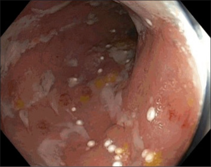
Colonoscopy showing moderate pan-colitis.
Figure 2.

Bronchoscopy showing tracheal ulcerations.
Discussion
Upper airway disease in IBD is rare and has been described in both ulcerative colitis (UC) and Crohn's disease, although cases occur predominantly in UC patients.4 Although clinically significant airway disease is rare, the most common pulmonary manifestations are bronchiectasis and chronic bronchitis, both of which are accompanied by large amounts of sputum production. Since patients with pulmonary manifestations of CD are usually asymptomatic, diagnoses are often made incidentally through abnormal screening tests.4 Chronic inflammation is common in the bronchi and alveoli of patients with CD, with 61% of asymptomatic CD patients exhibiting bronchoalveolar lavage features of overt lymphocytic alveolitis.5
The differential diagnosis for lung disease in the patient with active IBD still includes predominantly common lung and airway diseases such as reactive airway disease, infectious causes, and other inflammatory granulomatous diseases such as Wegener's granulomatosis. Specific to this population, mesalamine has also been reported to cause interstitial lung disease in rare occurrences.6 Airway disease in IBD patients usually does not present during an acute flare, but tends to occur during inactive phases or even after colectomies.4,6,7 Our patient is unique in that she presented with a non-productive cough during an acute CD flare.
Imaging is usually helpful for diagnosing patients with tracheobronchitis from CD. CT more frequently reveals airway changes than lung parenchymal findings. Such CT findings in tracheobronchitis associated with UC have been described as “circumferentially thickened tracheobronchial wall involving both the cartilaginous and membranous components.”8 Chest radiographs and chest CT may show circumferential or nodular narrowing of the trachea or the bronchi, although bronchoscopy is still the diagnostic procedure of choice.9 Pulmonary function testing shows an obstructive pattern.6 Our patient's squamous papilloma findings were likely an incidental finding unrelated to her lung pathology, as squamous cell papillomas are common benign tracheal neoplasms.10 Although the biopsy was non-diagnostic, the gross appearance of her trachea on bronchoscopy was consistent with Crohn's disease.
Treatment of upper airway disease in CD patients usually involves oral or inhaled corticosteroid therapy, which is effective in improving both respiratory and gastrointestinal symptoms. Our patient was treated initially with intravenous steroids given her concurrent gastrointestinal flare. Intravenous steroids are reserved for cases of subglottic stenosis, stridor, continued symptoms despite oral treatment, or those with concurrent illness that requires intravenous steroids. In her case, though she did not have true subglottic stenosis, the severity of her tracheobronchitis had escalated to a point where her cough was becoming stridorous. In patients experiencing only respiratory symptoms, good results have been reported from inhaled corticosteroids alone.11,12
Disclosures
Author contributions: V. Yeung wrote the manuscript. AG Govind, S. Arastu, and CH Henry wrote and edited the manuscript. CH Henry is the article guarantor.
Financial disclosure: None to report.
Informed consent was obtained for this case report.
References
- 1.Larsen S, Bendtzen K, Nielsen OH. Extraintestinal manifestations of inflammatory bowel disease: Epidemiology, diagnosis, and management. Ann Med. 2010;42(2):97–114. [DOI] [PubMed] [Google Scholar]
- 2.Godet PG, Cowie R, Woodman RC, Sutherland LR. Pulmonary function abnormalities in patients with ulcerative colitis. Am J Gastroenterol. 1997;92(7):1154–6. [PubMed] [Google Scholar]
- 3.Levine JS, Burakoff R. Extraintestinal manifestations of inflammatory bowel disease. Gastroenterol Hepatol. 2011;7(4):235–41. [PMC free article] [PubMed] [Google Scholar]
- 4.Papanikolaou I, Kagouridis K, Papiris SA. Patterns of airway involvement in inflammatory bowel diseases. World J Gastrointest Pathophysiol. 2014;5(4):560–9. [DOI] [PMC free article] [PubMed] [Google Scholar]
- 5.Wallaert B, Dugas M, Dansin E, et al. Subclinical alveolitis in immunological systemic disorders: Transition between health and disease? Eur Respir J. 1990;3(10):1206–16. [PubMed] [Google Scholar]
- 6.Ji XQ, Wang LX, Lu DG. Pulmonary manifestations of inflammatory bowel disease. World J Gastroenterol. 2014;20(37):13501–11. [DOI] [PMC free article] [PubMed] [Google Scholar]
- 7.Kuźniar T, Sleiman C, Brugière O, et al. Severe tracheobronchial stenosis in a patient with Crohn's disease. Eur Respir J. 2000;15(1):209–12. [DOI] [PubMed] [Google Scholar]
- 8.Acar T, Bayraktaroglu S, Ceylan N, Savas R. Computed tomography findings of tracheobronchial system diseases: A pictorial essay. Jpn J Radiol. 2014;33(2):51–8. [DOI] [PubMed] [Google Scholar]
- 9.Bayraktaroglu S, Basoglu O, Ceylan N, et al. A rare extraintestinal manifestation of ulcerative colitis: Tracheobronchitis associated with ulcerative colitis. J Crohns Colitis. 2010;4(6):679–82. [DOI] [PubMed] [Google Scholar]
- 10.Wu CC, Shepard JA. Tracheal and airway neoplasms. Semin Roentgenol. 2013;48(4):354–64. [DOI] [PubMed] [Google Scholar]
- 11.Asami T, Koyama S, Watanabe Y, et al. Tracheobronchitis in a patient with Crohn's disease. Intern Med Tokyo Jpn. 2009;48(16):1475–8. [DOI] [PubMed] [Google Scholar]
- 12.Kinebuchi S, Oohashi K, Takada T, et al. Tracheobronchitis associated with Crohn's disease improved on inhaled corticotherapy. Intern Med Tokyo Jpn. 2004;43(9):829–34. [DOI] [PubMed] [Google Scholar]