

Abstract
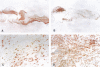
OBJECTIVE: To investigate the extent of plaque inflammation in culprit lesions of patients with chronic stable angina. DESIGN: Retrospective study. SETTING: Amsterdam reference centre. SUBJECTS: 89 consecutive patients who underwent directional coronary atherectomy, 58 of whom met the following inclusion criteria: chronic stable angina (Canadian Cardiovascular Society classification 1-3 (group 1, n = 28)); unstable angina (Braunwald class II (group 2, n = 18)); unstable angina (Braunwald class III (group 3, n = 12)). INTERVENTIONS: Directional atherectomy in patients with angina pectoris. MAIN OUTCOME MEASURES: Tissue areas of culprit lesions occupied by inflammatory cells and smooth muscle cells related to clinically defined ischaemic syndrome. RESULTS: Areas (% of total surface area (mean (SEM)) rich in smooth muscle cells were larger in patients with chronic stable angina (group 1, 51.2 (20.9)) than in those with unstable angina (group 2, 42.1 (20.5); group 3, 29.5 (19.4)) (1 v 2 and 2 v 3, NS; 1 v 3, P < 0.004). Macrophage rich areas were significantly smaller in patients with stable angina (group 1, 21.8 (11.9)) than in those with unstable angina (group 2, 31.5 (14.6); group 3, 46.4 (16.7)) (1 v 2, P < 0.02; 2 v 3, P < 0.02; 1 v 3, P < 0.001). Mean numbers of T cells per mm2 were as follows: group 1, 17 (9.4); group 2, 25 (15.9); group 3, 41 (30.6) (1 v 2, P 0.04; 2 v 3, P 0.07; 1 v 3, P < 0.001). Areas with HLA-DR positive cells showed the same pattern as macrophages and T cells and were smaller in stable (29.9 (12.4)) than in unstable angina (group 2, 40.4 (17.6); group 3, 52.4 (12.0)) (1 v 2, P < 0.02; 2 v 3, P < 0.05; 1 v 3, P < 0.001). CONCLUSION: The inverse relation between the extent of inflammatory activity in plaque tissues of culprit lesions and the clinical stability of the ischaemic syndrome supports the concept that reduction of inflammation favours plaque stabilisation. At the same time, the considerable overlap between groups indicates that patients with clinically stable angina do not all have histologically stable plaques.
Full text
PDF


Images in this article
Selected References
These references are in PubMed. This may not be the complete list of references from this article.
- Alexander R. W. Inflammation and coronary artery disease. N Engl J Med. 1994 Aug 18;331(7):468–469. doi: 10.1056/NEJM199408183310709. [DOI] [PubMed] [Google Scholar]
- Arbustini E., De Servi S., Bramucci E., Porcu E., Costante A. M., Grasso M., Diegoli M., Fasani R., Morbini P., Angoli L. Comparison of coronary lesions obtained by directional coronary atherectomy in unstable angina, stable angina, and restenosis after either atherectomy or angioplasty. Am J Cardiol. 1995 Apr 1;75(10):675–682. doi: 10.1016/s0002-9149(99)80652-3. [DOI] [PubMed] [Google Scholar]
- Arnold A. E., Simoons M. L., Van de Werf F., de Bono D. P., Lubsen J., Tijssen J. G., Serruys P. W., Verstraete M. Recombinant tissue-type plasminogen activator and immediate angioplasty in acute myocardial infarction. One-year follow-up. The European Cooperative Study Group. Circulation. 1992 Jul;86(1):111–120. doi: 10.1161/01.cir.86.1.111. [DOI] [PubMed] [Google Scholar]
- Braunwald E. Unstable angina. A classification. Circulation. 1989 Aug;80(2):410–414. doi: 10.1161/01.cir.80.2.410. [DOI] [PubMed] [Google Scholar]
- Brown B. G., Zhao X. Q., Sacco D. E., Albers J. J. Lipid lowering and plaque regression. New insights into prevention of plaque disruption and clinical events in coronary disease. Circulation. 1993 Jun;87(6):1781–1791. doi: 10.1161/01.cir.87.6.1781. [DOI] [PubMed] [Google Scholar]
- Brown D. L., Hibbs M. S., Kearney M., Loushin C., Isner J. M. Identification of 92-kD gelatinase in human coronary atherosclerotic lesions. Association of active enzyme synthesis with unstable angina. Circulation. 1995 Apr 15;91(8):2125–2131. doi: 10.1161/01.cir.91.8.2125. [DOI] [PubMed] [Google Scholar]
- Campeau L. Letter: Grading of angina pectoris. Circulation. 1976 Sep;54(3):522–523. [PubMed] [Google Scholar]
- Cheng G. C., Loree H. M., Kamm R. D., Fishbein M. C., Lee R. T. Distribution of circumferential stress in ruptured and stable atherosclerotic lesions. A structural analysis with histopathological correlation. Circulation. 1993 Apr;87(4):1179–1187. doi: 10.1161/01.cir.87.4.1179. [DOI] [PubMed] [Google Scholar]
- Davies M. J., Bland J. M., Hangartner J. R., Angelini A., Thomas A. C. Factors influencing the presence or absence of acute coronary artery thrombi in sudden ischaemic death. Eur Heart J. 1989 Mar;10(3):203–208. doi: 10.1093/oxfordjournals.eurheartj.a059467. [DOI] [PubMed] [Google Scholar]
- Davies M. J., Richardson P. D., Woolf N., Katz D. R., Mann J. Risk of thrombosis in human atherosclerotic plaques: role of extracellular lipid, macrophage, and smooth muscle cell content. Br Heart J. 1993 May;69(5):377–381. doi: 10.1136/hrt.69.5.377. [DOI] [PMC free article] [PubMed] [Google Scholar]
- Davies M. J., Thomas A. C. Plaque fissuring--the cause of acute myocardial infarction, sudden ischaemic death, and crescendo angina. Br Heart J. 1985 Apr;53(4):363–373. doi: 10.1136/hrt.53.4.363. [DOI] [PMC free article] [PubMed] [Google Scholar]
- Fuster V., Badimon L., Badimon J. J., Chesebro J. H. The pathogenesis of coronary artery disease and the acute coronary syndromes (1). N Engl J Med. 1992 Jan 23;326(4):242–250. doi: 10.1056/NEJM199201233260406. [DOI] [PubMed] [Google Scholar]
- Galis Z. S., Sukhova G. K., Kranzhöfer R., Clark S., Libby P. Macrophage foam cells from experimental atheroma constitutively produce matrix-degrading proteinases. Proc Natl Acad Sci U S A. 1995 Jan 17;92(2):402–406. doi: 10.1073/pnas.92.2.402. [DOI] [PMC free article] [PubMed] [Google Scholar]
- Galis Z. S., Sukhova G. K., Lark M. W., Libby P. Increased expression of matrix metalloproteinases and matrix degrading activity in vulnerable regions of human atherosclerotic plaques. J Clin Invest. 1994 Dec;94(6):2493–2503. doi: 10.1172/JCI117619. [DOI] [PMC free article] [PubMed] [Google Scholar]
- Hangartner J. R., Charleston A. J., Davies M. J., Thomas A. C. Morphological characteristics of clinically significant coronary artery stenosis in stable angina. Br Heart J. 1986 Dec;56(6):501–508. doi: 10.1136/hrt.56.6.501. [DOI] [PMC free article] [PubMed] [Google Scholar]
- Hansson G. K., Jonasson L., Seifert P. S., Stemme S. Immune mechanisms in atherosclerosis. Arteriosclerosis. 1989 Sep-Oct;9(5):567–578. doi: 10.1161/01.atv.9.5.567. [DOI] [PubMed] [Google Scholar]
- Jukema J. W., Bruschke A. V., van Boven A. J., Reiber J. H., Bal E. T., Zwinderman A. H., Jansen H., Boerma G. J., van Rappard F. M., Lie K. I. Effects of lipid lowering by pravastatin on progression and regression of coronary artery disease in symptomatic men with normal to moderately elevated serum cholesterol levels. The Regression Growth Evaluation Statin Study (REGRESS). Circulation. 1995 May 15;91(10):2528–2540. doi: 10.1161/01.cir.91.10.2528. [DOI] [PubMed] [Google Scholar]
- Kaski J. C., Chester M. R., Chen L., Katritsis D. Rapid angiographic progression of coronary artery disease in patients with angina pectoris. The role of complex stenosis morphology. Circulation. 1995 Oct 15;92(8):2058–2065. doi: 10.1161/01.cir.92.8.2058. [DOI] [PubMed] [Google Scholar]
- Libby P. Molecular bases of the acute coronary syndromes. Circulation. 1995 Jun 1;91(11):2844–2850. doi: 10.1161/01.cir.91.11.2844. [DOI] [PubMed] [Google Scholar]
- Loree H. M., Kamm R. D., Stringfellow R. G., Lee R. T. Effects of fibrous cap thickness on peak circumferential stress in model atherosclerotic vessels. Circ Res. 1992 Oct;71(4):850–858. doi: 10.1161/01.res.71.4.850. [DOI] [PubMed] [Google Scholar]
- Moreno P. R., Falk E., Palacios I. F., Newell J. B., Fuster V., Fallon J. T. Macrophage infiltration in acute coronary syndromes. Implications for plaque rupture. Circulation. 1994 Aug;90(2):775–778. doi: 10.1161/01.cir.90.2.775. [DOI] [PubMed] [Google Scholar]
- Richardson P. D., Davies M. J., Born G. V. Influence of plaque configuration and stress distribution on fissuring of coronary atherosclerotic plaques. Lancet. 1989 Oct 21;2(8669):941–944. doi: 10.1016/s0140-6736(89)90953-7. [DOI] [PubMed] [Google Scholar]
- Stemme S., Faber B., Holm J., Wiklund O., Witztum J. L., Hansson G. K. T lymphocytes from human atherosclerotic plaques recognize oxidized low density lipoprotein. Proc Natl Acad Sci U S A. 1995 Apr 25;92(9):3893–3897. doi: 10.1073/pnas.92.9.3893. [DOI] [PMC free article] [PubMed] [Google Scholar]
- Van der Loos C. M., Das P. K., Van den Oord J. J., Houthoff H. J. Multiple immunoenzyme staining techniques. Use of fluoresceinated, biotinylated and unlabelled monoclonal antibodies. J Immunol Methods. 1989 Feb 8;117(1):45–52. doi: 10.1016/0022-1759(89)90117-8. [DOI] [PubMed] [Google Scholar]
- van der Wal A. C., Becker A. E., van der Loos C. M., Das P. K. Site of intimal rupture or erosion of thrombosed coronary atherosclerotic plaques is characterized by an inflammatory process irrespective of the dominant plaque morphology. Circulation. 1994 Jan;89(1):36–44. doi: 10.1161/01.cir.89.1.36. [DOI] [PubMed] [Google Scholar]
- van der Wal A. C., Das P. K., Bentz van de Berg D., van der Loos C. M., Becker A. E. Atherosclerotic lesions in humans. In situ immunophenotypic analysis suggesting an immune mediated response. Lab Invest. 1989 Aug;61(2):166–170. [PubMed] [Google Scholar]