Multicellular organisms coordinate cell polarity and mitotic spindle orientation to develop and maintain tissue architecture throughout life. Through a series of carefully managed spatiotemporal cues, cells divide to produce daughter cells of either identical or differing fates. One pathway governing these oriented cell divisions centers on Partner of Inscuteable (Pins; LGN in mammals), whose polarized cortical localization and coordinated interactions with the proteins Mushroom Body Defect (Mud; NuMA in mammals) and Discs Large (Dlg) works to align the mitotic spindle and promote oriented divisions in diverse cell types.1 Recent evidence suggests that the cell cycle-promoting Hippo kinase pathway (Hpo; MST1/2 in mammals) is activated by cell polarity components and may participate in oriented cell divisions.2 Canonically, the Hippo pathway restricts cell proliferation through a kinase cascade first involving an activating phosphorylation of Warts kinase (Wts; LATS1/2 in mammals), in turn phosphorylating the transcription factor Yorkie (Yki; Yap1/Taz in mammals), preventing it from activating cell proliferation genes. We have recently demonstrated a link between the Hpo and Pins pathways in regulation of oriented cell divisions. Our results describe a novel, non-canonical role for the Hpo core complex in mitotic spindle positioning independent of Yki.3
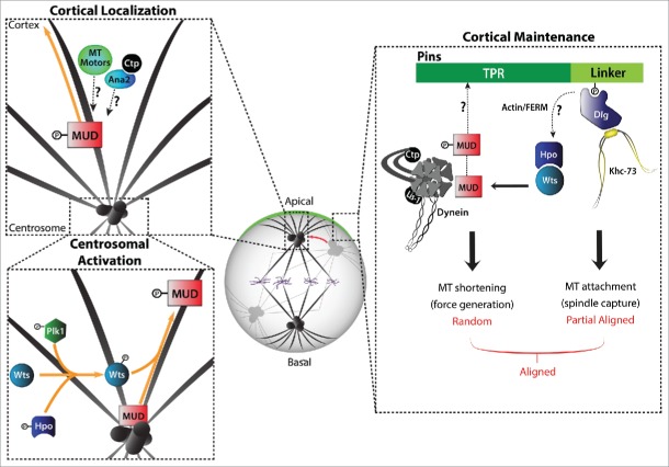
Through in silico analysis, we identified a predicted Wts phosphorylation site in the C-terminal coiled-coil domain of Mud and demonstrated that phosphorylation by Wts activated Pins/Mud interaction by relieving an autoinhibitory interaction between the coiled-coil and Pins-binding domains within Mud (Fig. 1). Using a reconstituted cell polarity approach in Drosophila S2 cells, we showed that loss of Wts prevents Pins-mediated cortical Mud association and spindle orientation. Unaffected, however, was Mud localization to spindle poles, a prominent site of Wts localization as well, suggesting Wts phosphorylates Mud to promote relocalization from spindle poles to the cell cortex. These findings were mirrored in vivo, with Mud failing to localize properly to the cell cortex of Drosophila wing disc epithelia following loss of Wts. Spindle positioning defects were also found in these wing disc cells, further implicating this pathway in the regulation of oriented cell divisions. Finally, we demonstrated the non-canonical, Yki-independent nature of Hpo function through use of a constitutively active Yki mutant. Overexpression of the Yki mutant alone produced overgrown discs, but did not induce spindle alignment defects. When combined with Mud knockdown, spindle orientation defects were evident along with overgrowth. Loss of Wts produced a similar phenotype as the Yki/Mud double mutant, consistent with Wts acting as a common upstream regulator of distinct cell growth and spindle orientation pathways. Our findings highlight a novel role for the Hpo complex in regulation of oriented cell divisions.
Figure 1.
Wts kinase can phosphorylate Mud to enhance its affinity for Pins and promote its cortical localization. We propose a model wherein Wts phosphorylates Mud at mitotic centrosomes to promote its association with proteins that involved in centrosome-to-cortex trafficking along astral microtubules, such as microtubule motors. The actin cytoskeleton and cortical polarity proteins (e.g. Dlg) may act as upstream Wts activators.
The discovery of a novel spindle orientation function stemming from a previously described cell-cycle regulator demonstrates remarkable multifunctionality of the Hpo tumor suppressor pathway. Inappropriate cellular delamination caused by defective oriented divisions may contribute to epithelial-mesenchymal transitions, thus this new role for Hpo could serve as an additional mechanism of tumor suppression. Many questions still remain regarding the molecular mechanism of Hpo-dependent spindle orientation, however. For instance, how is the Hpo complex recruited to centrosomes, and moreover activated in concert with cell cycle progression, to promote phosphorylation of Mud? Hpo complex components localize to spindle poles and are key regulators of centrosomal separation at the onset of mitosis.4 A recent study implicated Polo-like Kinase 1 (Plk1) in the phosphorylation of a Wts ortholog (NDR1), and illustrated this activity was key in promotion of proper Pins/Mud-mediated spindle orientation,5 offering a potential pathway for Wts-mediated Mud activation at the centrosome. Still, questions remain regarding the full nature of Hpo pathway regulation of Mud. Once phosphorylated, how does Mud become localized to the cell cortex? Given that Mud interacts with Dynein in its spindle orientation functionality,1 a microtubule motor could be an attractive candidate for cortical recruitment. Other candidates likely involved in this activity are the centriolar protein Anastral Spindle 2 (Ana2) and dynein light chain protein Cut Up (Ctp), which are implicated in cortical Mud localization in Drosophila neuroblasts.6 Are active Hpo components required at the cell cortex to maintain Mud phosphorylation and promote efficient Pins/Mud activity? If so, how is the Hpo complex recruited to the cortex? Hpo complex components could be recruited to the cortex through interactions with polarity proteins.2 An interesting candidate for this interaction is Dlg, a multidomain scaffold protein and second adapter protein of the Pins pathway. Dlg is also part of the Scribble polarity complex, a complex shown previously to interact with and activate canonical Hpo activity in epithelia.2 Another appealing candidate for cortical localization and activation of Hpo is the actin cytoskeleton, shown previously to activate Hpo components,2 and also to be cortically polarized in certain oriented divisions.7
Our results define a new regulator of oriented cell division and provide a novel link between mitotic spindle positioning and cell cycle progression. We are excited to see where research on Hippo-dependent spindle orientation will lead, and are hopeful it will provide better understanding of oriented cell division and its relation to disease.
References
- [1].Johnston CA, et al. Cell 2009; 138:1150-63; PMID:19766567; http://dx.doi.org/ 10.1016/j.cell.2009.07.041 [DOI] [PMC free article] [PubMed] [Google Scholar]
- [2].Yu FX, Guan KL. Genes Dev 2013; 27:355-71; PMID:23431053; http://dx.doi.org/ 10.1101/gad.210773.112 [DOI] [PMC free article] [PubMed] [Google Scholar]
- [3].Dewey EB, et al. Curr Biol 2015; 25:2751-62; PMID:26592339; http://dx.doi.org/ 10.1016/j.cub.2015.09.025 [DOI] [PMC free article] [PubMed] [Google Scholar]
- [4].Agircan FG, et al. Philos Trans R Soc Lond B Biol Sci 2014; 369; pii: 20130461; PMID:25047615; http://dx.doi.org/ 10.1098/rstb.2013.0461 [DOI] [PMC free article] [PubMed] [Google Scholar]
- [5].Yan M, et al.. Sci Rep 2015; 5:10449; PMID:26057687; http://dx.doi.org/ 10.1038/srep10449 [DOI] [PMC free article] [PubMed] [Google Scholar]
- [6].Wang C, et al.. Dev Cell 2011; 21:520-33; PMID:21920316; http://dx.doi.org/ 10.1016/j.devcel.2011.08.002 [DOI] [PubMed] [Google Scholar]
- [7].Kunda P, Baum B. Trends Cell Biol 2009; 19:174-9; PMID:19285869; http://dx.doi.org/ 10.1016/j.tcb.2009.01.006 [DOI] [PubMed] [Google Scholar]


