

Abstract
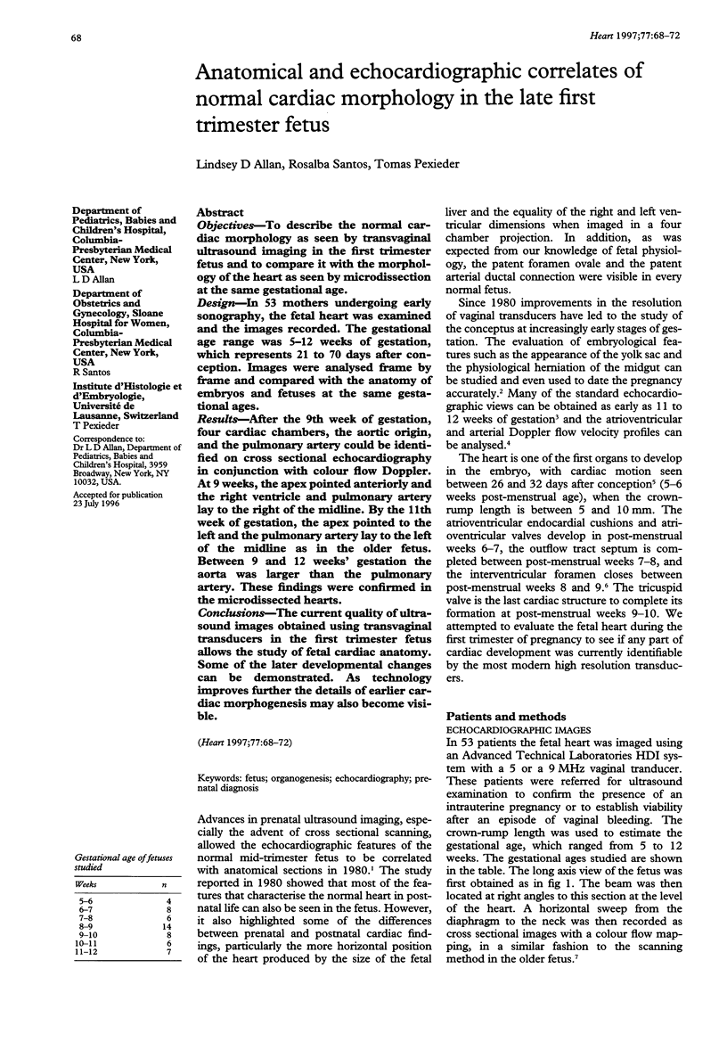
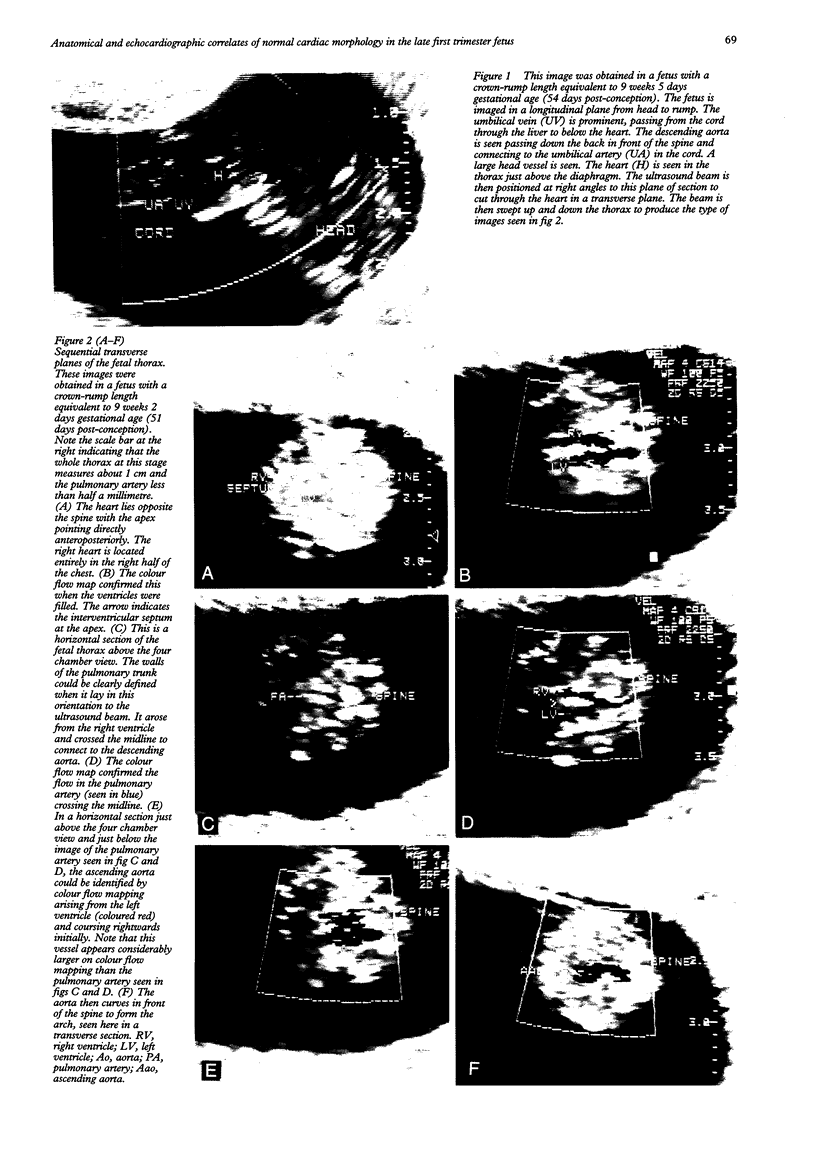
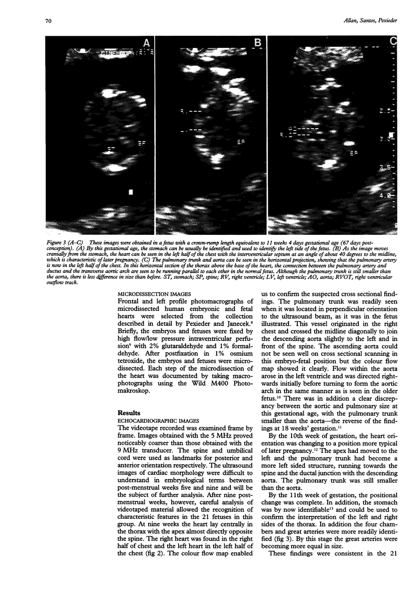
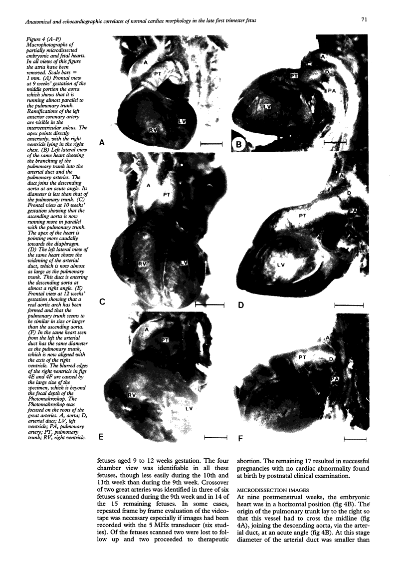
OBJECTIVES: To describe the normal cardiac morphology as seen by transvaginal ultrasound imaging in the first trimester fetus and to compare it with the morphology of the heart as seen by microdissection at the same gestational age. DESIGN: In 53 mothers undergoing early sonography, the fetal heart was examined and the images recorded. The gestational age range was 5-12 weeks of gestation, which represents 21 to 70 days after conception. Images were analysed frame by frame and compared with the anatomy of embryos and fetuses at the same gestational ages. RESULTS: After the 9th week of gestation, four cardiac chambers, the aortic origin, and the pulmonary artery could be identified on cross sectional echocardiography in conjunction with colour flow Doppler. At 9 weeks, the apex pointed anteriorly and the right ventricle and pulmonary artery lay to the right of the midline. By the 11th week of gestation, the apex pointed to the left and the pulmonary artery lay to the left of the midline as in the older fetus. Between 9 and 12 weeks' gestation the aorta was larger than the pulmonary artery. These findings were confirmed in the microdissected hearts. CONCLUSIONS: The current quality of ultrasound images obtained using transvaginal transducers in the first trimester fetus allows the study of fetal cardiac anatomy. Some of the later developmental changes can be demonstrated. As technology improves further the details of earlier cardiac morphogenesis may also become visible.
Full text
PDF
Images in this article
Selected References
These references are in PubMed. This may not be the complete list of references from this article.
- Allan L. D., Lockhart S. Intrathoracic cardiac position in the fetus. Ultrasound Obstet Gynecol. 1993 Mar 1;3(2):93–96. doi: 10.1046/j.1469-0705.1993.03020093.x. [DOI] [PubMed] [Google Scholar]
- Allan L. D., Tynan M. J., Campbell S., Wilkinson J. L., Anderson R. H. Echocardiographic and anatomical correlates in the fetus. Br Heart J. 1980 Oct;44(4):444–451. doi: 10.1136/hrt.44.4.444. [DOI] [PMC free article] [PubMed] [Google Scholar]
- Blaas H. G., Eik-Nes S. H., Kiserud T., Hellevik L. R. Early development of the abdominal wall, stomach and heart from 7 to 12 weeks of gestation: a longitudinal ultrasound study. Ultrasound Obstet Gynecol. 1995 Oct;6(4):240–249. doi: 10.1046/j.1469-0705.1995.06040240.x. [DOI] [PubMed] [Google Scholar]
- Bronshtein M., Siegler E., Eshcoli Z., Zimmer E. Z. Transvaginal ultrasound measurements of the fetal heart at 11 to 17 weeks of gestation. Am J Perinatol. 1992 Jan;9(1):38–42. doi: 10.1055/s-2007-994667. [DOI] [PubMed] [Google Scholar]
- Dolkart L. A., Reimers F. T. Transvaginal fetal echocardiography in early pregnancy: normative data. Am J Obstet Gynecol. 1991 Sep;165(3):688–691. doi: 10.1016/0002-9378(91)90310-n. [DOI] [PubMed] [Google Scholar]
- Hornberger L. K., Sanders S. P., Sahn D. J., Rice M. J., Spevak P. J., Benacerraf B. R., McDonald R. W., Colan S. D. In utero pulmonary artery and aortic growth and potential for progression of pulmonary outflow tract obstruction in tetralogy of Fallot. J Am Coll Cardiol. 1995 Mar 1;25(3):739–745. doi: 10.1016/0735-1097(94)00422-M. [DOI] [PubMed] [Google Scholar]
- Howe R. S., Isaacson K. J., Albert J. L., Coutifaris C. B. Embryonic heart rate in human pregnancy. J Ultrasound Med. 1991 Jul;10(7):367–371. doi: 10.7863/jum.1991.10.7.367. [DOI] [PubMed] [Google Scholar]
- Mcbride R. E., Moore G. W., Hutchins G. M. Development of the outflow tract and closure of the interventricular septum in the normal human heart. Am J Anat. 1981 Mar;160(3):309–331. doi: 10.1002/aja.1001600308. [DOI] [PubMed] [Google Scholar]
- Moscoso G., Pexieder T. Variations in microscopic anatomy and ultrastructure of human embryonic hearts subjected to three different modes of fixation. Pathol Res Pract. 1990 Dec;186(6):768–774. doi: 10.1016/S0344-0338(11)80268-2. [DOI] [PubMed] [Google Scholar]
- Sharland G. K., Chita S. K., Allan L. D. The use of colour Doppler in fetal echocardiography. Int J Cardiol. 1990 Aug;28(2):229–236. doi: 10.1016/0167-5273(90)90065-d. [DOI] [PubMed] [Google Scholar]
- Smith R. S., Comstock C. H., Kirk J. S., Lee W. Ultrasonographic left cardiac axis deviation: a marker for fetal anomalies. Obstet Gynecol. 1995 Feb;85(2):187–191. doi: 10.1016/0029-7844(94)00350-M. [DOI] [PubMed] [Google Scholar]
- Warren W. B., Timor-Tritsch I., Peisner D. B., Raju S., Rosen M. G. Dating the early pregnancy by sequential appearance of embryonic structures. Am J Obstet Gynecol. 1989 Sep;161(3):747–753. doi: 10.1016/0002-9378(89)90394-3. [DOI] [PubMed] [Google Scholar]
- Wladimiroff J. W., Huisman T. W., Stewart P. A. Fetal cardiac flow velocities in the late 1st trimester of pregnancy: a transvaginal Doppler study. J Am Coll Cardiol. 1991 May;17(6):1357–1359. doi: 10.1016/s0735-1097(10)80147-0. [DOI] [PubMed] [Google Scholar]
- Zimmer E. Z., Chao C. R., Santos R. Amniotic sac, fetal heart area, fetal curvature, and other morphometrics using first trimester vaginal ultrasonography and color Doppler imaging. J Ultrasound Med. 1994 Sep;13(9):685–690. doi: 10.7863/jum.1994.13.9.685. [DOI] [PubMed] [Google Scholar]