Abstract
The present study was designed to investigate the sperm damages occurring in acrosome, plasma membrane, mitochondrial activity, and DNA of fresh, equilibrated and frozen–thawed buffalo semen by fluorescent probes. The stability of sperm acrosome and plasma membrane stability, mitochondrial activity and DNA status were assessed by fluorescein conjugated lectin Pisum sativum agglutinin, Annexin–V/propidium iodide, JC-1 and TUNEL assay, respectively, under the fluorescent microscope. The damages percentage of acrosome integrity was significantly increased during equilibration and freezing–thawing process. The stability of sperm plasma membrane is dependent on stability of phosphatidylserine (PS) on the inner leaflet of plasma membrane. The frozen–thawed sperm showed externalization of PS leading to significant increase in apoptotic, early necrotic and necrotic changes and lowered high mitochondrial membrane potential as compared with the fresh sperm but all these parameters were not affected during equilibration. However, the DNA integrity was not affected during equilibration and freezing–thawing procedure. In conclusion, the present study revealed that plasma membrane and mitochondria of buffalo sperm are more susceptible to damage during cryopreservation. Furthermore, the use of fluorescent probes to evaluate integrity of plasma and acrosome membranes, as well as mitochondrial membrane potential and DNA status increased the accuracy of semen analyses.
Keywords: Apoptosis, Buffalo bull, Cryopreservation, DNA integrity, Semen
Introduction
Cryopreservation of sperm cells is a valuable tool for buffalo industry due to huge demand of high genetic merit bulls semen to breed the female available in country. Although, cryopreservation is a harmful process and induces many unfavorable changes in spermatozoa. Freezing and thawing result in structural change in plasma membrane permeability (Anzar et al. 2002; Khan et al. 2009; Kadirvel et al. 2012), and damage of mitochondria (Martin et al. 2004; Kadirvel et al. 2012). Furthermore, it is suggested that buffalo bull spermatozoa is more prone to oxidative stress due to high contents of polyunsaturated phospholipids (Sansone et al. 2000). Many recent studies have demonstrated that sperm fertilizing potential depend on the integrity and functionality of the different cellular structures. Acrosome integrity is essential for the occurrence of oocyte fertilization since the acrosome reaction is fundamental for sperm penetration into the zona pellucida and for oocyte plasma membrane fusion (Õura and Toshimori 1990; Flesch and Gadella 2000). The plasma membrane is responsible for the preservation of cellular homeostasis; in this way the plasma membrane integrity has a vital role on sperm survival in the female reproductive tract and on the preservation of its fertility (Õura and Toshimori 1990). The mitochondrial membrane potential, responsible for ATP production, is indispensable for the flagellar beat and sperm motility (Flesch and Gadella 2000). The evaluation of DNA integrity and apoptosis like events in ejaculated and cryopreserved spermatozoa has also shown the clinical importance (Boue et al. 2000; Gillan et al. 2005). Any DNA damage may cause morphological changes in spermatozoa leading to reduced embryonic survival or genetic defects, which results in cell death and resorption during early pregnancy (Oosterhuis and Vermes 2004; Rodriguez-Marinez et al. 2009).
Research on sperm damage has always been one of the hotspots in the field of reproductive biology. In the past, much attention has been paid to observe the conventional parameters of semen, e.g. sperm density, vitality, motility and morphology through optical microscope which partially discloses its functional ability to fertilize the egg cell. However, with the development of technology, fluorescent staining techniques are more widely applied to evaluate sperm characteristics including the sperm acrosome status, membrane stability, mitochondrial activity, and DNA integrity. The type and extent of sperm damage can be analysed more precisely through the application of fluorescent microscope, flow cytometry and computer analysis system (Celeghini et al. 2007; Kadirvel et al. 2012; Singh et al. 2013; Kumar et al. 2014). In buffalo, there are only few reports regarding the actual assessment of damages during semen cryopreservation. Therefore, the aim of the present study was the assessment of sperm damages occurring in acrosome, plasma membrane, mitochondria, and DNA in fresh, equilibrated and frozen–thawed buffalo semen by fluorescent probes which may help in making strategies to reduce the damage during cryopreservation in buffalo.
Materials and methods
Semen collection, evaluation, processing and cryopreservation
Five breeding Murrah buffalo bulls (3–5 years age) were used for semen collection under progeny testing program of the institute and maintained under identical milieu. All experiments were carried out in accordance with the approval of the Institute Animal Ethics Committee. A total of 50 ejaculates (ten ejaculates from each bull, twice in a week) were collected using artificial vagina and conventionally assessed for volume, color, sperm concentration by using Accucell bovine photometer (IMV, L’Aigla, France) and mass activity and percentage of motile spermatozoa. Sperm motility was subjectively assessed under phase contrast microscope equipped with a warm stage (37 °C) at 400× magnification and only ejaculates with ≥70 % sperm motility were used for cryopreservation. The fresh semen was extended in Tris-egg yolk extender containing Tris (3.02 % w/v), citric acid (1.67 % w/v), fructose (1 % w/v), egg yolk (20 % v/v), penicillin (500 IU/mL, Sigma-Aldrich, St. Louis, MO, USA) and streptomycin (500 µg/mL, Sigma-Aldrich, St. Louis, MO, USA) plus glycerol (6.4 % v/v, Thermo Fisher Scientific, Mumbai, India), to make a final concentration of 80 × 106 spermatozoa/mL. Thereafter, the extended semen was slowly cooled to 4 °C and kept for a period of 3–4 h for equilibration. The equilibrated semen was loaded into 0.25 mL plastic straws (IMV, France) and frozen into a programmable biological freezer (Mini Digi-cool, IMV Technologies, L’Aigle, France) for cooling down from 4 to −140 °C. Each semen sample was initially cooled at the rate of −5 °C/min from 4 to −10 °C. Between −10 and −100 °C, freezing rate was −40 °C/min and then from −100 to −140 °C, its rate was −20 °C/min. After reaching −140 °C, semen straws were immediately plunged into liquid nitrogen at −196 °C for storage.
Sperm evaluation
The semen was evaluated for acrosome integrity, plasma membrane stability, mitochondrial membrane potential (MMP) and DNA status at fresh, equilibrated and frozen–thawed stages of cryopreservation. For semen analysis, semen samples were washed twice in tris buffer to remove seminal plasma and extender. The whole experiment was repeated thrice and at least 200 sperm were counted for each analysis per bull.
Assessment of acrosome integrity
The acrosomal integrity was evaluated using fluorescein conjugated lectin Pisum sativum agglutinin (FITC-PSA, Sigma-Aldrich, St. Louis, MO, USA) staining method as reported by Mendoza et al. 1992, with minor modifications. Briefly, 20 µL of diluted semen was re-suspended in 500 µL PBS and centrifuged at 1,500 rpm for 10 min and supernatant was discarded. Finally, pellet of spermatozoa was re-suspended in 250 µL PBS and then one drop for smear was taken onto a pre-cleaned microscope slide. The smear was dried and fixed with paraformaldehyde (4 %) at room temperature for 45 min. Air dried slides were covered with FITC-PSA (50 µg/mL in PBS solution) in dark place for 20 min at room temperature. Subsequently, excess stain was removed by washing with Milli-Q water, then the slide was air dried and a cover slip was applied with glycerol and examined under fluorescent microscope (Ti eclapse, Nikon, Tokyo, Japan) fitted with an excitation filter of 365 nm and a barrier filter of 397 nm.
Assessment of plasma membrane stability
Sperm plasma membrane integrity was assessed with dual fluorescent probes, annexin V/propidium iodide (PI, Calbiochem, Darmstadt, Germany) as described earlier (Kumar et al. 2014). Briefly, the spermatozoa were washed (1,500 rpm for 10 min) twice in Tris-buffered saline (pH 7.0). The sperm pellet was resuspended in Annexin-V binding buffer (10 mM Hepes/NaOH, 140 mM NaCl, and 2.5 mM CaCl2) at room temperature to make a final concentration of 1–2 × 10⁵ spermatozoa/mL. The cell suspension (97.5 µL) was transferred to a clean tube and 2.5 µL Annexin V-FITC (Calbiochem, Darmstadt, Germany) was added, and incubated for 15 min at room temperature. After incubation, samples were washed with binding buffer and centrifuged at 1,000 rpm to pellet the cells and the supernatant was discarded. The cell pellet was re-suspended in 95 µL binding buffer and 5 µL PI (50 mg/mL) was added, gently mixed and incubated for 15 min at room temperature in the dark, then finally analyzed under fluorescence microscope. Apoptotic sperm labeled with Annexin-V only, gave green fluorescence, necrotic sperm labeled only with PI, showed red fluorescence, early necrotic sperm labeled with both Annexin-V and PI, appeared green and red fluorescence, whereas viable sperm did not label with any one, hence showed no fluorescence.
Assessment of mitochondrial membrane potential
The assessment of MMP was done as described earlier (Selvaraju et al. 2008). In brief, A stock solution 1.53 mM of JC-1 dye (5,5′,6,6′-tetrachloro-1,1′,3,3′-tetraethyl-benzimidazolyl carbocyanine iodide) (Molecular Probes®, Eugene, OR, USA), was prepared in dimethyl sulfoxide (DMSO). JC-1 (1.3 µL) was added to a 100 µL semen sample containing 25 × 106 sperm/mL and incubated at 37 °C for 30 min. After 20 min incubation, 10 µL of PI (0.27 mg/mL in phosphate buffer) was added to counter stain nuclei of spermatozoa. The sperm cells were washed, resuspended in PBS, smeared, and analyzed with fluorescence emission ratio at 590 and 530 nm epifluorescence microscope (Ti eclapse, Nikon) using an excitation wavelength of 485 nm.
Assessment of DNA integrity
An Apo-BrdU kit (Calbiochem, Darmstadt, Germany) was used for the detection of nicked DNA in spermatozoa as described by Anzar et al. 2002. Briefly, the semen samples were first centrifuged (800×g for 10 min) to remove seminal plasma and extender. In all subsequent washings, the sperm suspensions were centrifuged at 500×g for 10 min at room temperature. Fresh, equilibrated and frozen–thawed spermatozoa were diluted to 2 × 10⁶ cells/mL in PBS. The diluted spermatozoa were fixed by adding 10 ml of paraformaldehyde (1 % w/v) in PBS (pH 7.4) on ice for 15 min. After fixation, sperm were washed twice in PBS, and the pellet was resuspended in 0.5 mL of PBS followed by 5.0 mL of ice-cold ethanol (70 % v/v). The fixed sperm suspensions were stored at −20 °C until analyzed for nicked DNA. On the day of analysis, the fixed sperm were centrifuged to remove ethanol. The sperm suspensions were washed twice in 1 mL of wash buffer (provided with kit) and the supernatant was removed by aspiration. Then, the pellet was resuspended in 51 µL of freshly prepared DNA-labeling solution containing TdT enzyme (0.75 µL), BrdUTP (8.0 µL), reaction buffer (10.0 µL), and distilled water (32.25 µL). The sperm were incubated in the DNA labeling solution at 37 °C for 2 h. These suspensions were gently shaken every 30 min. At the end of the incubation time, the sperm were washed twice in 1 ml of rinse buffer, and the pellet was resuspended in 100 µL of freshly prepared antibody solution containing 5 µL monoclonal antibody (fluorescein-labeled PRB-1) and 95 µL of rinse buffer. The antibody PRB-1, rinse buffer, PI/RNase A solution all these materials are supplied with the Apo BrdU kit. The sperm suspensions were incubated in the dark for 30 min at room temperature and analyzed under fluorescence microscope after addition of 300 µL of PI/RNase A solution. The spermatozoa having nicked DNA exhibited green fluorescence.
Statistical analysis
Statistical analyses were performed using statistical analysis system (SAS) 9.2 for window (SAS Institute Inc. Cary, NC, USA). In this study, one-way ANOVA followed by Duncan multiple range post-test were used to assess differences among mean of different stages of cryopreservation on acrosome integrity, plasma membrane integrity, MMP and DNA integrity. The test was performed at 5 % level of significance and all data are reported as mean ± standard error (SE).
Results
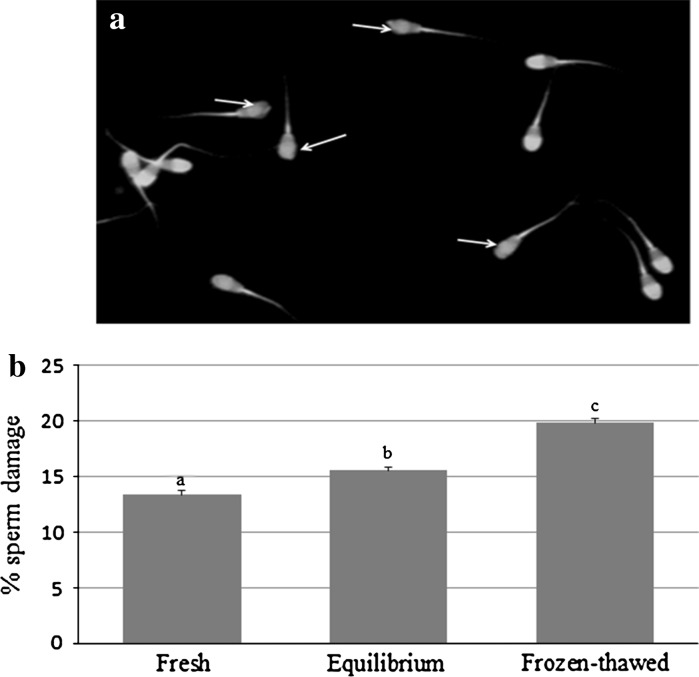
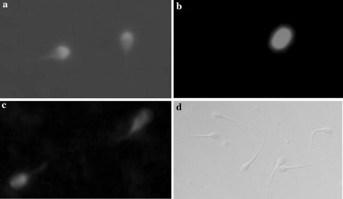
The acrosome integrity was evaluated by using fluorescently labeled lectin with P. sativum agglutinin staining. Sperm possessing an intact acrosome showed more intense fluorescence in the acrosome region with a distinct ring whereas damaged sperm heads had less intense fluorescence (Fig. 1a). The percentage damages of acrosome integrity were significantly (P < 0.05) increased during equilibration and freezing–thawing process as shown in Fig. 1b. The stability of plasma membrane was analysed through Annexin-V-FITC/PI assay which gave different types of fluorescent patters for translocation of PS, on basis of which these were classified as: apoptotic, early necrotic, necrotic and viable. These fluorescent patterns were confirmed by observation under fluorescence microscope as represented in Fig. 2. The percentages of four populations of sperm did not vary significantly between fresh and equilibrated semen (Table 1). The fresh semen sample having 10.62 ± 0.37 % apoptotic, 10.46 ± 0.37 % early necrotic, 9.86 ± 0.35 % necrotic and 70.06 ± 0.70 % viable sperm count, whereas, frozen–thawed samples resulted in increased percentages of apoptotic (19.38 ± 0.28 %), early necrotic (15.06 ± 0.28 %), necrotic (14.29 ± 0.38 %) and decreased percentage of viable (57.25 ± 0.48) sperm cells.
Fig. 1.
Evaluation of acrosome integrity through FITC-PSA staining. a Buffalo spermatozoa with intact (having distinct ring) and defected acrosome (indicated by arrow), b Impact on acrosome damages of the buffalo sperm at different stages of cryopreservation. Bars with different superscripts differ significantly (P < 0.05)
Fig. 2.
Plasma membrane stability of buffalo sperm evaluated under fluorescent microscope using Annexin-V/PI assay. a Apoptotic sperm (labeled Annexin-V only, green fluorescence), b Necrotic sperm (labeled only PI, red fluorescence). c Early necrotic sperm (labeled both Annexin-V and PI, green and red fluorescence), and d viable sperm (not labeled, no fluorescence)
Table 1.
Plasma membrane stability of buffalo sperm at different stages of cryopreservation (% ± SE)
| Parameters | Fresh | Equilibrium | Frozen–thawed |
|---|---|---|---|
| Apoptotic | 10.62a ± 0.37 | 11.70a ± 0.34 | 19.38b ± 0.28 |
| Early necrotic | 10.46a ± 0.37 | 11.35a ± 0.35 | 15.06b ± 0.28 |
| Necrotic | 9.86a ± 0.35 | 10.54a ± 0.37 | 14.29b ± 0.38 |
| Viable | 70.06a ± 0.70 | 68.39a ± 0.67 | 57.25b ± 0.48 |
Values with different superscripts within row differ significantly (P < 0.05)
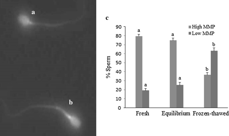
Mitochondrial activity was observed by fluorescence of JC-1, mainly detected over the mid-piece under fluorescence microscope. Two types of sperm populations were observed: (1) high green and low orange fluorescence, considered as high MMP (Fig. 3a), and (2) low or medium green and high orange fluorescence, considered as low MMP (Fig. 3b). The mean values of the MMP of sperm cells during the cryopreservation process are summarized in Fig. 3c. Cryopreservation induced a significant increase in the proportion of buffalo sperm cells with low MMP (63.23 ± 3.15 %) compared to fresh (19.26 ± 2.25 %) and equilibrated (25.13 ± 3.15 %) samples. The fresh and equilibrated semen had significantly higher percentage of sperm with high MMP than that of frozen–thawed semen samples. Furthermore, DNA integrity was assessed by TUNEL assays. The percentages of nicked DNA in fresh, equilibrated and frozen–thawed samples were 8.30 ± 0.44 %, 9.11 ± 0.44 % and 10.43 ± 0.47 %, respectively, which did not lead to any statistically significant difference among them.
Fig. 3.
MMP of buffalo spermatozoa evaluated by JC-1. a Sperm with low MMP (low green and high orange fluorescence), b sperm with high MMP (high green and low orange fluorescence), c MMP of buffalo spermatozoa at different stages of cryopreservation. Bars with different superscripts are significantly different (P < 0.05)
Discussion
The presence of intact acrosome (containing hydrolytic enzymes) is pre-requisite for fertilization process and it is highly correlated with fertility of frozen semen (Medeiros et al. 2002). The cryopreservation provokes reduction in the percentage of live spermatozoa and an increase in the proportion of spermatozoa with a reacted acrosome (Medeiros et al. 2002). In this study, the percentage of sperm acrosome damages were significantly (P < 0.05) increased during equilibration and freezing–thawing process. The acrosome integrity was negatively affected by thawing, it is speculated that acrosomal caps might be damaged during thawing of spermatozoa, as demonstrated in ram (Nur et al. 2010), pig (Jeong et al. 2009), bull (Leite et al. 2010) and buffalo (Rasul et al. 2001). In the current study, the overall damages in the percent of acrosome during cryopreservation was <20 %, mimicing previous reports for buffalo bulls sperm (Chinnaiya and Ganguli 1980; Rasul et al. 2001). In other species, acrosomal damages was observed during cryopreservation in ram to about 45–65 % (Soylu et al. 2007; Abdelhakeam et al. 1991), in goat to about 38–43 % (Chauhan et al. 1994) and in cattle to 26 % (Azam et al. 1998). Therefore, acrosome of buffalo sperm is more resistant to the damage during cryopreservation than the other species cited above but actual reasons are unknown.
The intactness of the plasma membrane of the sperm cells is one of the sound parameters for quality assessment. In this study, we found that the percentage of apoptotic, early necrotic, necrotic and viable sperm cells did not differ during equilibration, but after freezing and thawing the number of these cells increased significantly due to damage of plasma membrane during cryopreservation and ultimately reduced the viable cells by 13 % in the population. Similarly, increase in apoptotic and early necrotic and decrease in viable count in the population after cryopreservation were observed in buffalo (Kadirvel et al. 2012), cattle (Martin et al. 2004) and human (Paasch et al. 2004). During cryopreservation, sperm plasma membranes are destabilized due to low temperature and high salt concentration (Holt and North 1991). Annexin V conjugated with fluorescein isothiocyanate is the protein that binds phosphatidylserine, enabling the detection of early apoptosis. The additional use of propidium iodide allows to examine the stability of cell membrane (Koonjaenak et al. 2007) and to differentiate the cells dying through apoptosis or necrosis (Kadirvel et al. 2012). This method is useful for evaluation of apoptotic changes in spermatozoa demonstrated by fluorescence microscope, which were undetectable during quantitative analysis of semen parameters using traditional methods (Gillan et al. 2005).
The sperm motility subjected to cryopreservation is reduced by some changes in the active transport and the permeability of the plasma membrane in the tail region (Watson 1995; Long and Guthrie 2006). A reduction of spermatozoa motility may also be triggered by a change in the availability of energy or an injury of the axonemal elements. Moreover, it has been noted that the alterations in the ultra structure of mitochondria occurring during cryopreservation are followed by a loss of the internal mitochondrial structure of frozen–thawed spermatozoa (Long and Guthrie 2006). This study observed a significant decrease in high MMP in cryopreserved sperm. Similar finding was observed by various workers in different species (Ly et al. 2003; Martin et al. 2007; Kadirvel et al. 2012). Decrease in MMP lead to lack of energy, which may be responsible for the reduced sperm motility. Hence, reduced sperm motility induced by cryopreservation is believed to be mainly associated with mitochondrial damage (Januskauskas and Zillinskas 2002; Ruiz-Pesini et al. 2001).
The routine analysis of buffalo semen does not include evaluation of DNA fragmentation, but nowadays in clinical practice tests are used to determine the occurrence of sperm DNA fragmentation. However, DNA integrity assessment is important because sperm with damaged DNA may not be able to fertilize oocytes, which potentially disturb genetic regulation of the early embryo and block further development (Fatehi et al. 2006). In the present study, no significant changes were observed in DNA fragmentation during cryopreservation in buffalo sperm. Our result has been supported by previous observation of maintenance of DNA integrity during cryopreservation process in buffalo (Koonjaenak et al. 2007; Kadirvel et al. 2012), and cattle (Anzar et al. 2002). On the other hand, sperm DNA and chromatin structure are to be altered or damaged during cryopreservation in human (Donnelly et al. 2001), boar (Fraser and Strzezek 2004) and ram (Peris et al. 2004) sperm. Thus, buffalo sperm DNA seems to be unaffected by cryopreservation, which could be due to the compact nature of the sperm nucleus and DNA, and reduction in their accessibility to endonucleases during cryopreservation.
In conclusion, the present study revealed that plasma membrane and mitochondria of buffalo sperm are more susceptible to damage during cryopreservation. However, the findings of reduction in MMP and translocation of PS inside the plasma membrane which occur in cryopreserved buffalo sperm signify that these cells could follow a unique mode of cell death process upon freezing–thawing, which needs to be investigated further. Furthermore, the use of fluorescent probes to evaluate integrity of plasma and acrosome membranes, as well as mitochondrial membrane potential and DNA status increase the accuracy of semen analyses. In future, studies may be designed to reduce the plasma membrane and mitochondrial damages by supplementing plasma membrane stabilizing agents in the extender or modifying protocol for buffalo semen cryopreservation.
Acknowledgments
The authors thank the Director of the institute for provided necessary facilities to conduct this work. Authors thank Dr PP Dubey, Assistant Professor, GADVASU, Ludhiana for assisting in data analysis, and the lab staff of semen freezing lab for helping in semen collection and cryopreservation.
Conflict of interest
The authors have declared that no conflict of interest exists among them.
References
- Abdelhakeam AA, Graham EF, Vazquez IA. Studies on the absence of glycerol in unfrozen and frozen ram semen: fertility trials and the effect of dilution methods on freezing ram semen in the absence of glycerol. Cryobiology. 1991;28:36–42. doi: 10.1016/0011-2240(91)90005-9. [DOI] [Google Scholar]
- Anzar M, He L, Buhr MM, Kroetsch TG, Pauls KP. Sperm apoptosis in fresh and cryopreserved bull semen detected by flow cytometry and its relationship with fertility. Biol Reprod. 2002;66:354–360. doi: 10.1095/biolreprod66.2.354. [DOI] [PubMed] [Google Scholar]
- Azam M, Anzar M, Arslan M. Assessment of post-thaw semen quality of buffalo and Sahiwal bulls using new semen assays. Pak Vet J. 1998;18:74–80. [Google Scholar]
- Boue F, Delhomme A, Chaffaux S. Reproductive manage-ment of silver foxes (Vulpes vulpes) in captivity. Theriogenology. 2000;53:1717–1728. doi: 10.1016/S0093-691X(00)00310-1. [DOI] [PubMed] [Google Scholar]
- Celeghini ECC, Arruda RP, Andrade AFC. Practical techniques for bovine sperm simultaneous fluorimetric assessment of plasma, acrosomal and mitochondrial membranes. Reprod Domest Anim. 2007;42:479–488. doi: 10.1111/j.1439-0531.2006.00810.x. [DOI] [PubMed] [Google Scholar]
- Chauhan MS, Kapila R, Gandhi KK, Anand SR. Acrosome damage and enzyme leakage of goat spermatozoa during dilution, cooling and freezing. Andrologia. 1994;26:21–26. doi: 10.1111/j.1439-0272.1994.tb00748.x. [DOI] [PubMed] [Google Scholar]
- Chinnaiya GP, Ganguli NC. Acrosomal damage of buffalo spermatozoa during freezing in extenders. Zentralbl Veterinarmed A. 1980;27:339–342. doi: 10.1111/j.1439-0442.1980.tb02013.x. [DOI] [PubMed] [Google Scholar]
- Donnelly ET, McClure N, Lewis SE. Cryopreservation of human semen and prepared sperm: effects on motility parameters and DNA integrity. Fertil Steril. 2001;76:892–900. doi: 10.1016/S0015-0282(01)02834-5. [DOI] [PubMed] [Google Scholar]
- Fatehi AN, Bevers MM, Schoevers E. DNA damage in bovine sperm does not block fertilization and early embryonic development but induces apoptosis after the first cleavages. J Androl. 2006;27:176–188. doi: 10.2164/jandrol.04152. [DOI] [PubMed] [Google Scholar]
- Flesch FM, Gadella BM. Dynamics of the mammalian sperm membrane in the process of fertilization. Biochim Biophys Acta. 2000;1469:197–235. doi: 10.1016/S0304-4157(00)00018-6. [DOI] [PubMed] [Google Scholar]
- Fraser L, Strzezek J. The use of comet assay to assess DNA integrity of boar spermatozoa following liquid preservation at 5 and 16 °C. Folia Histochem Cytobiol. 2004;42:49–55. [PubMed] [Google Scholar]
- Gillan L, Evans GW, Maxwell MC. Flow cytometric evaluation of sperm parameters in relation to fertility potential. Theriogenology. 2005;63:445–457. doi: 10.1016/j.theriogenology.2004.09.024. [DOI] [PubMed] [Google Scholar]
- Holt WV, North RD. Cryopreservation, actin localization and thermotropic phase transitions in ram sperm. J Reprod Fertil. 1991;91:451–461. doi: 10.1530/jrf.0.0910451. [DOI] [PubMed] [Google Scholar]
- Januskauskas A, Zillinskas H. Bull semen evaluation post-thaw and relation of semen characteristics to bull’s fertility. Veterinarija ir zootechnika. 2002;17:39. [Google Scholar]
- Jeong YJ, Kim M, Song HJ, Kang EJu, Ock SA, Kumar BM, Balasubramanian S, Rho GJ. Effect of α-tocopherol supplementation during boar semen cryopreservation on sperm characteristics and expression of apoptosis related genes. Cryobiology. 2009;58:181–189. doi: 10.1016/j.cryobiol.2008.12.004. [DOI] [PubMed] [Google Scholar]
- Kadirvel G, Periasamy S, Kumar S. Effect of cryopreservation on apoptotic-like events and its relationship with cryocapacitation of Buffalo (Bubalus bubalis) sperm. Reprod Domest Anim. 2012;47:143–150. doi: 10.1111/j.1439-0531.2011.01818.x. [DOI] [PubMed] [Google Scholar]
- Khan DR, Ahmad N, Anzar M, Channa AA. Apotosis in fresh and cryopreserved sperm. Theriogenology. 2009;71:872–876. doi: 10.1016/j.theriogenology.2008.09.056. [DOI] [PubMed] [Google Scholar]
- Koonjaenak S, Chanatinart V, Aiumlamai S, Pinyopumontr T, Rodriguez-Martinez H. Seasonal variation in semen quality of swamp buffalo bulls (Bubalus bubalis) in Thailand. Asian J Androl. 2007;9:92–101. doi: 10.1111/j.1745-7262.2007.00230.x. [DOI] [PubMed] [Google Scholar]
- Kumar D, Kumar P, Singh P, Yadav SP, Sarkar SK, Bharadwaj A, Yadav PS. Characteristics of frozen thawed semen in predicting the fertility of buffalo bulls. Indian J Anim Sci. 2014;84:389–392. [Google Scholar]
- Leite TG, do Vale Filhoa VR, Arruda RP, Andrade AFC, Emericka LL, Zaffalon FG, Zaffalon JAM, Martinsa JAM, de Andrade VJ. Effects of extender and equilibration time on post-thaw motility and membrane integrity of cryopreserved Gyr bull semen evaluated by CASA and flow cytometry. Anim Reprod Sci. 2010;120:31–38. doi: 10.1016/j.anireprosci.2010.04.005. [DOI] [PubMed] [Google Scholar]
- Long JA, Guthrie HD. Validation of a rapid, large-scale assay to quantify ATP concentration in spermatozoa. Theriogenology. 2006;65:1620–1630. doi: 10.1016/j.theriogenology.2005.06.020. [DOI] [PubMed] [Google Scholar]
- Ly J, Grubb DR, Lawen A. The mitochondrial membrane potential (ψm) in apoptosis; an update. Apoptosis. 2003;8:115–128. doi: 10.1023/A:1022945107762. [DOI] [PubMed] [Google Scholar]
- Martin G, Sabido O, Durand P, Levy R. Cryopreservation induces an apoptosis-like mechanism in bull sperm. Biol Reprod. 2004;71:28–37. doi: 10.1095/biolreprod.103.024281. [DOI] [PubMed] [Google Scholar]
- Martin G, Cagnon N, Sabido O, Sion B, Grizard G, Durand P, Levy R. Kinetics of occurrence of some features of apoptosis during the cryopreservation process of bovine spermatozoa. Hum Reprod. 2007;22:380–388. doi: 10.1093/humrep/del399. [DOI] [PubMed] [Google Scholar]
- Medeiros CMO, Forell F, Oliveira ATD, Rodrigues JL. Current status of sperm cryopreservation: why isn’t it better? Theriogenology. 2002;57:327–344. doi: 10.1016/S0093-691X(01)00674-4. [DOI] [PubMed] [Google Scholar]
- Mendoza C, Carreras A, Moos J, Tesarik J. Distinction between true acrosome reaction and degenerative acrosome loss by a one-step staining method using Pisum sativum agglutinin. J Reprod Fertil. 1992;95:755–763. doi: 10.1530/jrf.0.0950755. [DOI] [PubMed] [Google Scholar]
- Nur Z, Zik B, Ustuner B, Sagirkaya H, Ozguden CG. Effects of different cryoprotective agents on ram sperm morphology and DNA integrity. Theriogenology. 2010;73:1267–1275. doi: 10.1016/j.theriogenology.2009.12.007. [DOI] [PubMed] [Google Scholar]
- Oosterhuis GJE, Vermes I. Apoptosis in human ejaculatedspermatozoa. J Biol Regul Homeost Agents. 2004;18:115–119. [PubMed] [Google Scholar]
- Õura C, Toshimori K. Ultrastructural studies on the fertilization of mammalian gametes. Int Rev Cytol. 1990;122:105–151. doi: 10.1016/S0074-7696(08)61207-3. [DOI] [PubMed] [Google Scholar]
- Paasch U, Sharma RK, Gupta AK, Grunewald S, Mascha EJ, Thomas AJ, Glander HJ, Agarwal A. Cryopreservation and thawing is associated with varying extent of activation of apoptotic machinery in subsets of ejaculated human sperm. Biol Reprod. 2004;71:1828–1837. doi: 10.1095/biolreprod.103.025627. [DOI] [PubMed] [Google Scholar]
- Peris SI, Morrier A, Dufour M, Bailey JL. Cryopreservation of ram semen facilitates sperm DNA damage: relationship between sperm andrological parameters and the sperm chromatin structure assay. J Androl. 2004;25:224–233. doi: 10.1002/j.1939-4640.2004.tb02782.x. [DOI] [PubMed] [Google Scholar]
- Rasul Z, Ahmad N, Anzar M. Changes in motion characteristics, plasma membrane integrity, and acrosome morphology during cryopreservation of buffalo spermatozoa. J Androl. 2001;22:278–283. [PubMed] [Google Scholar]
- Rodriguez-Marinez H, Tapia JA, Pena FJ. Apoptotic markerscan be used to forecast freezeability of stallion spermatozoa. Anim Reprod Sci. 2009;114:393–403. doi: 10.1016/j.anireprosci.2008.10.005. [DOI] [PubMed] [Google Scholar]
- Ruiz-Pesini E, Alvarez E, Enriquez J, Lopez-Perez M. Association between seminal plasma carnitine and sperm mitochondrial enzymatic activities. Int J Androl. 2001;24:335–340. doi: 10.1046/j.1365-2605.2001.00311.x. [DOI] [PubMed] [Google Scholar]
- Sansone G, Nastri MJ, Fabbrocini A. Storage of buffalo (Bubalus bubalis) semen. Anim Reprod Sci. 2000;62:55–76. doi: 10.1016/S0378-4320(00)00154-8. [DOI] [PubMed] [Google Scholar]
- Selvaraju S, Ravindra JP, Ghosh J, Gupta PSP, Suresh KP. Evaluation of sperm functional attributes in relation to in vitro sperm-zona pellucida binding ability and cleavage rate in assessing frozen thawed buffalo (Bubalus bubalis) semen quality. Anim Reprod Sci. 2008;106:311–321. doi: 10.1016/j.anireprosci.2007.05.005. [DOI] [PubMed] [Google Scholar]
- Singh P, Kumar D, Kumar P, Singh I, Yadav PS. Cryopreservation and quality assessment of buffalo bull semen collected from farmer’s doorstep. Agric Res. 2013;2:148–152. doi: 10.1007/s40003-013-0056-8. [DOI] [Google Scholar]
- Soylu MK, Nur Z, Ustuner B, Dogan I, Sagirkaya H, Gunay U, Ak K. Effects of various cryoprotective agents and extender osmolality on post-thaw ram semen. Bull Vet Inst Pulawy. 2007;51:241–246. [Google Scholar]
- Watson PF. Recent developments and concepts in the cryopreservation of spermatozoa and the assessment of their postthawing function. Reprod Fertil Dev. 1995;7:871–891. doi: 10.1071/RD9950871. [DOI] [PubMed] [Google Scholar]


