Abstract
Granulomatosis with polyangiitis (GPA) is a primary, systemic small vessel vasculitis. The respiratory tract is typically involved in the course of the disease. Abnormalities on the chest radiograph are noted in more than 70% patients at some point during their disease history. In some clinical situations it is difficult to distinguish whether symptoms result from the underlying disease or are a symptom of infection. In these clinical situations, chest computed tomography (CT) can be very useful. We present a patient with GPA localized mainly in the respiratory tract with sudden deterioration of the general state and new abnormalities revealed in the CT of the chest.
Keywords: granulomatosis with polyangiitis, computed tomography, infectious complications, respiratory tract stenosis
Introduction
Granulomatosis with polyangiitis (GPA) is a systemic vasculitis of the medium and small arteries, as well as the venules and arterioles, usually associated with ANCA (anti-neutrophil cytoplasmic antibodies). It typically produces granulomatous inflammation of the upper and lower respiratory tracts and necrotizing, pauci-immune glomerulonephritis in the kidneys. Pulmonary involvement is observed in the majority of patients. Abnormalities on the chest radiograph are noted in more than 70% of patients at the some point of the disease history. The clinical pulmonary manifestations in course of GPA include cough, hemoptysis (due to alveolar hemorrhage and/or tracheobronchial disease), dyspnea, and less commonly pleuritic pain [1]. Infections are frequent in vasculitis patients andstudies have shown that in patients with GPA more often Staphylococcus aureus colonization is found than in the healthy population. Also the carriers of Staphylococcus aureus are an independent factor contributing to recurrence of the GPA.
It was shown that infectious agents (specially Staphylococcus aureus) may trigger vasculitis and may stimulate the progression of the disease [2]. It is believed that the microorganisms may in different ways be responsible for the development of vasculitis – both the damage to the walls of the endothelial cells, effect of immune complexes and the effect of superantigens (SAgs) highly stimulating lymphocytes. As sources of superantigens beyond Staphylococcus aureus may be for e.g. Mycoplasma, Pseudomonas aeruginosa, Yersinia or Mycobacterium tuberculosis [3].
Infections are also well known complications of immunosuppressive treatment. In some clinical situations, it is difficult to distinguish whether symptoms are due to exacerbation of the disease whether they are a symptom of infection. Clinical presentation, laboratory findings as well as chest radiographs, especially high resolution computed tomography (HRCT), are necessary for differential diagnosis.
Case report
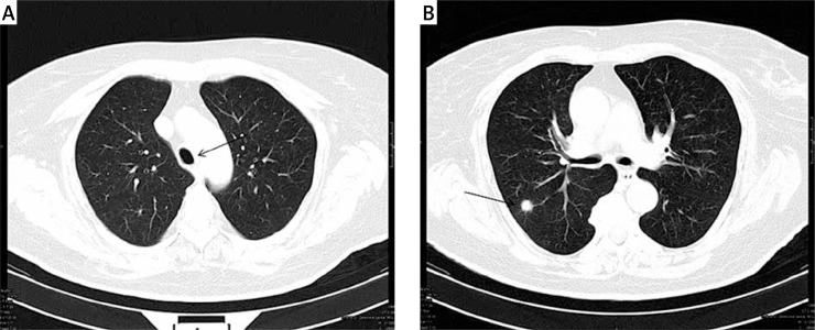
A 57-year-old woman with a history of hypertension, chronic obstructive pulmonary disease, hypothyroidism, diabetes mellitus type 2, was admitted to the hospital with cough, progressive shortness of breath, low-grade fever, joint pain, leakage of both ears with left ear hearing loss, bloody discharge and obstruction of the nose with severe crusting and peripheral left facial nerve paralysis. Physical examination revealed bloody discharge from the nose, nasal crusting, hearing loss, left facial nerve paralysis, dyspnea, cough and wheezing at auscultation, pulse oxygen saturation 89%. Laboratory test have shown leukocytosis (14.4 G/l), thrombocytosis (PLT 606 G/l), marked elevation of inflammatory markers (C-reactive protein – CRP: 100 mg/l, erythrocyte sedimentation rate – ESR: 90 mm/h), proteinuria, and an active urine sediment with red cells and white cells. The plasma creatinine was normal. Immunological tests shown presence of cytoplasmic anti-neutrophil cytoplasmic antibodies (cANCA 1 : 40, n < 1 : 10 U/ml), and anti-proteinase 3 antibodies (anti-PR3 27 U/ml, n < 20 U/ml). Antinuclear antibodies (ANA) were negative. Nasal swab culture showed growth of Staphylococcus aureus. Bronchofiberoscopy was performed in which no pathological lesions were found in the trachea or large bronchi. CT scan of the chest revealed a focal lesion with a diameter of 9 mm in the right lung (Fig. 1).
Fig. 1.
Computed tomography scan of the chest in the diagnosis of GPA. The trachea of normal size just above the bifurcation (A). Right lung nodule with a diameter of 9 mm (B).
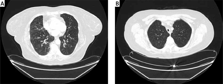
Also during the diagnosis the nasal mucosa biopsy was performed. Histopathological examination revealed purulent exudate, fibrinoid necrosis, polynuclear giant cells, infiltration of T and B lymphocytes as well as neutrophils and eosinophils. Considering the clinical course, CT imaging, laboratory results and histopathological findings confirmed the diagnosis of GPA. The activity of the disease was measured using Birmingham Vasculitis Activity Score (BVAS/GPA = 20 points). The patient received steroid therapy (oral prednisone 1 mg per kilogram of body weight per day) and cyclophosphamide (CF, 15 mg per kilogram of body weight i.v. every 3 weeks). Pneumocystis jiroveci prophylaxis with trimethoprim-sulfamethoxazole (160 mg/800 mg three times per week) was administrated. After 3 months of therapy the patient's complaints included persistent inspiratory dyspnea, very tiring cough with sputum difficult to expectorate and persisting left ear hearing loss. Auscultation revealed symmetrical rales at the base of both lungs and between the shoulder blades, inspiratory stridor audible over the trachea, small ulcers on the neck and legs like pyodermia gangrenosum (biopsy wasn't performed). Laboratory tests revealed only slightly elevated ESR (24 mm/h) and CRP (7.65 mg/l), leukocytosis (13.8 G/l) and thrombocytosis (420 G/l), but no abnormalities in urine analysis. Also creatinine level was normal. The patient got the fourth pulse of CF (1 g i.v., the total dose administered 4 g). Since then, she reported malaise, severe weakness and nausea. Significant increase of CRP (112 mg/l), pulse oxygen saturation 92% was found in laboratory results. Chest CT revealed conglomerates of small nodules in both lungs, reticulo-nodular lesions in stromal throughout the lungs (Fig. 2A) and a segmental, significant narrowing of the trachea up to 7 mm in diameter at a distance of approximately 1.5 cm with a concentric thickening of its walls (Fig. 2B).
Fig. 2.
Computed tomography scan of the chest performed after 3 months of immunosuppressive therapy while worsening of clinical symptoms. CT scans show a conglomerate of small nodules in both lungs, reticulo- nodular lesions in stromal throughout the lungs (A) and narrowing of the trachea up to 7 mm in diameter at a distance of approximately 1.5 cm with a concentric thickening of its walls (B).
The differential diagnosis of clinical deterioration included: the progression of the underlying disease, infectious complication of immunosuppressive therapy, tuberculosis, sarcoidosis or spread of the cancer of unknown origin. During bronchofiberoscopy, the narrowing of the trachea was found with the retention of purulent contents in the bronchi. Microbiologic analysis of sputum and bronchial aspirate excluded active tuberculosis (direct smear), but revealed growth of mixed hospital's bacteria: Acinetobacter baumannii, Streptococcus pneumoniae, methicyllin-resistant Staphylococcus aureus (MRSA) and Candida albicans. Targeted antibiotic therapy was applied – vancomycin, imipenem, fluconazole. Gradually, the patient's clinical condition was improving and normalization of CRP (1.1 mg/l) was noted.
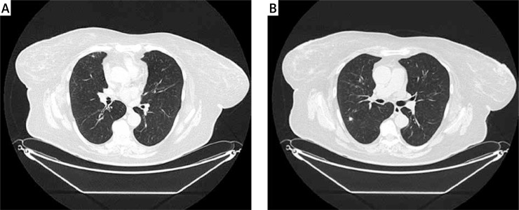
The control chest CT found almost complete regression of reticulo-nodular lesions and interstitial changes (Fig. 3A). Initially described nodule in the right lung was reduced to the size of 7.5 mm (Fig. 3B).
Fig. 3.
Computed tomography scan of the chest after anti-bacterial treatment. Small nodules disappeared (A). Primary right lung nodule is smaller compared to pre-treatment CT scans (B).
Therefore it was decided to continue the treatment of CF and prednisone. After 6 months of immunosuppressive treatment (total dose of cyclophosphamide 6 g, prednisone 10 mg/d) disease activity was assessed using Birmingham Vasculitis Activity Score (BVAS/GPA = 12 points).
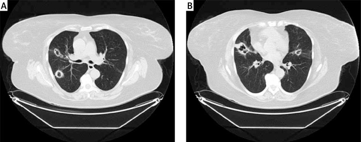
The patient reported dyspnea of constant intensity, limited exercise tolerance, periodic dry cough. She denied fever. Clinical examination revealed hearing impairment, perforation of the nasal septum, mild left eye stare, small skin ulcers on the neck, back and limbs, pulse oxygen saturation 93%. Mild normocytic anemia (Hgb 11.7 g/dl), leukocytosis (WBC 12.6 G/l), thrombocytosis (PLT 469 G/l), increased ESR (43 mm/h) and CRP (14 mg/l) were found in laboratory tests. Immunological tests revealed also the presence of cANCA antibodies (cANCA 1 : 80, normal < 1 : 40), but ANCA-PR3 antibodies were negative. Nasal swab culture was negative. CT of the chest revealed the progression of the main disease (Fig. 4).
Fig. 4.
Computed tomography scan of the chest reveals a progression of the main disease – numerous cavitating lung nodules typical for GPA on both sides.
The patient was repeatedly referred to thoracic surgeons, but further bronchoscopy showed no significant stenosis of trachea so it was not decided on surgery. Pulmonary function tests (spirometry, body plethysmography, single-breath diffusing capacity for carbon monoxide – DLCO) revealed a very severe airway obstruction with significant air trapping (RV – residual volume – 197%) and a slight decrease in gas diffusion capacity in the lung (DLCO – 67.2%). The patient initially refused on biological treatment, so CF therapy was continued to a total dose 13.0 g. Steroids were also given in tapering doses. Finally because of lack of remission, after obtaining the consent of the patient, intravenous infusion of rituximab in total dose of 2 g was applied (2 × 1 g every two weeks). Three months later the patient reported a partial improvement in general state. Dyspnea, cough and permanent deterioration in exercise tolerance as well as skin changes were still present but less intense. Unfortunately, four months after the initiation of rituximab therapy the patient choked on food and died.
Discussion
Granulomatosis with polyangiitis is a systemic vasculitis of the medium and small arteries, as well as the venules and arterioles, usually associated with ANCA. It typically produces granulomatous inflammation of the upper and lower respiratory tracts and necrotizing, pauci-immune glomerulonephritis in the kidneys. The most common lower respiratory tract symptoms in GPA are cough, hemoptysis (due to alveolar hemorrhage and/or tracheobronchial disease), dyspnea, and pleuritic pain [1]. The severity of symptoms and signs varies considerably from asymptomatic (one-third of patients) to acute and fulminant alveolar hemorrhage with respiratory failure. The specific clinical manifestations vary depending on whether the patient has tracheobronchial disease, lung parenchymal nodules, or alveolar hemorrhage. Initial lung presentation in described patient was single nodule, but in the course of disease the radiological image has changed and significant narrowing of trachea appeared. Also deterioration of general condition was associated with the emergence of numerous new conglomerates of small nodules in the lungs.
The differential diagnosis of multiple pulmonary nodules includes metastatic solid organ malignancy, non-Hodgkin lymphoma, septic embolism, granulomatous infections (e.g., Mycobacteria, histoplasmosis, coccidioidomycosis, cryptococcosis, aspergillosis), sarcoidosis, rheumatoid arthritis, lymphomatoid granulomatosis, inflammatory myofibroblastic tumor, silicosis, and coal-worker's pneumoconiosis. Differentiation is based on history of exposures, presence of other features (e.g., erosive arthritis, cutaneous lesions), serology, special stains and culture, and histopathology. Sudden deterioration of the patients state strongly suggested infectious complication of immunosuppressive treatment. Infections are frequent in vasculitis patients and can be severe. They are sometimes life threatening and are one of the major causes of deaths [4–6]. They require rapid diagnosis and targeted therapy. Patients with clinical deterioration raise a differential diagnosis of infection, drug toxicity, disease relapse or a new, unrelated disease. A study by Bradley et al. [7] found that 10% of vasculitis patients who had been treated with cyclophosphamide developed clinically important infections (sepsis and pneumonia) even when patients with drug-induced leucopenia were excluded. Fungal infections, suchas aspergillosis or candidiasis, although not very frequent, remain a concern for clinicians. Bronchial aspirate culture revealed mixed hospital's bacterial and fungal flora in our patient, which were considered the etiological agents of her clinical deterioration.
High-resolution computed tomography (HRCT) is an optimal diagnostic tool to assess pulmonary changes in many connective tissue diseases (e.g. systemic lupus erythematosus) [8]. It makes it possible to detect lesions significantly earlier and much more precisely than can be done with other imaging tests. Imaging studies in small-vessel vasculitides are important for determining disease extent and activity. Ground-glass opacities, cavitating nodules and masses measuring > 3 cm represent active disease. One should remember, that patchy ground glass attenuation can be due to cyclophosphamide-induced acute alveolar damage as well as Pneumocystis jiroveci pneumonia. Because the Pneumocystis jiroveci infection can be very severe in patients during immunosuppressive treatment, prophylaxis with trimethoprim-sulfamethoxazole is recommended. For GPA patients with a persistent lung nodule, the risk of developing aspergillosis must be always considered. Candidiasis should be systematically sought in patients with chronic fever particularly with chronic neutropenia. But these complications are not observed in most patients, so systematic prophylaxis is not recommended [6].
Granulomatosis with polyangiitis may also involve the tracheobronchial tree in 12–23% of cases [9]. Tracheobronchial involvement has several manifestations, including subglottic stenosis, tracheal and bronchial stenosis, mass lesions (inflammatory pseudotumors) and tracheoesophageal fistulae [10, 11].
It is often asymptomatic initially, but becomes apparent as hoarseness, pain, cough, wheezing or stridor. As the airway caliber narrows, mucous plugging becomes a greater concern, as it can cause acute stridulous exacerbations and airway obstruction. CT scans are often useful, but the most accurate means of assessing tracheal stenosis is by direct laryngoscopy. Patients should be carefully assessed for critical airway obstruction and treated with medical treatment especially since the course of bronchial stenosis, as well as subglottic stenosis, seems to be independent of systemic disease activity. Our patient had regular endoscopic examinations performed, but the narrowing seemed stable, so it was decided not to perform surgery.
Up to 80% of patients may require surgical management of subglottic stenosis, and the remaining 20% will respond to systemic medical therapy (conventional immunosuppressive therapy, endoscopic dilation, endoscopic or laser excision, and surgical resection of the stenotic segment followed by reconstruction). The use of local steroid injections is restricted to subglottic stenosis whereas stenting represent a therapeutic option in patients with bronchial stenosis. The optimal endoscopic interventions providing the best efficacy and the best timing for such interventions remain unclear.
Terrier et al. [9] observed that a shorter time from GPA diagnosis to endoscopic procedure was associated with a higher cumulative incidence of treatment failure. Authors suggested that endoscopic procedures should be performed long after maximal inflammatory activity of tracheobronchial stenosis [9]. There are two main parameters that should be taken into consideration in the assessment of indications for treatment: patients complaints and physical signs [12].
Our patients complaints were still on the same level and there was no progression in narrowing of the trachea. Also the location of stenosis in our patient significantly limited the possibility of endoscopic treatment (only stenting was taken into account). The question whether earlier surgical intervention would prevent the fatal outcome in our patient, remains open. It was planned for a period of remission which she has not reached.
The current standards in the treatment of anti-neutrophil cytoplasmic antibody associated vasculitis (AAV) have been optimized on the basis of a number of randomized studies conducted over the past 20 years and adjusted to the duration and severity of the disease [13]. Two randomized clinical trials, RITUXVAS (Randomised Trial of Rituximab versus Cyclophosphamide for ANCA Associated Renal Vasculitis) and RAVE (Rituximab for ANCA-associated Vasculitis), categorically confirmed the equivalent efficacyof rituximab (RTX) and cyclophosphamide in the induction treatment of AAV. RTX should be taken into account particularly in refractory and relapsing forms of the disease as in the present case. It can be administered in two schemes: 4 × 375 mg/m2 BS (RITUXVAS trial, RAVE trial) or 2 × 1 g every two weeks [14]. Our patient received RTX according to the second scheme.
Summary
In conclusion it's worth to mention that careful monitoring for complications is absolutely necessary to minimize the morbidity and mortality of the disease and its therapies. In the assessment of pulmonary changes CT in connection with clinical presentation and laboratory tests allows the diagnosis of organ damage in an early stage and the introduction of appropriate treatment.
The authors declare no conflict of interest.
References
- 1.Madej M, Matuszewska A, Białowąs K, Wiland P. Clinical forms of granulomatosis with polyangiitis. Reumatologia. 2014;52:332–338. [Google Scholar]
- 2.Tadema H, Heeringa P, Kallenberg CG. Bacterial infections in Wegener's granulomatosis: mechanisms potentially involved in autoimmune pathogenesis. Curr Opin Rheumatol. 2011;23:366–371. doi: 10.1097/BOR.0b013e328346c332. [DOI] [PubMed] [Google Scholar]
- 3.Wiatr E, Gawryluk D. Nowe aspekty patogenezy ziarniniakowatości Wegenera. Pneumonol Alergol Pol. 2002;70:326–333. [PubMed] [Google Scholar]
- 4.Goupil R, Brachemi S, Nadeau-Fredette AC, et al. Lymphopenia and treatment-related infectious complications in ANCA-associated vasculitis. Clin J Am Soc Nephrol. 2013;8:416–423. doi: 10.2215/CJN.07300712. [DOI] [PMC free article] [PubMed] [Google Scholar]
- 5.Hoffman GS, Kerr GS, Leavitt RY, et al. Wegener granulomatosis: An analysis of 158 patients. Ann Intern Med. 1992;116:488–498. doi: 10.7326/0003-4819-116-6-488. [DOI] [PubMed] [Google Scholar]
- 6.Guillevin L. Infections in vasculitis. Best Prac Res Clin Rheumatol. 2013;27:19–31. doi: 10.1016/j.berh.2013.01.004. [DOI] [PubMed] [Google Scholar]
- 7.Bradley JD, Brandt KD, Katz BP. Infectious complications of cyclophosphamide treatment for vasculitis. Arthritis Rheum. 1989;32:45–53. doi: 10.1002/anr.1780320108. [DOI] [PubMed] [Google Scholar]
- 8.Jeka S, Żuchowski P, Dura M, et al. Assessment of pulmonary changes in the course of systemic lupus erythematosus – application of high resolution computed tomography. Reumatologia. 2014;52:339–343. [Google Scholar]
- 9.Terrier B, Dechartres A, Girard C, et al. Granulomatosis with polyangiitis: endoscopic management of tracheobronchial stenosis: results from a multicenter experience. Rheumatology (Oxford) 2015;54:1852–1857. doi: 10.1093/rheumatology/kev129. [DOI] [PubMed] [Google Scholar]
- 10.Langford C, Sneller M, Hallahan C, et al. Clinical features and therapeutic management of subglottic stenosis in patients with Wegener's granulomatosis. Arthritis Rheum. 1996;39:1754–1760. doi: 10.1002/art.1780391020. [DOI] [PubMed] [Google Scholar]
- 11.Lebovics R, Hoffman G, Leavitt R, et al. The management of subglottic stenosis in patients with Wegener's granulomatosis. Laryngoscope. 1992;102:1341–1345. doi: 10.1288/00005537-199212000-00005. [DOI] [PubMed] [Google Scholar]
- 12.Schokkenbroek A, Franssen C, Dikkers F. Dilatation tracheoscopy for laryngeal and tracheal stenosis in patients with Wegener's granulomatosis. Eur Arch Otorhinolaryngol. 2008;265:549–555. doi: 10.1007/s00405-007-0518-3. [DOI] [PMC free article] [PubMed] [Google Scholar]
- 13.Zdrojewski Z. Individualization of the ANCA-associated vasculitis treatment. Reumatologia. 2014;52:221–223. [Google Scholar]
- 14.Jones RB, Ferraro AJ, Chaudhry AN, et al. A multicenter survey of rituximab therapy for refractory antineutrophil cytoplasmic antibody-associated vasculitis. Arthritis Rheum. 2009;60:2156–2168. doi: 10.1002/art.24637. [DOI] [PubMed] [Google Scholar]


