

Abstract
BACKGROUND: OCT can image plaque microstructure at a level of resolution not previously demonstrated with other imaging techniques because it uses infrared light rather than acoustic waves. OBJECTIVES: To compare optical coherence tomography (OCT) and intravascular ultrasound (IVUS) imaging of in vitro atherosclerotic plaques. METHODS: Segments of abdominal aorta were obtained immediately before postmortem examination. Images of 20 sites from five patients were acquired with OCT (operating at an optical wavelength of 1300 nm which was delivered to the sample through an optical fibre) and a 30 MHz ultrasonic transducer. After imaging, the microstructure of the tissue was assessed by routine histological processing. RESULTS: OCT yielded superior structural information in all plaques examined. The mean (SEM) axial resolution of OCT and IVUS imaging was 16 (1) and 110 (7), respectively, as determined by the point spread function from a mirror. Furthermore, the dynamic range of OCT was 109 dB compared with 43 dB for IVUS imaging. CONCLUSIONS: OCT represents a promising new technology for intracoronary imaging because of its high resolution, broad dynamic range, and ability to be delivered through intravascular catheters.
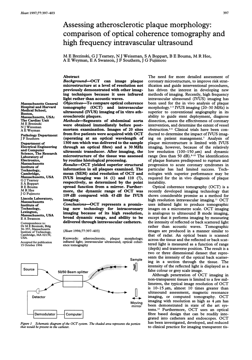
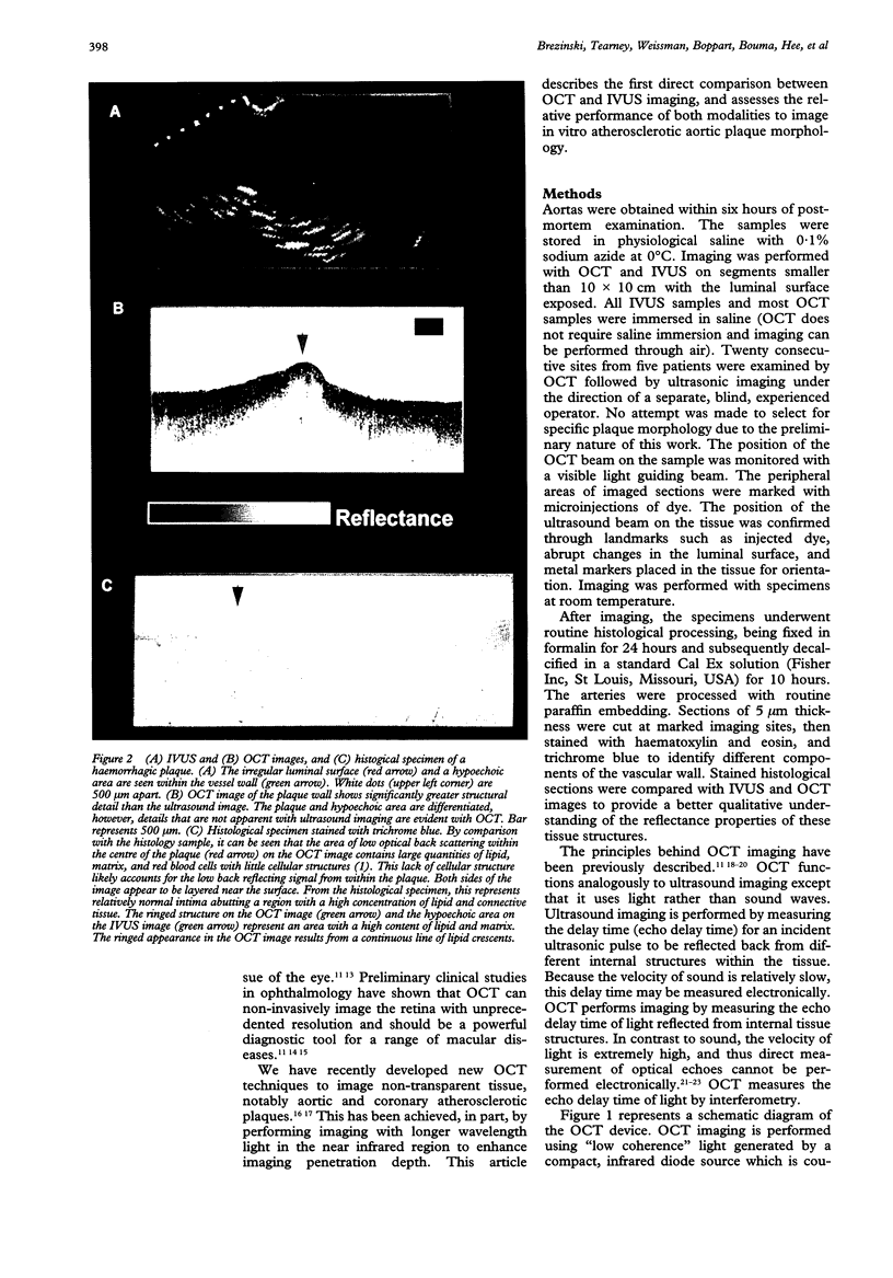
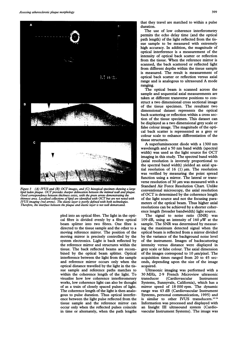
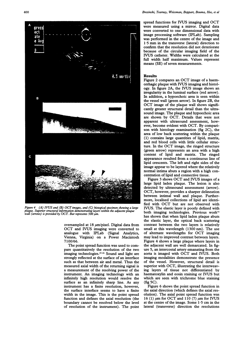
Full text
PDF
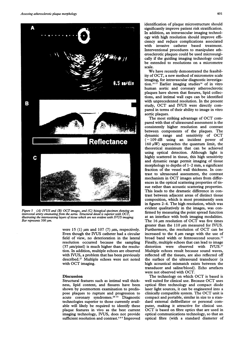
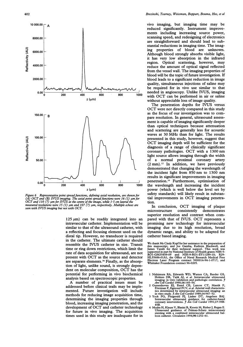
Images in this article
Selected References
These references are in PubMed. This may not be the complete list of references from this article.
- Ambrose J. A., Tannenbaum M. A., Alexopoulos D., Hjemdahl-Monsen C. E., Leavy J., Weiss M., Borrico S., Gorlin R., Fuster V. Angiographic progression of coronary artery disease and the development of myocardial infarction. J Am Coll Cardiol. 1988 Jul;12(1):56–62. doi: 10.1016/0735-1097(88)90356-7. [DOI] [PubMed] [Google Scholar]
- Benkeser P. J., Churchwell A. L., Lee C., Abouelnasr D. M. Resolution limitations in intravascular ultrasound imaging. J Am Soc Echocardiogr. 1993 Mar-Apr;6(2):158–165. doi: 10.1016/s0894-7317(14)80486-0. [DOI] [PubMed] [Google Scholar]
- Brezinski M. E., Tearney G. J., Bouma B. E., Boppart S. A., Hee M. R., Swanson E. A., Southern J. F., Fujimoto J. G. Imaging of coronary artery microstructure (in vitro) with optical coherence tomography. Am J Cardiol. 1996 Jan 1;77(1):92–93. doi: 10.1016/s0002-9149(97)89143-6. [DOI] [PubMed] [Google Scholar]
- Brezinski M. E., Tearney G. J., Bouma B. E., Izatt J. A., Hee M. R., Swanson E. A., Southern J. F., Fujimoto J. G. Optical coherence tomography for optical biopsy. Properties and demonstration of vascular pathology. Circulation. 1996 Mar 15;93(6):1206–1213. doi: 10.1161/01.cir.93.6.1206. [DOI] [PubMed] [Google Scholar]
- Davies M. J., Thomas A. C. Plaque fissuring--the cause of acute myocardial infarction, sudden ischaemic death, and crescendo angina. Br Heart J. 1985 Apr;53(4):363–373. doi: 10.1136/hrt.53.4.363. [DOI] [PMC free article] [PubMed] [Google Scholar]
- Falk E. Plaque rupture with severe pre-existing stenosis precipitating coronary thrombosis. Characteristics of coronary atherosclerotic plaques underlying fatal occlusive thrombi. Br Heart J. 1983 Aug;50(2):127–134. doi: 10.1136/hrt.50.2.127. [DOI] [PMC free article] [PubMed] [Google Scholar]
- Finet G., Maurincomme E., Tabib A., Crowley R. J., Magnin I., Roriz R., Beaune J., Amiel M. Artifacts in intravascular ultrasound imaging: analyses and implications. Ultrasound Med Biol. 1993;19(7):533–547. doi: 10.1016/0301-5629(93)90077-2. [DOI] [PubMed] [Google Scholar]
- Fitzgerald P. J., St Goar F. G., Connolly A. J., Pinto F. J., Billingham M. E., Popp R. L., Yock P. G. Intravascular ultrasound imaging of coronary arteries. Is three layers the norm? Circulation. 1992 Jul;86(1):154–158. doi: 10.1161/01.cir.86.1.154. [DOI] [PubMed] [Google Scholar]
- Fuster V., Badimon L., Badimon J. J., Chesebro J. H. The pathogenesis of coronary artery disease and the acute coronary syndromes (1). N Engl J Med. 1992 Jan 23;326(4):242–250. doi: 10.1056/NEJM199201233260406. [DOI] [PubMed] [Google Scholar]
- Gussenhoven E. J., Essed C. E., Lancée C. T., Mastik F., Frietman P., van Egmond F. C., Reiber J., Bosch H., van Urk H., Roelandt J. Arterial wall characteristics determined by intravascular ultrasound imaging: an in vitro study. J Am Coll Cardiol. 1989 Oct;14(4):947–952. doi: 10.1016/0735-1097(89)90471-3. [DOI] [PubMed] [Google Scholar]
- Hee M. R., Izatt J. A., Swanson E. A., Huang D., Schuman J. S., Lin C. P., Puliafito C. A., Fujimoto J. G. Optical coherence tomography of the human retina. Arch Ophthalmol. 1995 Mar;113(3):325–332. doi: 10.1001/archopht.1995.01100030081025. [DOI] [PubMed] [Google Scholar]
- Hibberd M. G., Vuille C., Weyman A. E. Intravascular ultrasound: basic principles and role in assessing arterial morphology and function. Am J Card Imaging. 1992 Dec;6(4):308–324. [PubMed] [Google Scholar]
- Huang D., Swanson E. A., Lin C. P., Schuman J. S., Stinson W. G., Chang W., Hee M. R., Flotte T., Gregory K., Puliafito C. A. Optical coherence tomography. Science. 1991 Nov 22;254(5035):1178–1181. doi: 10.1126/science.1957169. [DOI] [PMC free article] [PubMed] [Google Scholar]
- Lee D. Y., Eigler N., Luo H., Nishioka T., Tabak S. W., Forrester J. S., Siegel R. J. Effect of intracoronary ultrasound imaging on clinical decision making. Am Heart J. 1995 Jun;129(6):1084–1093. doi: 10.1016/0002-8703(95)90387-9. [DOI] [PubMed] [Google Scholar]
- Mudra H., Klauss V., Blasini R., Kroetz M., Rieber J., Regar E., Theisen K. Ultrasound guidance of Palmaz-Schatz intracoronary stenting with a combined intravascular ultrasound balloon catheter. Circulation. 1994 Sep;90(3):1252–1261. doi: 10.1161/01.cir.90.3.1252. [DOI] [PubMed] [Google Scholar]
- Nishimura R. A., Edwards W. D., Warnes C. A., Reeder G. S., Holmes D. R., Jr, Tajik A. J., Yock P. G. Intravascular ultrasound imaging: in vitro validation and pathologic correlation. J Am Coll Cardiol. 1990 Jul;16(1):145–154. doi: 10.1016/0735-1097(90)90472-2. [DOI] [PubMed] [Google Scholar]
- Peters R. J., Kok W. E., Havenith M. G., Rijsterborgh H., van der Wal A. C., Visser C. A. Histopathologic validation of intracoronary ultrasound imaging. J Am Soc Echocardiogr. 1994 May-Jun;7(3 Pt 1):230–241. doi: 10.1016/s0894-7317(14)80393-3. [DOI] [PubMed] [Google Scholar]
- Puliafito C. A., Hee M. R., Lin C. P., Reichel E., Schuman J. S., Duker J. S., Izatt J. A., Swanson E. A., Fujimoto J. G. Imaging of macular diseases with optical coherence tomography. Ophthalmology. 1995 Feb;102(2):217–229. doi: 10.1016/s0161-6420(95)31032-9. [DOI] [PubMed] [Google Scholar]
- Richardson P. D., Davies M. J., Born G. V. Influence of plaque configuration and stress distribution on fissuring of coronary atherosclerotic plaques. Lancet. 1989 Oct 21;2(8669):941–944. doi: 10.1016/s0140-6736(89)90953-7. [DOI] [PubMed] [Google Scholar]