Abstract
The vermiform appendix is a tubular, narrow, worm-shaped part of the alimentary canal that lies near the ileocecal junction and communicates with the caecum. Duplication of the vermiform appendix is rare, with a reported incidence of 0.004 %. Till now, fewer than 100 cases have been reported. We present a case of an 8-year-old male child with duplex appendix who presented to the emergency department of our institution with features of acute appendicitis.
Keywords: Duplication, Appendix, Caecum, Anomalies
Introduction
The vermiform appendix is a tubular, narrow, worm-shaped part of the alimentary canal that lies near the ileocecal junction and communicates with the caecum [1, 2]. It develops as a conical extension from the apex of the caecal diverticulum which arises from the antimesenteric border of the proximal part of the post arterial segment of the mid gut [3]. In the fetal period, the caecum is tube shaped and gradually changes its shape to quadrilateral due to the asymmetrical growth of the wall which makes the vermiform appendix to occupy different positions; most common is the sub-caecal position, with changing peritoneal relations [4].
Duplication of the vermiform appendix is rare, with a reported incidence of 0.004 % [5]. Till now, fewer than 100 cases have been reported. Picoli in 1892 reported the first case of duplex appendix in a female patient who had associated anomalies of duplication of the entire large bowel, two uteri with two vaginae, ectopia vesicae, and exomphalos [6].
Wallbridge [7] modified Cave’s original classification [8] of duplicated vermiform appendix which was again modified by Biermann in 1993 as follows:
-
Type A
Single caecum with one appendix exhibiting partial duplication.
-
Type BSingle caecum with two obviously separate appendices.
- The two appendices arise on either side of the ileocaecal valve in a “bird-like” manner.
- In addition to a normal appendix arising from the caecum at the usual site, there is also a second, usually rudimentary, appendix arising from the caecum along the lines of the taenia at a varying distance from the first.
- The second appendix is located along the taenia of the hepatic flexure of the colon.
- The location of the second appendix is along the taenia of the splenic flexure of the colon.
-
Type C
Double caecum, each bearing its own appendix and associated with multiple duplication anomalies of the intestinal tract as well as the urinary tract.
Case Report
We present a case of an 8-year-old male child who presented to the emergency department of our institution with chief complaints of pain in the peri-umbilical region which shifted to the RIF for 2 days. There was also history of nausea, vomiting, and anorexia. On examination, the pulse rate was 104 beats per minute and temperature was normal.
Abdominal examination showed tenderness and rebound tenderness in the right iliac fossae. Laboratory investigations showed leukocytosis predominately polymorphs. A diagnosis of acute appendicitis was made. Ultrasonographic examination was suggestive of acute appendicitis.
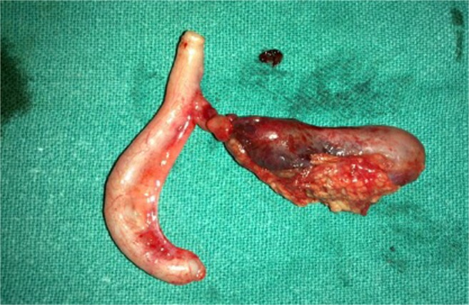
On exploration via a grid iron incision, two appendices with common base were found (Fig. 1). One moiety was inflamed more so near the tip where another was gangrenous throughout its length. The base and caecum were healthy. Histopathological examination showed gangrenous appendicitis in one moiety and acute inflammation in another.
Fig. 1.

Specimen showing double appendix with common base
Discussion
The overall incidence of duplex appendix is about 0.004 % and may be associated with the duplication of other organs or with other anomalies. It is usually asymptomatic; the majority of them are diagnosed at surgery or on postmortem examination. Sometimes can be picked up on barium enema. Symptoms arise due to acute inflammation.
Compliance with Ethical Standards
Conflict of Interest
This is to certify that this article is not under consideration for publication elsewhere.
This is to certify that all the authors mentioned below have participated in person in this study and in the submission of this journal for consideration for publication.
This is to certify that there is no conflict of interest between the authors or any other person, association, or journal.
Contributor Information
Gulzar Ahmad Bhat, Email: drbhatgulzar@gmail.com.
Tarooq Ahmad Reshi, Email: drtariqreshi@gmail.com.
Asiya Rashid, Email: asiya7rashid@gmail.com.
References
- 1.Gray’s Anatomy (2009) 40th Edition, The anatomical basis of clinical practice, Expert Consult—Online and Print By Susan Standring, PhD, DSc. Churchill Livingstone, 2009
- 2.Last’s anatomy: regional and applied by Chummy S. Sinnatamby and R. J (2011) Last: Elsevier-Health Sciences Division 2011
- 3.The developing human: (2011) clinically oriented embryology with student consult online access, 9th Edition by Keith L. Moore. Saunders; 9th edition, 2011
- 4.Malas MA, Gökçimen A, Sulak O. Growing of caecum and vermiform appendix during the fetal period. Fetal Diagn Ther. 2001;16(3):173–177. doi: 10.1159/000053904. [DOI] [PubMed] [Google Scholar]
- 5.Majid Mushtaque1, Asif Mehraj1, Samina Ali Khanday2, Rayees A. Dar1 (2012) International Journal of Clinical Medicine, 2012, 3, 61–62 Doi:10.4236/ijcm.2012.31013. Published Online January 2012 (http://www.SciRP.org/journal/ijcm)
- 6.Khanna AK. Appendix vermiformis duplex. Postgrad Med J. 1983;59:69–70. doi: 10.1136/pgmj.59.687.69. [DOI] [PMC free article] [PubMed] [Google Scholar]
- 7.Wallbridge PH. Double appendix. Br J Surg. 1963;50(221):346–347. doi: 10.1002/bjs.18005022124. [DOI] [PubMed] [Google Scholar]
- 8.Cave AJE. Appendix vermiformis duplex. J Anat. 1936;70:283–292. [PMC free article] [PubMed] [Google Scholar]


