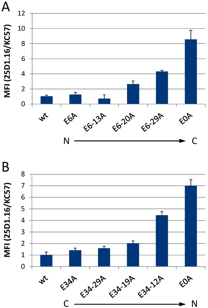
Figure 7.
Successive mutation of the glutamic acids within p6 progressively increases the MHC-I antigen presentation of Gag-derived epitopes. HeLa-Kb cells were transiently transfected with pNLenv1-SL expression plasmids coding for wt or the sequential Glu mutants of p6 from (A) the N- to the C-terminus or (B) the C- to the N-terminus, respectively. H2-Kb-SL complexes presented on the surface of Gag-positive cells were quantified by flow cytometry using the mAb 25D1.16-APC [58]. After fixation and permeabilization, intracellular Gag was stained with anti-Gag Ab KC57-FITC. The mean fluorescence intensity (MFI) of the 25D1.16 staining, normalized to the MFI of the intracellular anti-Gag staining (see Figure S2) is shown. Bars represent mean values ± SD from three independent experiments.

