Abstract
Aripiprazole, a dopamine D2 receptor (D2R) partial agonist, possesses a unique clinical profile. Glycogen synthase kinase 3β (GSK3β)-dependent signalling pathways have been implicated in the pathophysiology of schizophrenia and antipsychotic drug actions. The present study examined whether aripiprazole differentially affects the GSK3β-dependent signalling pathways in the prefrontal cortex (PFC), nucleus accumbens (NAc), and caudate putamen (CPu), in comparison with haloperidol (a D2R antagonist) and bifeprunox (a D2R partial agonist). Rats were orally administrated aripiprazole (0.75 mg/kg), bifeprunox (0.8 mg/kg), haloperidol (0.1 mg/kg) or vehicle three times per day for one week. The levels of protein kinase B (Akt), p-Akt, GSK3β, p-GSK3β, dishevelled (Dvl)-3, and β-catenin were measured by Western Blots. Aripiprazole increased GSK3β phosphorylation in the PFC and NAc, respectively, while haloperidol elevated it in the NAc only. However, Akt activity was not changed by any of these drugs. Additionally, both aripiprazole and haloperidol, but not bifeprunox, increased the expression of Dvl-3 and β-catenin in the NAc. The present study suggests that activation of GSK3β phosphorylation in the PFC and NAc may be involved in the clinical profile of aripiprazole; additionally, aripiprazole can increase GSK3β phosphorylation via the Dvl-GSK3β-β-catenin signalling pathway in the NAc, probably due to its relatively low intrinsic activity at D2Rs.
Keywords: antipsychotics, aripiprazole, β-catenin, bifeprunox, Dvl-3, GSK3β, haloperidol
1. Introduction
Aripiprazole is an atypical antipsychotic drug with therapeutic effects on both positive and negative symptoms of schizophrenia, but reduced extrapyramidal side-effects (EPS) compared with typical antipsychotics (e.g., haloperidol) [1]. The exact mechanisms of aripiprazole remain unclear. Glycogen synthase kinase 3β (GSK3β) has been implicated in the pathophysiology of schizophrenia and the actions of antipsychotic drugs [2]. GSK3β is a major downstream regulator of dopamine D2 receptors (D2Rs), which is targeted by most antipsychotics (including aripiprazole) [3]. Activation of D2Rs facilitates the formation of the β-arrestin2-protein phosphatase 2A-protein kinase B (PKB or Akt) complex, resulting in dephosphorylation of Akt (inactivation), followed by dephosphorylation (activation) of GSK3β [4,5,6]. Aripiprazole has been shown to have effects on regulating the Akt-GSK3β signalling pathway [2]. For example, Seo et al. [7] have revealed that aripiprazole altered GSK3β activity in the frontal cortex. However, whether aripiprazole can affect GSK3β activity in other schizophrenia-related brain regions has not yet been studied. Our previous acute study [8] has found that acute administration of aripiprazole increased the phosphorylation levels of GSK3β in various brain regions, including the prefrontal cortex (PFC), caudate putamen (CPu), and nucleus accumbens (NAc). However, it is interesting that Akt did not show parallel changes with GSK3β after acute administration [8]. One possibility is that aripiprazole might affect GSK3β activity via alternative pathway(s) that is independent of Akt. One candidate pathway is the dishevelled (Dvl)-GSK3β-β-catenin signalling pathway. In vitro evidence has suggested that various antipsychotics (e.g., clozapine, haloperidol) increase the cellular levels of Dvl and β-catenin via affecting D2Rs [9,10]. In vivo studies have reported that antipsychotic drug administration (including aripiprazole and haloperidol) promoted phosphorylation of GSK3β and expression of Dvl and β-catenin in various brain regions [10,11,12,13,14]. It has been also revealed that administration of aripiprazole attenuated the decreased phosphorylation of GSK3β and reduced expression of β-catenin in the frontal cortex and hippocampus caused by immobilisation stress [7,15]. It should be noted that all these previous studies used intramuscular or subcutaneous injections to deliver aripiprazole. The effects of oral administration that mimic the clinical situation is of importance. Therefore, in this study we examined the Dvl-GSK3β-β-catenin signalling pathway after sub-chronic oral administration of aripiprazole.
Aripiprazole is a D2R partial agonist. Researchers have attributed the unique clinical profile of aripiprazole to its partial agonism at D2Rs [16,17]. However, the role that D2R partial agonism plays in the regulation of the Dvl-GSK3β-β-catenin signalling pathway by aripiprazole is not clear. To investigate this issue, we chose a potent D2R partial agonist—bifeprunox [18] to compare with aripiprazole. Therefore, the present study examined the different effects of one-week oral administration of aripiprazole on the Akt-GSK3β and Dvl-GSK3β-β-catenin signalling pathways in three schizophrenia-related brain regions in comparison with a D2R antagonist—haloperidol and a D2R partial agonist—bifeprunox.
2. Results
2.1. Effects of Antipsychotics in the Prefrontal Cortex
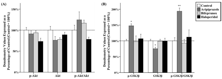
Antipsychotic drug administration had significant effects on the expression of total GSK3β (F3,20 = 3.656, p < 0.05), p-GSK3β (F3,20 = 3.722, p < 0.05) and the ratio of p-GSK3β/GSK3β (F3,20 = 9.207, p < 0.01) in the PFC, but had no effect on Akt, p-Akt, or the ratio of p-Akt/Akt (Figure 1A,D). Post hoc tests demonstrated that administration of aripiprazole significantly increased the protein levels of p-GSK3β by 47.7% ± 6.4% (p < 0.05), but reduced total GSK3β expression by 24.9% ± 4.7% (p < 0.05) compared with the control; the ratio of p-GSK3β/GSK3β was also increased by administration of aripiprazole (p < 0.01) (Figure 1B,D). Furthermore, the protein levels of Dvl-3 and β-catenin in the PFC were not significantly altered by any antipsychotic drug administration (Figure 1C,D).
Figure 1.
Effects of three antipsychotics in the prefrontal cortex. The effects of aripiprazole (ARI), bifeprunox (BIF), haloperidol (HAL), and control (CON) on the activity of protein kinase B (Akt) (A); glycogen synthase kinase 3β (GSK3β) (B); and the expression of dishevelled (Dvl)-3 and β-catenin (C) were measured in the prefrontal cortex (* p ≤ 0.05, ** p < 0.01 vs. the control). All data were expressed as mean ± S.E.M. The representative bands of Western blot are shown in (D).
2.2. Effects of Antipsychotics in the Caudate Putamen
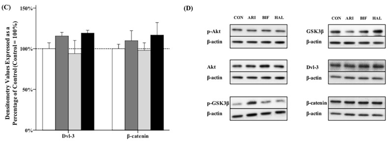
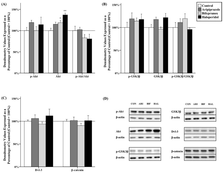
One-way analysis of variance (ANOVA) tests indicated significant effects of antipsychotics on the protein levels of total Akt (F3,20 = 9.707, p < 0.01) in the CPu. Post hoc tests showed that the levels of total Akt were significantly increased by administration of bifeprunox (+18.7% ± 4.8%, p < 0.05) and haloperidol (+37.0% ± 4.0%, p < 0.01) in the CPu (Figure 2A,D); however, they did not affect the levels of p-Akt, nor the ratio of p-Akt/Akt. Additionally, the protein levels of Dvl-3 and β-catenin were not significantly affected by any antipsychotic drug administration in the CPu (Figure 2B–D).
Figure 2.
Effects of three antipsychotics in the caudate putamen. The effects of aripiprazole (ARI), bifeprunox (BIF), haloperidol (HAL), and control (CON) on the activity of Akt (A); GSK3β (B); and the expression of Dvl-3 and β-catenin (C) were measured in the caudate putamen (* p ≤ 0.05, ** p < 0.01 vs. the control). All data were expressed as mean ± S.E.M. The representative bands of Western blot are shown in (D).
2.3. Effects of Antipsychotics in the Nucleus Accumbens
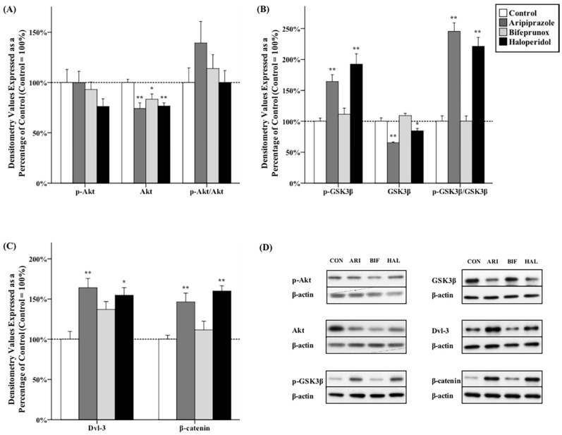
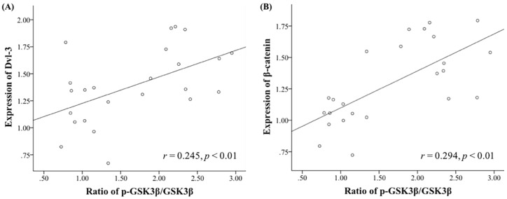
ANOVA tests revealed that antipsychotic drug administration had significant effects on the protein levels of Akt (F3,20 = 6.792, p < 0.01), GSK3β (F3,20 = 25.381, p < 0.01), p-GSK3β (F3,20 = 11.817, p < 0.01), the ratio of p-GSK3β/GSK3β (F3,20 = 42.603, p < 0.01), Dvl-3 (F3,20 = 4.121, p < 0.01), and β-catenin (F3,20 = 10.718, p < 0.01) in the NAc. Post hoc tests indicated that administration of all three chemicals was shown to be able to reduce the protein levels of total Akt (aripiprazole, −25.9% ± 5.9%, p < 0.01; bifeprunox, −16.5% ± 5.0%, p < 0.05; haloperidol, −23.4% ± 3.2%, p < 0.01) in the NAc; however, no antipsychotic drug administration significantly affected the protein levels of p-Akt, nor the ratios of p-Akt/Akt (Figure 3A,D). Additionally, the expression of total GSK3β was reduced by both aripiprazole and haloperidol administration (aripiprazole, −34.5% ± 1.2%, p < 0.01; haloperidol, −15.3% ± 7.8%, p < 0.05). Moreover, both aripiprazole and haloperidol administration was able to elevate the levels of p-GSK3β (aripiprazole, +64.4% ± 11.0%, p < 0.05; haloperidol, +92.4% ± 16.7%, p < 0.01) and the ratios of p-GSK3β/GSK3β (aripiprazole, p < 0.01; haloperidol, p < 0.01) (Figure 3B,D). Furthermore, it was shown that administration of aripiprazole was able to promote the expression of both Dvl-3 (+64.1% ± 11.5%, p < 0.01) and β-catenin (+46.5% ± 10.7%, p < 0.01); haloperidol administration also had a positive effect on the protein levels of both Dvl-3 (+54.8% ± 9.4%, p < 0.05) and β-catenin (+59.9% ± 6.6%, p < 0.01) (Figure 3C,D). Lastly, we found that the ratio of p-GSK3β/GSK3β is positively correlated with the expression of Dvl-3 in the NAc (r = 0.245, p < 0.01) (Figure 4A); the ratio of p-GSK3β/GSK3β is also positively correlated with the expression of β-catenin (r = 0.294, p < 0.01) (Figure 4B).
Figure 3.
Effects of three antipsychotics in the nucleus accumbens. The effects of aripiprazole (ARI), bifeprunox (BIF), haloperidol (HAL), and control (CON) on the activity of Akt (A); GSK3β (B); and the expression of Dvl-3 and β-catenin (C) were measured in the nucleus accumbens (* p ≤ 0.05, ** p < 0.01 vs. the control). All data were expressed as mean ± S.E.M. The representative bands of Western blot are shown in (D).
Figure 4.
Correlations between the ratio of p-GSK3β/GSK3β and the expression of Dvl-3 and β-catenin in the NAc. The ratio of p-GSK3β/GSK3β is positively correlated with the expression of Dvl-3 (A); and with the expression of β-catenin in the NAc (B).
3. Discussion
The present study has examined the effects of aripiprazole on the Akt-GSK3β and Dvl-GSK3β-β-catenin signalling pathways in three key brain regions that are related to the pathophysiology of schizophrenia, in comparison with bifeprunox and haloperidol. Our findings have provided in vivo evidence that aripiprazole is able to alter the activity of GSK3β in the PFC and NAc. We also found that both aripiprazole and haloperidol, but not bifeprunox, activated the Dvl-GSK3β-β-catenin signalling pathway in the NAc.
A wide range of evidence has identified reduced phosphorylation levels and elevated GSK3β protein levels in the brains of schizophrenic patients, indicating hyper-activity of GSK3β in schizophrenia [19,20]. In addition, antipsychotics, including aripiprazole and haloperidol, have been shown to be able to induce inhibition of GSK3β function in various brain regions [8,11,12,13,21]. In the present study, both aripiprazole and haloperidol were able to increase the phosphorylation levels of GSK3β (the ratio of p-GSK3β/GSK3β) in the NAc and PFC (only for aripiprazole), which is not completely consistent with the findings in previous studies [7,8,11,12,13,15,21]. It should be noted that the present study used oral administration to deliver the drugs (for one week) to mimic the clinical situations, which is different from the methods of other previous studies (e.g., intraperitoneal and subcutaneous injection); the dosages of antipsychotics used in this study are transferred from recommended clinical dosages, which are lower than those in previous studies [7,11,12,13,15,21]. Therefore, the results of the present study might be of more significance for clinic. However, whether these discrepancies are caused by different drug delivering methods requires further investigations.
However, the effects of aripiprazole and haloperidol on GSK3β were not completely consistent in every brain region in the present study. Therefore, by comparing the effects of aripiprazole with those of haloperidol, we may further understand the mechanisms of aripiprazole and elucidate its unique clinical profile. The present study has demonstrated that aripiprazole, but not haloperidol, increased the phosphorylation levels of GSK3β in the PFC. This effect is consistent with the result of our previous acute study [8] and another chronic in vivo study [7]. Since prefrontal dysfunction is linked to the negative symptoms of schizophrenia [22,23], it is suggested that suppression of GSK3β function in the PFC is very likely to contribute to the effects of aripiprazole on the negative symptoms of schizophrenia, which cannot be achieved by haloperidol [7,8]. Moreover, we have observed that aripiprazole increased GSK3β phosphorylation levels in the NAc in the present and previous acute study [8]; haloperidol also showed similar effects in the NAc presently and previously [8,12,21]. It is suggested that dysfunction of the NAc is related to the positive symptoms of schizophrenia [24]. Therefore, our finding further indicates that inhibition of GSK3β function in the NAc may contribute to the effects of antipsychotics on the positive symptoms of schizophrenia.
It is worth noting that Akt did not change in parallel with GSK3β, which is not consistent with previous reports [12,21,25]. This might be explained by following reasons. First, Roh et al. [25] have reported that the phosphorylation of Akt induced by antipsychotics were much shorter in duration than those of GSK3β. In the present study, the animals were sacrificed several hours after the last administration. Therefore, the phosphorylation levels of Akt might have already decreased to undetectable levels. This might be the major reason that we only observed the altered p-GSK3β levels, but not p-Akt. Second, there are two phosphorylating sites of Akt—Thr308 and Ser473, both could be affected by antipsychotic drug administration [4,21,25,26,27]. The present study has examined the Thr308 site of Akt only, since phospho-Thr308-Akt was involved in the D2Rs-mediated Akt-GSK3β signalling [4,27]. However, Akt phosphorylated with either site induces phosphorylation of GSK3β at Ser9 that was examined in the current study. Thereby, it is possible that the elevated p-GSK3β levels in this study might be induced by phospho-Ser473-Akt from other signalling pathway(s), and further investigations are needed to study this issue. Lastly, GSK3β is a multi-targeted regulator. Antipsychotics might affect GSK3β via alternative pathway(s) rather than the D2Rs-mediated signalling pathway, such as the Dvl-GSK3β-β-catenin signalling pathway.
We have examined the effects of antipsychotics on the Dvl-GSK3β-β-catenin signalling pathway. It was observed that both aripiprazole and haloperidol administration increased the expression of Dvl-3 and β-catenin in accordance with the enhanced phosphorylation of GSK3β in the NAc, suggesting that antipsychotics is very likely to affect GSK3β activity via Dvl-GSK3β-β-catenin pathway in this study. However, further studies (e.g., pharmacological or genetic intervention) are required to confirm this suggestion. In addition, it has been reported that antipsychotics (e.g., aripiprazole, haloperidol, clozapine, and risperidone) increased the expression of Dvl-3 and/or β-catenin in various brain regions, including the PFC and striatum [10,12,14]. It is worth noting that the studies by Alimohamad et al. [12] and Sutton et al. [14] have mixed NAc and CPu together, thus preventing identification of the sub-region(s) in which the levels of Dvl-3 and β-catenin were increased by antipsychotic drug administration. This study has separated NAc and CPu, and demonstrated that antipsychotics affect Dvl-GSK3β-β-catenin signalling specifically in the NAc. Taken together, it suggested that activation of Dvl-GSK3β-β-catenin signalling in the NAc is a common route, through which different classes of antipsychotics exert their effects. Lastly, our results do not show any alteration in the expression of Dvl-3 and β-catenin in the PFC, which is inconsistent with the findings of previous studies [10,12,14]. The exact reason remains unclear. This may be because the previous studies [10,12,14] used intramuscular or subcutaneous injection to deliver the drugs, whereas the current study used oral treatment with different dosages to mimic the clinical situation. Therefore, whether the effect of antipsychotics on Dvl-GSK3β-β-catenin signalling is treatment method-dependent requires further validation. Furthermore, one limitation of this study is that the samples have been investigated by only Western blots method, it is also worthy to further validate these findings using other methods such as qPCR and immunohistochemistry.
Previously, Min and colleagues [9] have investigated the interaction between the dopaminergic nervous system and Dvl-GSK3β-β-catenin signalling, and found that only D2Rs directly affected β-catenin distribution in the cell nucleus. The present study used aripiprazole, haloperidol and bifeprunox, all of which have strong affinity with D2Rs [18,28]. Haloperidol is a potent D2R antagonist, whereas aripiprazole and bifeprunox are D2R partial agonists. Previous studies have revealed that the intrinsic activity of aripiprazole at D2Rs is weaker than that of bifeprunox (intrinsic activity at D2Rs: aripiprazole vs. bifeprunox vs. dopamine = 86.0% vs. 95.1% vs. 100%) [22,29]. Our results have demonstrated that administration of both aripiprazole and haloperidol, but not bifeprunox, had significant effects on altering the expression of Dvl-3 and β-catenin in the NAc. Therefore, first, blockade of D2Rs is (indirectly) linked to the activation of the Dvl-GSK3β-β-catenin signalling pathway. Second, it is very possible that aripiprazole competes with endogenous dopamine in the normal brain due to its relatively low intrinsic activity to reduce significantly the activity of endogenous dopamine, displaying an overall antagonising effect like haloperidol. In contrast, bifeprunox cannot achieve such effects, probably because of its relatively stronger intrinsic activity at D2Rs. Taken together, our study suggests that a relatively low intrinsic activity at D2Rs might be essential for a D2R partial agonist to achieve meaningful effects via affecting the Dvl-GSK3β-β-catenin signalling pathway.
4. Materials and Methods
4.1. Animals and Drug Administration
Male Sprague–Dawley rats (aged eight weeks) were obtained from the Animal Resource Centre (Perth, Australia). After arrival, all rats were housed in individual cages under environmentally controlled conditions (temperature 22 °C, light cycle from 07:00 a.m. to 07:00 p.m.), with ad libitum access to water and a standard laboratory chow diet. All experimental procedures were approved by the Animal Ethics Committee (Application #AE11/02, 02/2011), University of Wollongong, and complied with the Australian Code of Practice for the Care and Use of Animals for Scientific Purposes (2004). All efforts were made to minimise animal distress and prevent suffering.
Before drug administration commenced, the rats were trained for self-administration of the cookie dough pellets without drugs. After 1-week training, rats were randomly assigned into one of the following four groups (n = 6/group): aripiprazole (0.75 mg/kg, t.i.d. (ter in die), Otsuka, Tokyo, Japan); bifeprunox (0.8 mg/kg, t.i.d., Otava, Kiev, Ukraine); haloperidol (0.1 mg/kg, t.i.d., Sigma, Castle Hill, Australia); or vehicle for one week. Rats were offered cookies with drugs three times a day (at 06:00 a.m., 02:00 p.m. and 10:00 p.m.) and observed to ensure complete consumption of each pellet. The dosages were translated from recommended clinical dosages based on body surface area according to the FDA guidelines [30,31]. This drug administration method has been well established in our laboratory [32,33]. Specifically, a 0.75 mg/kg aripiprazole, 0.8 mg/kg bifeprunox and 0.1 mg/kg haloperidol dosage in rats is equivalent to ~7.5, ~8, and ~1 mg in humans (60 kg body weight), respectively, all of which are within the used/recommended clinical dosages [34,35,36]. It is worth noting that aripiprazole and bifeprunox induced over 90% D2 receptor occupancy in rat brains at these dosages [18], and haloperidol reached approximately 70% occupancy [37], all of which can display physiological and behavioural effects in rodents, without inducing EPS side-effects [18,38,39,40]. After one-week drug administration, all rats were sacrificed between 10:00 a.m. and 12:00 p.m. to minimise possible circadian-induced variation of protein expression. All animals were euthanised by using carbon dioxide. Brains were immediately dissected, frozen in liquid nitrogen and stored at −80 °C until further use.
4.2. Micro-Dissection of Brain Samples
Following a standard procedure used in our group [8], discrete brain regions were collected using brain microdissection puncture according to the brain atlas [41]. Briefly, three sections through the forebrain (Bregma 3.30 to 4.20 mm) were collected for the PFC; and three sections through the striatum (Bregma 1.00 to 2.20 mm) were collected for the CPu and NAc, respectively. Tissue dissected was kept at −80 °C.
4.3. Western Blots
The Western blot experiments were performed following standard procedures repeated in our previous studies [8,33]. Briefly, frozen tissue was homogenised with 9.8 mL NP-40 cell lysis buffer (Invitrogen, Camarillo, CA, USA) containing 100 μL Protease Inhibitor Cocktail (Sigma-Aldrich, St. Louis, MO, USA), 100 μL β-Glycerophosphate (Invitrogen) and 33.3 μL phenylmethylsulfonylfluoride (Sigma-Aldrich). The homogenised samples were centrifuged, and the supernatants were collected. Protein concentration of each homogenising solution was measured by using the DC Protein Assay (Bio-Rad, #500-0111). After denaturing proteins, samples containing 10 μg of protein were loaded into 4%–20% Criterion™ TGX™ Precast Gels (Bio-Rad, Hercules, CA, USA, #5671095) in a Criterion™ Vertical Electrophoresis Cell (Bio-rad, #1656001) at 200 V voltage for 50 min, and then transferred electrophoretically to a polyvinylidene difluoride membrane in a Criterion™ Blotter (Bio-rad, #1704071) at 100 V voltage for 60 min. All membranes were blocked by 5% bovine serum albumin (BSA) for 60 min and incubated in primary antibodies (diluted in 1% BSA) over night. Amersham Hyperfilm ECL (GE Healthcare, Chicago, IL, USA, #28-9068-36) and Luminata Classico Western HRP substrate (Millipore, Billerica, MA, USA, #WBLUC0500) were used to visualise the immunoreactive bands. The immunoreactive signals were quantified using Bio-Rad Quantity One software. The data of each targeted protein were then corrected based on their corresponding actin levels. Experiments were performed in duplicate to ensure consistency.
The antibodies used in the present study to examine the GSK3β-involved pathways were anti-Akt (1:2000; Cell Signalling, Danvers, MA, USA, #4691), anti-phosphor-Akt (Thr308) (1:1000; Cell Signalling, #13038), anti-GSK3β (1:2000; Cell Signalling, #5676), anti-phospho-GSK3β (Ser9) (1:1000; Cell Signalling, #9322), anti-Dvl-3 (1:1000; Santa Cruz Biotechnology, Dallas, TX, USA, #SC-8027) and anti-β-catenin (1:1000; Santa Cruz Biotechnology, #SC-7963). Mouse anti-actin primary polyclonal antibody (1:10000; Millipore, #MAB1501) was used to determine the actin levels. The secondary antibodies were HRP-conjugated anti-rabbit IgG antibody (1:3000; Cell Signalling, #7074) and HRP-conjugated anti-mouse IgG antibody (1:3000; Cell Signalling, #7076).
4.4. Statistics
All data was analysed using SPSS Statistics V22.0 program (IBM, New York, NY, USA). Data normality was tested using histograms and a Kolmogorov–Smirnov Z test. For statistical evaluation, one-way analysis of variance (ANOVA) was performed if the data was normally distributed. The post hoc Dunnett t test was then conducted to compare each drug treatment group with the control group. The results of Western blots were normalised by taking the average value of the control group as 100%. The phosphorylation to total signal was calculated using the data from the same blot. Pearson’s correlation test was used to analyse the relationships. A p-value of less than 0.05 was considered as statistically significant.
5. Conclusions
The present study explored the in vivo effects of one-week oral administration of aripiprazole on the GSK3β-dependent signalling pathways in three brain regions that are associated with schizophrenia and the actions of antipsychotics, in comparison with haloperidol and bifeprunox. The current study provides in vivo evidence that inhibition of GSK3β activity in the PFC and NAc might be linked to the clinical profile of aripiprazole. This study further suggests that, like haloperidol, aripiprazole can activate Dvl-GSK3β-β-catenin signalling in the NAc, which is probably due to the relatively low intrinsic activity at D2Rs.
Acknowledgments
This study was supported by the Australian National Health and Medical Research Council project grant (APP1008473) to Chao Deng. The funding source had no role in study design; in data analysis and interpretation; in writing of the report; or in the decision to submit the manuscript for publication. We would like to thank Jiamei Lian and Michael De-Santis for their technical support for the animal treatment.
Abbreviations
- Akt
protein kinase B
- CPu
caudate putamen
- D2R
dopamine D2 receptor
- Dvl
dishevelled
- EPS
extrapyramidal side-effects
- GSK3β
glycogen synthase kinase 3β
- NAc
nucleus accumbens
- PFC
prefrontal cortex
Author Contributions
Chao Deng and Bo Pan designed the study. Bo Pan performed the animal treatment. Bo Pan conducted experiments and analysed data. Bo Pan prepared the initial draft of the manuscript. Bo Pan, Chao Deng and Xu-Feng Huang revised the manuscript and interpreted the data. All of the authors approved the final manuscript.
Conflicts of Interest
The authors declare no conflict of interest.
References
- 1.Mailman R.B., Murthy V. Third generation antipsychotic drugs: Partial agonism or receptor functional selectivity? Curr. Pharm. Des. 2010;16:488–501. doi: 10.2174/138161210790361461. [DOI] [PMC free article] [PubMed] [Google Scholar]
- 2.Emamian E.S. AKT/GSK3 signaling pathway and schizophrenia. Front. Mol. Neurosci. 2012;5 doi: 10.3389/fnmol.2012.00033. [DOI] [PMC free article] [PubMed] [Google Scholar]
- 3.Howes O., McCutcheon R., Stone J. Glutamate and dopamine in schizophrenia: An update for the 21st century. J. Psychopharmacol. 2015;29:97–115. doi: 10.1177/0269881114563634. [DOI] [PMC free article] [PubMed] [Google Scholar]
- 4.Beaulieu J.M., Sotnikova T.D., Yao W.D., Kockeritz L., Woodgett J.R., Gainetdinov R.R., Caron M.G. Lithium antagonizes dopamine-dependent behaviors mediated by an AKT/glycogen synthase kinase 3 signaling cascade. Proc. Natl. Acad. Sci. USA. 2004;101:5099–5104. doi: 10.1073/pnas.0307921101. [DOI] [PMC free article] [PubMed] [Google Scholar]
- 5.Beaulieu J.M., Gainetdinov R.R., Caron M.G. The Akt-GSK-3 signaling cascade in the actions of dopamine. Trends Pharmacol. Sci. 2007;28:166–172. doi: 10.1016/j.tips.2007.02.006. [DOI] [PubMed] [Google Scholar]
- 6.Beaulieu J.M., Gainetdinov R.R., Caron M.G. Akt/GSK3 signaling in the action of psychotropic drugs. Annu. Rev. Pharmacol. Toxicol. 2009;49:327–347. doi: 10.1146/annurev.pharmtox.011008.145634. [DOI] [PubMed] [Google Scholar]
- 7.Seo M.K., Lee C.H., Cho H.Y., You Y.S., Lee B.J., Lee J.G., Park S.W., Kim Y.H. Effects of antipsychotic drugs on the expression of synapse-associated proteins in the frontal cortex of rats subjected to immobilization stress. Psychiatry Res. 2015;229:968–974. doi: 10.1016/j.psychres.2015.05.098. [DOI] [PubMed] [Google Scholar]
- 8.Pan B., Chen J., Lian J., Huang X.F., Deng C. Unique effects of acute aripiprazole treatment on the dopamine D2 receptor downstream cAMP-PKA and Akt-GSK3β signalling pathways in rats. PLoS ONE. 2015;10:459. doi: 10.1371/journal.pone.0132722. [DOI] [PMC free article] [PubMed] [Google Scholar]
- 9.Min C., Cho D.I., Kwon K.J., Kim K.S., Shin C.Y., Kim K.M. Novel regulatory mechanism of canonical Wnt signaling by dopamine D2 receptor through direct interaction with β-catenin. Mol. Pharmacol. 2011;80:68–78. doi: 10.1124/mol.111.071340. [DOI] [PubMed] [Google Scholar]
- 10.Sutton L.P., Honardoust D., Mouyal J., Rajakumar N., Rushlow W.J. Activation of the canonical Wnt pathway by the antipsychotics haloperidol and clozapine involves dishevelled-3. J. Neurochem. 2007;102:153–169. doi: 10.1111/j.1471-4159.2007.04527.x. [DOI] [PubMed] [Google Scholar]
- 11.Alimohamad H., Sutton L., Mouyal J., Rajakumar N., Rushlow W.J. The effects of antipsychotics on β-catenin, glycogen synthase kinase-3 and dishevelled in the ventral midbrain of rats. J. Neurochem. 2005;95:513–525. doi: 10.1111/j.1471-4159.2005.03388.x. [DOI] [PubMed] [Google Scholar]
- 12.Alimohamad H., Rajakumar N., Seah Y.H., Rushlow W. Antipsychotics alter the protein expression levels of β-catenin and GSK-3 in the rat medial prefrontal cortex and striatum. Biol. Psychiatry. 2005;57:533–542. doi: 10.1016/j.biopsych.2004.11.036. [DOI] [PubMed] [Google Scholar]
- 13.Li X., Rosborough K.M., Friedman A.B., Zhu W., Roth K.A. Regulation of mouse brain glycogen synthase kinase-3 by atypical antipsychotics. Int. J. Neuropsychopharmacol. 2007;10:7–19. doi: 10.1017/S1461145706006547. [DOI] [PubMed] [Google Scholar]
- 14.Sutton L.P., Rushlow W.J. The effects of neuropsychiatric drugs on glycogen synthase kinase-3 signaling. Neuroscience. 2011;199:116–124. doi: 10.1016/j.neuroscience.2011.09.056. [DOI] [PubMed] [Google Scholar]
- 15.Park S.W., Phuong V.T., Lee C.H., Lee J.G., Seo M.K., Cho H.Y., Fang Z.H., Lee B.J., Kim Y.H. Effects of antipsychotic drugs on BDNF, GSK-3β, and β-catenin expression in rats subjected to immobilization stress. Neurosci. Res. 2011;71:335–340. doi: 10.1016/j.neures.2011.08.010. [DOI] [PubMed] [Google Scholar]
- 16.Hirose T., Kikuchi T. Aripiprazole, a novel antipsychotic agent: Dopamine D2 receptor partial agonist. J. Med. Investig. 2005;52:284–290. doi: 10.2152/jmi.52.284. [DOI] [PubMed] [Google Scholar]
- 17.Burris K.D. Aripiprazole, a novel antipsychotic, is a high-affinity partial agonist at human dopamine D2 receptors. J. Pharmacol. Exp. Ther. 2002;302:381–389. doi: 10.1124/jpet.102.033175. [DOI] [PubMed] [Google Scholar]
- 18.Wadenberg M.-L.G. Bifeprunox: A novel antipsychotic agent with partial agonist properties at dopamine D2 and serotonin 5-HT1A receptors. Future Neurol. 2007;2:153–165. doi: 10.2217/14796708.2.2.153. [DOI] [Google Scholar]
- 19.Beasley C., Cotter D., Khan N., Pollard C., Sheppard P., Varndell I., Lovestone S., Anderton B., Everall I. Glycogen synthase kinase-3β immunoreactivity is reduced in the prefrontal cortex in schizophrenia. Neurosci. Lett. 2001;302:117–120. doi: 10.1016/S0304-3940(01)01688-3. [DOI] [PubMed] [Google Scholar]
- 20.Koros E., Dorner-Ciossek C. The role of glycogen synthase kinase-3β in schizophrenia. Drug News Perspect. 2007;20:437–445. doi: 10.1358/dnp.2007.20.7.1149632. [DOI] [PubMed] [Google Scholar]
- 21.Emamian E.S., Hall D., Birnbaum M.J., Karayiorgou M., Gogos J.A. Convergent evidence for impaired AKT1-GSK3β signaling in schizophrenia. Nat. Genet. 2004;36:131–137. doi: 10.1038/ng1296. [DOI] [PubMed] [Google Scholar]
- 22.Tadori Y., Miwa T., Tottori K., Burris K.D., Stark A., Mori T., Kikuchi T. Aripiprazole’s low intrinsic activities at human dopamine D2L and D2S receptors render it a unique antipsychotic. Eur. J. Pharmacol. 2005;515:10–19. doi: 10.1016/j.ejphar.2005.02.051. [DOI] [PubMed] [Google Scholar]
- 23.Benkert O., Muller-Siecheneder F., Wetzel H. Dopamine agonists in schizophrenia: A review. Eur. Neuropsychopharmacol. 1995;5:43–53. doi: 10.1016/0924-977X(95)00022-H. [DOI] [PubMed] [Google Scholar]
- 24.Mikell C.B., McKhann G.M., Segal S., McGovern R.A., Wallenstein M.B., Moore H. The hippocampus and nucleus accumbens as potential therapeutic targets for neurosurgical intervention in schizophrenia. Stereotact. Funct. Neurosurg. 2009;87:256–265. doi: 10.1159/000225979. [DOI] [PMC free article] [PubMed] [Google Scholar]
- 25.Roh M.S., Seo M.S., Kim Y., Kim S.H., Jeon W.J., Ahn Y.M., Kang U.G., Juhnn Y.S., Kim Y.S. Haloperidol and clozapine differentially regulate signals upstream of glycogen synthase kinase 3 in the rat frontal cortex. Exp. Mol. Med. 2007;39:353–360. doi: 10.1038/emm.2007.39. [DOI] [PubMed] [Google Scholar]
- 26.Smith G.C., McEwen H., Steinberg J.D., Shepherd P.R. The activation of the Akt/PKB signalling pathway in the brains of clozapine-exposed rats is linked to hyperinsulinemia and not a direct drug effect. Psychopharmacology. 2014;231:4553–4560. doi: 10.1007/s00213-014-3608-0. [DOI] [PubMed] [Google Scholar]
- 27.Beaulieu J.M., Sotnikova T.D., Marion S., Lefkowitz R.J., Gainetdinov R.R., Caron M.G. An Akt/β-arrestin 2/PP2A signaling complex mediates dopaminergic neurotransmission and behavior. Cell. 2005;122:261–273. doi: 10.1016/j.cell.2005.05.012. [DOI] [PubMed] [Google Scholar]
- 28.DeLeon A., Patel N.C., Crismon M.L. Aripiprazole: A comprehensive review of its pharmacology, clinical efficacy, and tolerability. Clin. Ther. 2004;26:649–666. doi: 10.1016/S0149-2918(04)90066-5. [DOI] [PubMed] [Google Scholar]
- 29.Tadori Y., Kitagawa H., Forbes R.A., McQuade R.D., Stark A., Kikuchi T. Differences in agonist/antagonist properties at human dopamine D(2) receptors between aripiprazole, bifeprunox and SDZ 208-912. Eur. J. Pharmacol. 2007;574:103–111. doi: 10.1016/j.ejphar.2007.07.031. [DOI] [PubMed] [Google Scholar]
- 30.U.S. Department of Health and Human Services. Food and Drug Administration. Center for Drug Evaluation and Research . Guidance for Industry. Food and Drug Administration; Rockville, MD, USA: 2005. Food and Drug Administration. Estimating the maximum safe starting dose in initial clinical trials for therapeutics in adult healthy volunteers. [Google Scholar]
- 31.Reagan-Shaw S., Nihal M., Ahmad N. Dose translation from animal to human studies revisited. FASEB J. 2008;22:659–661. doi: 10.1096/fj.07-9574LSF. [DOI] [PubMed] [Google Scholar]
- 32.De Santis M., Pan B., Lian J., Huang X.F., Deng C. Different effects of bifeprunox, aripiprazole, and haloperidol on body weight gain, food and water intake, and locomotor activity in rats. Pharmacol. Biochem. Behav. 2014;124:167–173. doi: 10.1016/j.pbb.2014.06.004. [DOI] [PubMed] [Google Scholar]
- 33.Deng C., Pan B., Hu C.H., Han M., Huang X.F. Differential effects of short- and long-term antipsychotic treatment on the expression of neuregulin-1 and ErbB4 receptors in the rat brain. Psychiatry Res. 2015;225:347–354. doi: 10.1016/j.psychres.2014.12.014. [DOI] [PubMed] [Google Scholar]
- 34.Mace S., Taylor D. Aripiprazole: Dose-response relationship in schizophrenia and schizoaffective disorder. CNS Drugs. 2009;23:773–780. doi: 10.2165/11310820-000000000-00000. [DOI] [PubMed] [Google Scholar]
- 35.Casey D.E., Sands E.E., Heisterberg J., Yang H.M. Efficacy and safety of bifeprunox in patients with an acute exacerbation of schizophrenia: Results from a randomized, double-blind, placebo-controlled, multicenter, dose-finding study. Psychopharmacology. 2008;200:317–331. doi: 10.1007/s00213-008-1207-7. [DOI] [PubMed] [Google Scholar]
- 36.Emsley R. Drugs in development for the treatment of schizophrenia. Expert Opin. Investig. Drugs. 2009;18:1103–1118. doi: 10.1517/13543780903066756. [DOI] [PubMed] [Google Scholar]
- 37.Kapur S., VanderSpek S.C., Brownlee B.A., Nobrega J.N. Antipsychotic dosing in preclinical models is often unrepresentative of the clinical condition: A suggested solution based on in vivo occupancy. J. Pharmacol. Exp. Ther. 2003;305:625–631. doi: 10.1124/jpet.102.046987. [DOI] [PubMed] [Google Scholar]
- 38.Han M., Huang X.F., Deng C. Aripiprazole differentially affects mesolimbic and nigrostriatal dopaminergic transmission: Implications for long-term drug efficacy and low extrapyramidal side-effects. Int. J. Neuropsychopharmacol. 2009;12:941–952. doi: 10.1017/S1461145709009948. [DOI] [PubMed] [Google Scholar]
- 39.Assie M.B., Dominguez H., Consul-Denjean N., Newman-Tancredi A. In vivo occupancy of dopamine D2 receptors by antipsychotic drugs and novel compounds in the mouse striatum and olfactory tubercles. Naunyn Schmiedebergs Arch. Pharmacol. 2006;373:441–450. doi: 10.1007/s00210-006-0092-z. [DOI] [PubMed] [Google Scholar]
- 40.Natesan S., Reckless G.E., Nobrega J.N., Fletcher P.J., Kapur S. Dissociation between in vivo occupancy and functional antagonism of dopamine D2 receptors: Comparing aripiprazole to other antipsychotics in animal models. Neuropsychopharmacology. 2006;31:1854–1863. doi: 10.1038/sj.npp.1300983. [DOI] [PubMed] [Google Scholar]
- 41.Paxinos G., Watson C. The Rat Brain in Stereotaxic Coordinates. Elsevier Academic Press; San Diego, CA, USA: 2005. [Google Scholar]


