
Figure 3.
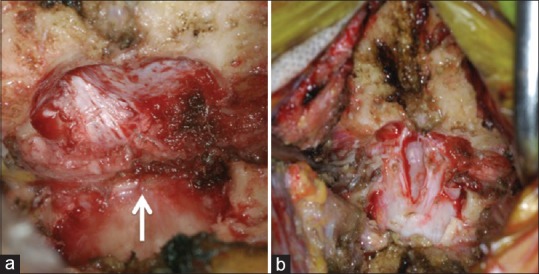
Intraoperative photographs after resection of the posterior arch and part of the occipital bone (a) the thickened reactive fibrous tissue band compressing the dural mater (arrow). Following the removal of the fibrous tissue band with opening of the dura mater, decompression of the spinal cord was achieved (b)