Abstract
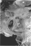
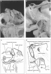
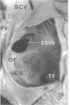
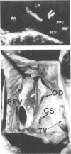
BACKGROUND: The diagnosis of sinus venosus defects remains a matter of debate. It is crucial to provide solid anatomical criteria, by identifying the very nature of the atrial septum relative to sinus venosus defects, to diagnose and differentiate them from other interatrial communications. OBJECTIVE: This study was designed to reestablish the anatomical criteria for the diagnosis of sinus venosus defects. METHODS: Five specimens with sinus venosus defects from the cardiopathological museum were examined. Study of the abnormal hearts was supplemented by examining the extent and structure of the atrial septum in 10 normal hearts. The echocardiograms and surgical notes were reviewed from 18 patients seen between July 1991 and August 1996 at the Royal Brompton Hospital in London diagnosed preoperatively to have a sinus venosus defect. RESULTS: The nature of the oval fossa and its muscular borders were identified in the normal hearts. In all three autopsied specimens of the superior variety of sinus venosus defect, the mouth of the superior caval vein was overriding the intact muscular anterosuperior border of the oval fossa. Two specimens thought initially to have the inferior variety of sinus venosus defect were re-classified as having defects within the oval fossa as it was the deficient oval fossa itself, rather than its intact muscular border, that was overridden by the mouth of the inferior caval vein. Sixteen patients had been diagnosed echocardiographically as exhibiting the superior variant of the defect. Retrospective review showed overriding of the superior caval vein across the upper rim of the oval fossa in 12 patients. These findings were confirmed by surgery in 11 patients with the 12th awaiting operation. Overriding of the fossa by the caval vein was not found in the other four patients. Surgery in all of these showed the defect to be within the oval fossa. In two patients diagnosed echocardiographically as having inferior defects, the surgical findings confirmed a biatrial connection of the inferior caval vein in one patient, the findings in the second were equivocal. CONCLUSIONS: The key anatomical criterion for the diagnosis of sinus venosus defects is overriding of the mouth of the superior or inferior caval vein across the intact muscular border of the oval fossa. The interatrial communication is then formed within the mouth of the overriding vein, and is outside the confines of the oval fossa.
Full text
PDF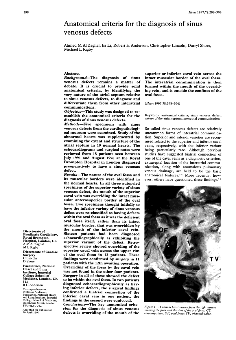
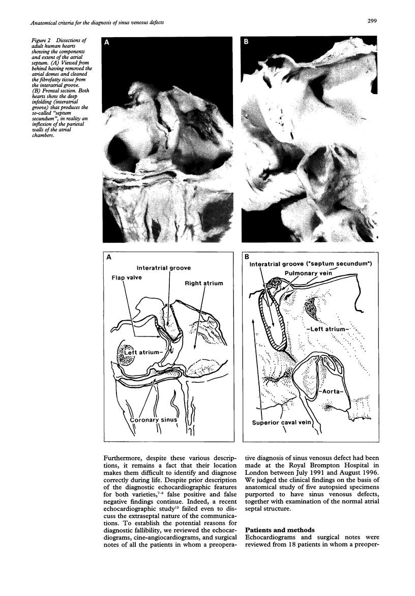
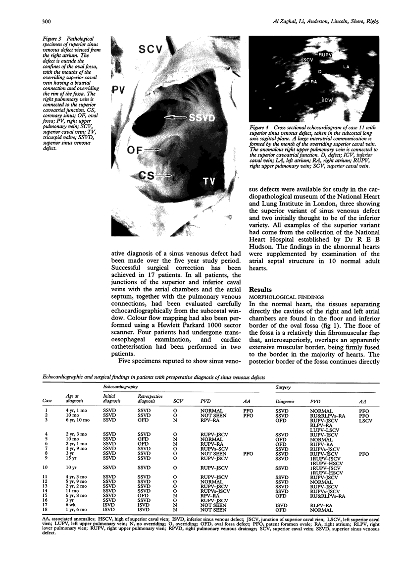
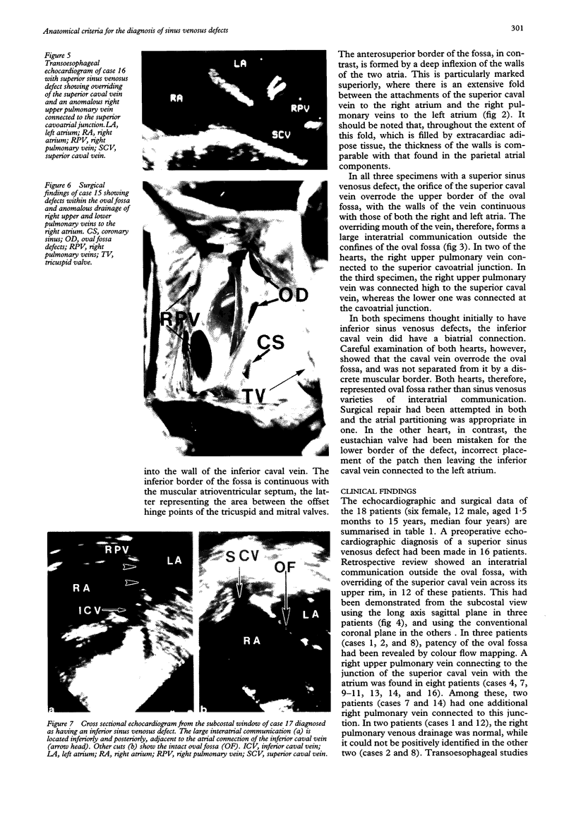
Images in this article
Selected References
These references are in PubMed. This may not be the complete list of references from this article.
- Anderson R. H., Brown N. A. The anatomy of the heart revisited. Anat Rec. 1996 Sep;246(1):1–7. doi: 10.1002/(SICI)1097-0185(199609)246:1<1::AID-AR1>3.0.CO;2-Y. [DOI] [PubMed] [Google Scholar]
- DeRuiter M. C., Gittenberger-De Groot A. C., Wenink A. C., Poelmann R. E., Mentink M. M. In normal development pulmonary veins are connected to the sinus venosus segment in the left atrium. Anat Rec. 1995 Sep;243(1):84–92. doi: 10.1002/ar.1092430110. [DOI] [PubMed] [Google Scholar]
- Ettedgui J. A., Siewers R. D., Anderson R. H., Park S. C., Pahl E., Zuberbuhler J. R. Diagnostic echocardiographic features of the sinus venosus defect. Br Heart J. 1990 Nov;64(5):329–331. doi: 10.1136/hrt.64.5.329. [DOI] [PMC free article] [PubMed] [Google Scholar]
- Kyger E. R., 3rd, Frazier O. H., Cooley D. A., Gillette P. C., Reul G. J., Jr, Sandiford F. M., Wukasch D. C. Sinus venosus atrial septal defect: early and late results following closure in 109 patients. Ann Thorac Surg. 1978 Jan;25(1):44–50. doi: 10.1016/s0003-4975(10)63485-6. [DOI] [PubMed] [Google Scholar]
- LEWIS F. J., TAUFIC M., VARCO R. L., NIAZI S. The surgical anatomy of atrial septal defects: experiences with repair under direct vision. Ann Surg. 1955 Sep;142(3):401–415. doi: 10.1097/00000658-195509000-00009. [DOI] [PMC free article] [PubMed] [Google Scholar]
- Mühler E. G., Engelhardt W., von Bernuth G. Detection of sinus venosus atrial septal defect by two-dimensional echocardiography. Eur Heart J. 1992 Apr;13(4):453–456. doi: 10.1093/oxfordjournals.eurheartj.a060196. [DOI] [PubMed] [Google Scholar]
- Pascoe R. D., Oh J. K., Warnes C. A., Danielson G. K., Tajik A. J., Seward J. B. Diagnosis of sinus venosus atrial septal defect with transesophageal echocardiography. Circulation. 1996 Sep 1;94(5):1049–1055. doi: 10.1161/01.cir.94.5.1049. [DOI] [PubMed] [Google Scholar]
- ROSS D. N. The sinus venosus type of atrial septal defect. Guys Hosp Rep. 1956;105(4):376–381. [PubMed] [Google Scholar]
- SWAN H. J., KIRKLIN J. W., BECU L. M., WOOD E. H. Anomalous connection of right pulmonary veins to superior vena cava with interatrial communications; hemodynamic data in eight cases. Circulation. 1957 Jul;16(1):54–66. doi: 10.1161/01.cir.16.1.54. [DOI] [PubMed] [Google Scholar]
- Sweeney L. J., Rosenquist G. C. The normal anatomy of the atrial septum in the human heart. Am Heart J. 1979 Aug;98(2):194–199. doi: 10.1016/0002-8703(79)90221-7. [DOI] [PubMed] [Google Scholar]
- Tasaka H., Krug E. L., Markwald R. R. Origin of the pulmonary venous orifice in the mouse and its relation to the morphogenesis of the sinus venosus, extracardiac mesenchyme (spina vestibuli), and atrium. Anat Rec. 1996 Sep;246(1):107–113. doi: 10.1002/(SICI)1097-0185(199609)246:1<107::AID-AR12>3.0.CO;2-T. [DOI] [PubMed] [Google Scholar]
- Van Praagh S., Carrera M. E., Sanders S. P., Mayer J. E., Van Praagh R. Sinus venosus defects: unroofing of the right pulmonary veins--anatomic and echocardiographic findings and surgical treatment. Am Heart J. 1994 Aug;128(2):365–379. doi: 10.1016/0002-8703(94)90491-x. [DOI] [PubMed] [Google Scholar]


