Abstract
Purpose
Several behavioral studies have shown that the reaction times of visually guided movements are slower in people with amblyopia, particularly during amblyopic eye viewing. Here, we tested the hypothesis that the initiation of smooth pursuit eye movements, which are responsible for accurately keeping moving objects on the fovea, is delayed in people with anisometropic amblyopia.
Methods
Eleven participants with anisometropic amblyopia and 14 visually normal observers were asked to track a step-ramp target moving at ±15°/s horizontally as quickly and as accurately as possible. The experiment was conducted under three viewing conditions: amblyopic/nondominant eye, binocular, and fellow/dominant eye viewing. Outcome measures were smooth pursuit latency, open-loop gain, steady state gain, and catch-up saccade frequency.
Results
Participants with anisometropic amblyopia initiated smooth pursuit significantly slower during amblyopic eye viewing (206 ± 20 ms) than visually normal observers viewing with their nondominant eye (183 ± 17 ms, P = 0.002). However, mean pursuit latency in the anisometropic amblyopia group during binocular and monocular fellow eye viewing was comparable to the visually normal group. Mean open-loop gain, steady state gain, and catch-up saccade frequency were similar between the two groups, but participants with anisometropic amblyopia exhibited more variable steady state gain (P = 0.045).
Conclusions
This study provides evidence of temporally delayed smooth pursuit initiation in anisometropic amblyopia. After initiation, the smooth pursuit velocity profile in anisometropic amblyopia participants is similar to visually normal controls. This finding differs from what has been observed previously in participants with strabismic amblyopia who exhibit reduced smooth pursuit velocity gains with more catch-up saccades.
Keywords: amblyopia, smooth pursuit, eye movements, initiation latency
Smooth pursuit eye movements serve to stabilize the image of continuously moving objects close to the fovea, and hence maintain visual acuity. Smooth pursuit initiation in humans is delayed by at least 100 ms subsequent to the onset of a target moving at a constant velocity (ramp stimulus) due to the latency of sensory processing of visual information.1 The use of a target step in the opposite direction prior to a velocity ramp (the step-ramp stimulus2) increases the pursuit initiation latency to approximately 150 ms,1 but it prevents the participant from initiating pursuit eye movements with a saccade.
The first 100 ms of smooth pursuit movement constitute the “open-loop” phase1 when visual target motion feedback is not yet available given the processing delay of the visual system. The open-loop velocity gains (the ratio of eye velocity to target velocity) in humans range from 0.3 to 0.7 depending on target velocity.3,4 After the open-loop phase, the eye velocity usually matches the target velocity as target motion feedback becomes available.1 This latter period is known as the “steady state” phase of smooth pursuit. Meyer and colleagues5 have reported that steady state velocity gains in humans can reach 0.90 for target velocities of less than 100°/s.
Amblyopia is a neurodevelopmental disorder characterized by a reduction in vision of one or both eyes that is not attributable only to a structural abnormality of the eye, and cannot be fixed with optical correction alone. Clinically, amblyopia is defined as greater or equal to a two line difference in visual acuity between the two eyes as measured by a Snellen chart after refractive correction.6 Amblyopia arises subsequent to the suppression of neural signals in the visual pathway of the amblyopic eye during the critical period of visual development.7 This monocular suppression of neurologic signals is associated with childhood strabismus (misalignment of visual axes), anisometropia (unequal refractive error in the two eyes), a combination of the two conditions, or other forms of visual deprivation such as cataracts.
Amblyopia is characterized by a loss of visual acuity,8 contrast sensitivity,8–10 and global motion detection11 in the amblyopic eye as well as deficits in stereoacuity.12 Amblyopia also impacts aspects of visuomotor control: individuals with amblyopia have altered spatiotemporal eye-hand coordination during reaching movements,13–16 perform poorly on motor tasks that require three-dimensional (3D) vision,17,18 and show deficits in day-to-day sensorimotor activities such as reading.19 These deficits are observable in the oculomotor domain as well. People with amblyopia have poorer fixation stability,20,21 have prolonged saccadic latencies,22 and exhibit reduced and more variable saccadic gain adaptation23 in their amblyopic eyes compared with visually healthy people.
Von Noorden and Mackensen24 were the first to record smooth pursuit eye movements in participants with strabismic amblyopia. They reported that the smooth sinusoidal eye position profiles were superseded by quick saccadic jumps at lower pursuit frequencies (i.e., lower pursuit velocities) in strabismic amblyopia participants. Schor25 studied smooth pursuit eye movements in five individuals with strabismic amblyopia using either triangular or sinusoidal wave stimuli. He noted a marked reduction in the steady state pursuit eye velocity and a corresponding increase in the frequency of error-correcting saccades toward temporalward target motion compared with nasalward target motion with the amblyopic eye viewing.
Though the aforementioned studies investigated smooth pursuit eye movements in amblyopia, they only included participants with strabismic amblyopia. Ciuffreda and colleagues26 examined smooth pursuit in participants with anisometropic and strabismic amblyopia, and found that the steady state velocity gains under amblyopic eye viewing in all participants ranged from 0.4 to 0.7. They also reported increased error-correcting saccadic substitution during amblyopic eye pursuit in four of their five strabismic amblyopia participants. In comparison, only one of their three anisometropic amblyopia participants demonstrated abnormal saccadic substitution and only for the smaller target amplitudes (≤2°). None of the above studies quantified the response latencies or open-loop gains of smooth pursuit movements in amblyopia.
In this study, we investigated the initiation of smooth pursuit eye movements in participants with anisometropic amblyopia. The smooth pursuit and the saccadic eye movements have common neural correlates,27 with their sensorimotor processing sharing several cortical and brainstem pathways.28,29 Thus, we hypothesized that similar to our previous observations of increased latency in the initiation of saccadic eye movements,22,23 people with anisometropic amblyopia would exhibit longer smooth pursuit initiation latencies when viewing with the amblyopic eye. We found that smooth pursuit initiation was delayed during amblyopic eye viewing in participants with anisometropic amblyopia. Mean steady state gains were normal in anisometropic amblyopia, but showed high variability across all viewing conditions.
Methods
Participants
Eleven participants with anisometropic amblyopia (1 male; age: 27 ± 8 years) and 14 visually normal controls (5 males; age: 26 ± 7 years) participated (see Table for clinical characteristics). A standard ophthalmic assessment was carried out on all participants by a certified orthoptist. It included an Early Treatment Diabetic Retinopathy Study (ETDRS) visual acuity test, measurement of refractive errors, prism cover test, Worth 4 Dot/Bagolini test for sensory fusion, and the Randot stereoacuity test. Amblyopia was defined as an interocular visual acuity difference of greater than or equal to 0.18 logMAR. Anisometropic amblyopia was defined as amblyopia in the presence of an interocular refractive error difference of greater than or equal to 1 diopter (D) of spherical or cylindrical power.
Table.
Clinical Characteristics of Participants With Anisometropic Amblyopia
All participants with amblyopia except numbers 9, 10, and 11 had participated in a previous study on saccadic adaptation (see Methods in Raashid et al.23). Amblyopic participant number 9 had a phoria but no visible manifest deviation on the prism cover test, and she met the criteria for monofixation syndrome.30 Eight participants had moderate amblyopia (amblyopic eye visual acuity of 0.2–0.7 logMAR31) and three had severe amblyopia (amblyopic eye visual acuity of 0.8–1.0 logMAR32). Eye dominance for visually normal observers was determined using the Dolman method.33 Exclusion criteria were any ocular cause for reduced visual acuity, prior intraocular surgery, or any neurologic disease. Individuals with strabismic amblyopia (tropia > 8 prism D) were also excluded because the misalignment of the eyes results in a known nasal-temporal asymmetry in motion sensitivity as well as the potential for the use of a nonfoveal locus for tracking.34 This study was approved by the Research Ethics Board at The Hospital for Sick Children (Toronto, Canada), and all protocols followed the guidelines of the Declaration of Helsinki. Written informed consent was obtained from each participant prior to the experiment.
Apparatus
The visual target was a red dot (0.5° diameter) produced by a laser galvanometer (GSI Group, Bedford, MA, USA) on a rear projection tangent screen in a dimly lit room. All eye movements were recorded by a head-mounted video-based eye tracker (Chronos Vision GmbH, Berlin, Germany). The stimulus position was updated every 4.6 ms (≈217 Hz), and the real-time laser position signal was digitized concurrently and synchronously with the eye position data from the Chronos eye tracker at 200 Hz. The participant was seated in a chair 80 cm from the screen with their head stabilized on a chin rest. Prior to each experiment, the eye movements of each participant were calibrated with fixation targets at center, leftward, rightward, upward, and downward directions (±10° eccentricities) on the screen.
Procedure
A step-ramp paradigm was used to elicit smooth pursuit eye movements.2 Each trial began with central fixation. After a random fixation duration of 750 to 1250 ms, the visual target stepped by ±4° horizontally before initiating motion at a constant velocity of 15°/s for one second in the horizontal direction opposite the step. A total of 40 trials were included in each experiment and the direction in which the target moved at a constant speed was presented randomly (20 trials in each horizontal direction). The experiment was conducted under three viewing conditions for each participant in the following sequence: monocular amblyopic (AE)/nondominant eye (NDE), binocular (BE), and monocular fellow (FE)/dominant eye (DE). Participants were instructed to follow the movement of the visual target as quickly and as accurately as possible.
Outcome Measures
Eye position data were differentiated using a 9-point Savitzky-Golay differentiator35 to yield the eye velocity data. Individual eye movement traces were inspected visually, and all saccades executed during pursuit movement were counted and eliminated from the calculations of the open-loop/steady state pursuit gains. The outcome measures included smooth pursuit latency, variability in smooth pursuit latency, open-loop gain, variability in open-loop gain, steady state gain, variability in steady state gain, and the frequency of catch-up saccades.
Smooth pursuit latencies were determined from the eye velocity traces based on the method of Kimmig and colleagues.36 The eye velocity traces were first inspected for the presence of a corrective saccade after pursuit initiation. When the first corrective saccade was found, a custom-written program looked backward in time from the point of the saccade onset to determine the pursuit onset time. A running average of the eye velocity preceding the saccade was calculated in bins of 20 ms, which were shifted backward iteratively 5 ms at a time. The time when the average eye velocity (before the first corrective saccade onset) fell to 12% of the maximum target velocity was defined as the pursuit onset time. The latency was then calculated by subtracting the target onset time from the pursuit onset time.
The open-loop gain was defined as the mean eye velocity divided by the target velocity during the first 100 ms after pursuit onset (i.e., during the period of pursuit before visual feedback is available). The steady state gain represented the ratio of mean eye velocity to target velocity during the constant eye velocity phase of movement. The variability in the smooth pursuit latency, open-loop gain, and steady state gain was defined as the within-subject standard deviation (SD) of these measures over all 40 trials. Catch-up saccades were defined as rapid eye movements that were executed in the same direction as the target movement during smooth pursuit. Trials with pursuit latencies of less than 80 ms and/or open-loop gain durations of less than 75 ms were excluded from analysis. Less than 7% of the overall data were excluded due to saccadic interruptions at pursuit onset, noisy recordings, blinks, and the above-mentioned criteria. The proportion of rejected trials was uniform across both groups, and was not represented disproportionately in one specific viewing condition (e.g., amblyopic eye viewing). Means and SDs for all outcome measures are reported in the results section.
Data Analysis
All statistical analyses were performed using the SAS 9.4 software package (SAS Institute, Inc., Cary, NC, USA). The statistical significance value was set at P less than 0.05. A preliminary analysis was performed to determine whether there was an effect of target horizontal direction on smooth pursuit latency, open-loop gain, and steady state gain. This test was run using a 3-way repeated measures ANOVA with one between-subjects factor: Group (2 levels: control and amblyopia), and two within-subjects factors; Viewing Condition (3 levels: BE, FE/DE, and AE/NDE); and Target Direction (2 levels: rightward and leftward). All outcome measures were examined using 2-way repeated measures ANOVA with one between-subjects factor: Group; and one within-subjects factor: Viewing Condition. For the binocular (BE) viewing condition only, left eye data were compared with the right data using paired Student's t-tests for smooth pursuit latency, open-loop gain, and steady state gain to assess potential differences between the data obtained from the two eyes.
To evaluate the potential nasal-/temporalward (N/T) asymmetry in the smooth pursuit latency, open-loop gain, and steady state gain measures, a separate 3-way repeated measures ANOVA was carried out only on the data from the monocular viewing conditions with one between-subjects factor: Group, and two within-subjects factors; Viewing Condition (2 levels: FE/DE, and AE/NDE); and Target N/T Direction (2 levels: nasalward and temporalward). A Pearson Product Moment correlation was computed to determine whether the smooth pursuit latency, open-loop gain, and steady state gain from the monocular AE viewing condition were associated with the visual acuity of the amblyopic eye. χ2 tests of goodness-of-fit and independence were used to assess for a preference for a certain Target Direction, and the association between Group and Viewing Condition, respectively, for the frequency of catch-up saccades measure.
Results
Preliminary analysis revealed no effect of Target Direction on smooth pursuit latency (F(1,23) = 0.3, P = 0.57) or open-loop gain (F(1,23) = 0.2, P = 0.67). There was a main effect of Target Direction on steady state gain (F(1,23) = 7.9, P = 0.01), where movements executed in the rightward direction (0.88 ± 0.07) had slightly higher gains than those in the leftward direction (0.85 ± 0.09). Therefore, the data from both horizontal target directions were pooled for the subsequent analyses of smooth pursuit latency and open-loop gain measures. For the binocular viewing condition, there was no difference between the values obtained from the right eye as compared with the left eye for smooth pursuit latency (t(20) = −0.8, P = 0.45), open-loop gain (t(20) = 1.0, P = 0.31), or steady state gain (t(20) = −1.3, P = 0.20). Accordingly, we only submitted the right eye data to the subsequent binocular viewing condition analyses.
Smooth Pursuit Latency
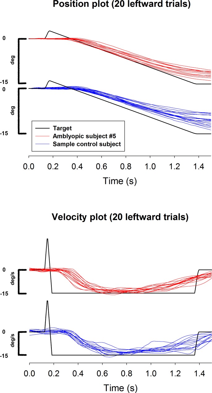
Figure 1 shows the desaccaded velocity and position traces of 20 leftward pursuit movements for an amblyopic participant and a representative control during the AE/NDE viewing condition. Both groups of participants elicited smooth pursuit in response to the constant speed target motion. Analysis of the mean pursuit latency revealed a significant interaction between Group and Viewing Condition (F(2,45) = 7.5, P = 0.002, Fig. 2). Participants with amblyopia had significantly longer pursuit latencies when they viewed with their amblyopic eye (206 ± 20 ms) compared with controls viewing with their nondominant eye (183 ± 17 ms, P = 0.009). Within amblyopic participants, mean smooth pursuit latency was significantly longer during the amblyopic eye viewing (206 ± 20 ms) compared with both binocular (171 ± 19 ms, P < 0.0001) and fellow eye (184 ± 29 ms, P < 0.0001) viewing. The increase in latency during amblyopic eye viewing did not correlate with the visual acuity of the amblyopic eye (r = −0.02, P = 0.95). Within control participants, mean smooth pursuit latency during the nondominant eye viewing (183 ± 17 ms) was significantly longer compared with the dominant eye viewing (169 ± 15 ms, P = 0.003) but not binocular viewing (175 ± 21 ms, P = 0.06). No statistically significant results were found for the monocular N/T analysis of smooth pursuit latency.
Figure 1.
Desaccaded position (top graph) and velocity (bottom graph) plots of 20 leftward pursuit movements shown for the anisometropic amblyopia participant number 5 (red) and a representative visually normal participant (blue) during the amblyopic/nondominant eye viewing condition. The target trace is shown in black.
Figure 2.
Mean smooth pursuit latencies of 11 participants with anisometropic amblyopia (black) and 14 visually normal individuals (white) shown for all three viewing conditions. The amblyopic eye latency was significantly longer than the nondominant eye latency in controls (*P = 0.002). Error bars indicate SEMs.
Mean variability in latency had a significant main effect of Group (F(1,23) = 5.3, P = 0.03) and Viewing Condition (F(2,45) = 4.8, P = 0.01). Averaged over all viewing conditions, the variability in latency was higher in participants with amblyopia (30 ± 8 ms) compared with controls (25 ± 6 ms, P = 0.03). The variability in latency was highest during the AE/NDE (30 ± 7 ms) viewing condition, compared with the BE (26 ± 7 ms, P = 0.008) and FE/DE (26 ± 6 ms, P = 0.01) viewing conditions for both participants with amblyopia and controls.
Open-Loop Gain
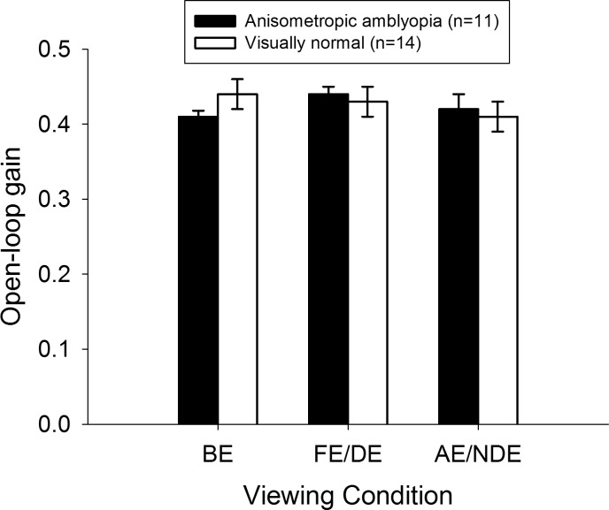
Mean open-loop gains were not significantly different between the two groups during AE/NDE (amblyopia: 0.42 ± 0.07, control: 0.41 ± 0.07), BE (amblyopia: 0.41 ± 0.03, control: 0.44 ± 0.09), or FE/DE (amblyopia: 0.44 ± 0.04, control: 0.43 ± 0.06) viewing conditions (F(2,45) = 1.5, P = 0.2, Fig. 3). The open-loop gains in the nasalward direction were comparable to those in the temporalward direction for both participants with amblyopia (nasal: 0.43 ± 0.06, temporal: 0.42 ± 0.07) and controls (nasal: 0.43 ± 0.07, temporal: 0.42 ± 0.07) during the monocular viewing conditions (F(1,23) = 0.6, P = 0.5).
Figure 3.
Mean open-loop smooth pursuit gains of 11 participants with anisometropic amblyopia (black) and 14 visually normal individuals (white) shown for all three viewing conditions. No comparisons were significant. Error bars indicate SEMs.
Mean variability in open-loop gain did not differ significantly between the two groups (F(1,23) = 1.7, P = 0.2) or between the three viewing conditions (F(2,45) = 0.1, P = 0.95).
Steady State Gain
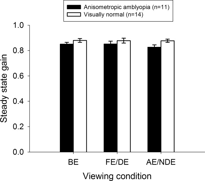
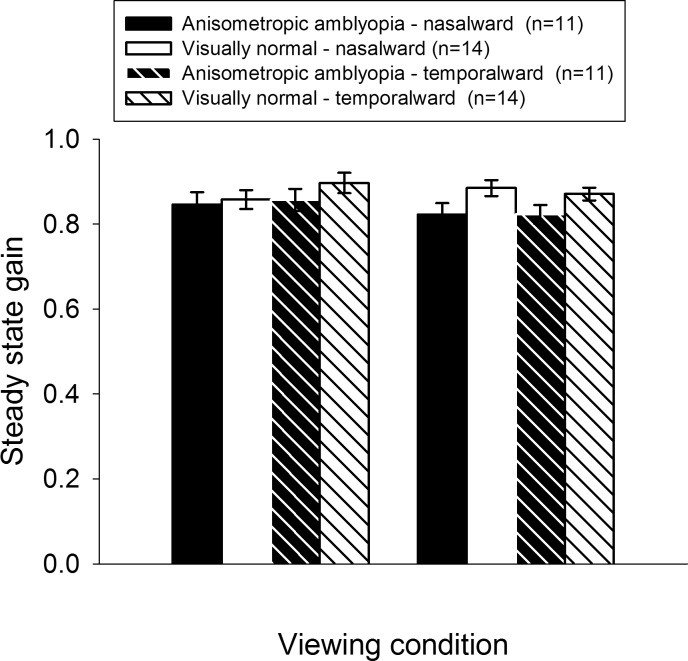
Mean steady state gains were not significantly different between the two groups during AE/NDE (amblyopia: 0.82 ± 0.08, control: 0.88 ± 0.06), BE (amblyopia: 0.85 ± 0.07, control: 0.87 ± 0.09), or FE/DE (amblyopia: 0.85 ± 0.09, control: 0.88 ± 0.09) viewing conditions (F(2,45) = 0.81, P = 0.45, Fig. 4). Further analysis revealed that the steady state gains in the nasalward direction were comparable to those in the temporalward direction for both participants with amblyopia (nasal: 0.83 ± 0.09, temporal: 0.84 ± 0.08) and controls (nasal: 0.87 ± 0.08, temporal: 0.88 ± 0.07) during the monocular viewing conditions (F(1,23) = 0.44, P = 0.52, Fig. 5).
Figure 4.
Mean steady state smooth pursuit gains of 11 participants with anisometropic amblyopia (black) and 14 visually normal individuals (white) shown for all three viewing conditions. No comparisons were significant. Error bars indicate SEMs.
Figure 5.
Mean steady state smooth pursuit gains of 11 participants with anisometropic amblyopia (black) and 14 visually normal individuals (white) shown for the nasalward (solid bars) and temporalward (striped bars) pursuit movements during the two monocular viewing conditions. No comparisons were significant. Error bars indicate SEMs.
There was a significant main effect of Group for the variability in steady state gain (F(1,23) = 4.5, P = 0.045), with the steady state gains being more variable in participants with amblyopia (0.11 ± 0.05) compared with controls (0.08 ± 0.04, P = 0.045). No other comparisons were statistically significant.
Catch-Up Saccades
On average, participants in both groups executed two saccades per trial independent of the horizontal pursuit direction. The χ2 goodness-of-fit test found no preference for the rightward (n = 34) or the leftward (n = 39) direction for the frequency of catch-up saccades (χ2(df=1) = 0.34, P = 0.56). Averaged across both target directions, the χ2 test of independence found no association between Group and Viewing Condition for the frequency of catch-up saccades (χ2(df=2) = 0.04, P = 0.98). Participants in both groups elicited an equivalent number of saccades during the AE/NDE (amblyopia: n = 33, control: n = 41), BE (amblyopia: n = 37, control: n = 43), and FE/DE (amblyopia: n = 34, control: n = 40) viewing conditions.
Discussion
This is the first study to examine the initiation of smooth pursuit eye movements in participants with anisometropic amblyopia and compare them to the eye movements of visually normal individuals. We found that: (1) participants with anisometropic amblyopia exhibited significantly longer pursuit initiation latency (by ∼23 ms) when viewing with the amblyopic eye, which was not correlated with the severity of amblyopia. The variability in latency was also higher in participants with amblyopia compared with controls during all viewing conditions, (2) the open-loop gain, steady state gain, and the frequency of catch-up saccades did not differ between participants with amblyopia and controls under any viewing condition; however, participants with amblyopia had more variable steady state gains, and (3) the characteristics of smooth pursuit did not differ between the nasalward and temporalward directions for either participants with amblyopia or controls.
Prolonged Pursuit Latencies During Amblyopic Eye Viewing
To our knowledge, no study has investigated smooth pursuit initiation latencies in anisometropic amblyopia. It is known that the visually-related reaction times in individuals with amblyopia are prolonged during both manual button-press37 and saccadic movement22,23,38–40 tasks. These latter studies tested mostly reflexive22,23,38,39 or overlap40 saccades, and have shown that when viewing with the amblyopic eye, the mean saccadic latencies are delayed by 27 to 57 ms compared with the nondominant eye viewing in controls; specifically, in participants with anisometropic amblyopia, the mean saccadic latency delay during amblyopic eye viewing ranges from 32 to 44 ms22,23,38,40 In this study, we found that the smooth pursuit initiation latencies were 23 ms longer on average during amblyopic eye viewing compared with nondominant eye viewing in controls. This increased delay with amblyopic eye viewing is consistent with the results reported in the aforementioned studies on saccades, but the magnitude of the delay is smaller (∼23 ms compared with 32–44 ms).
The smooth pursuit and saccadic brain circuitry share several cortico-ponto-cerebellar structures; however, there are notable differences between the two.27 For both smooth pursuit/saccades, the respective visual velocity/position signals are processed first in the primary visual cortex (V1).27 The difference between the pursuit and saccadic processing pathways arises from the extent of contributions from the frontal lobe, parietal lobe, and superior colliculus. Pharmacologic inactivation of the smooth eye movement subregion in the frontal eye fields reduces the steady state pursuit velocities, but has negligible impact on pursuit initiation latencies.41 The pursuit region of the posterior parietal cortex (or lateral intraparietal area in monkeys) monitors the extraretinal pursuit signals42 but does not play a major role in determining pursuit initiation latencies.43 The superior colliculus, which contains a spatial retinotopic map, plays a limited role in smooth pursuit movements: it may be involved in goal selection for eye movements in general44 and in modifying pursuit metrics,45 but it has been speculated that it does not play a direct role in initiating pursuit movements (see fig. 2 in Krauzlis27). In contrast, the frontal/parietal lobes and the superior colliculus play instrumental roles in the initiation of saccadic eye movements. It is known that the lesions of the human posterior parietal cortex delay the initiation of reflexive saccades.46 Also, the pharmacologic inactivation of superior colliculus in monkeys causes saccadic latencies to increase.47
We speculate that the longer latency reported previously in participants with anisometropic amblyopia for reflexive and overlap saccades (32–44 ms22,23,38,40) compared with that observed here for smooth pursuit (∼23 ms) could be attributable to further signal processing delays at the posterior parietal cortex or superior colliculus. Because these two structures are involved marginally in the initiation of smooth pursuit, they are possible loci where the additional temporal delay for saccadic initiation could occur. Alternatively, the difference between the smooth pursuit and saccadic latency delay could reflect a difference in how the retinal velocity and retinal position signals are processed in anisometropic amblyopia. Smooth pursuit is driven by a retinal velocity signal, whereas saccades are driven by a retinal position signal. It is possible that the processing of velocity error signals by the amblyopic visual system is not as delayed as the processing of position error signals, which could explain why the prolonged latency for pursuit initiation is less than that for saccades. For both kinds of eye movements, it is unlikely that a simple visual acuity loss is associated with the prolonged latency we report here for pursuit and previously for saccades22,23 because the latencies did not correlate with the severity of visual acuity deficit in the amblyopic eye.
It is known that target contrast can also modulate smooth pursuit latencies. Previous studies have shown that reducing the contrast of the visual stimulus increases the smooth pursuit latencies in visually normal observers.48,49 Individuals with amblyopia have known contrast sensitivity deficits, and their visual cortical areas under-sample high spatial frequency content. It is therefore not surprising that the smooth pursuit latencies in amblyopic participants, who have inherently reduced contrast sensitivity, are prolonged when compared to visually normal observers. Future investigations should test whether modulating the contrast of the visual target has any effect on smooth pursuit latencies in people with amblyopia.
Normal Pursuit Gains and Absence of Saccadic Interruptions
The smooth pursuit movements in people with strabismic amblyopia are frequently interrupted by quick bursts of saccades.24-26 These saccades are usually in response to a marked reduction in the steady state gain of the smooth pursuit, especially if the saccades executed were either higher in frequency or larger in amplitude. In contrast, participants with anisometropic amblyopia exhibited a very different response than in participants with strabismic amblyopia. First, the frequency of catch-up saccades executed by anisometropic amblyopia participants and visually normal individuals were not statistically different, indicating that unlike strabismic amblyopia, the smooth pursuit movements made by participants with anisometropic amblyopia do not have the same pattern of abnormal saccadic substitution. This observation is in agreement with Ciuffreda and colleagues26 who also reported little to no saccadic substitution for 4° and 8° amplitude stimuli in their sample of three participants with anisometropic amblyopia. Second, the mean open-loop and steady state velocity gains in anisometropic amblyopia were high, and similar to those observed in visually normal participants during all viewing conditions. Ciuffreda et al.26 reported that the amblyopic eye steady state gains in all their participants were between 0.4 and 0.7, which is low compared with the amblyopic eye steady state gain range in our study (0.75–0.95), and the normal range of 0.80 to 1.00.5 It is likely that the lower gains reported in the Ciuffreda et al.26 study were attributable to the presence of strabismus in five of their nine participants with amblyopia.
People with amblyopia exhibit deficits in global motion processing.11,50,51 Typically, global motion tasks require participants to ascertain the direction of moving stimuli over a broad area of the visual field, and in most cases the coherently-moving “signal” stimuli are spatially comingled with randomly moving “noise” stimuli (random-dot kinematogram). People with amblyopia show a weaker global motion response to contrast-defined (second-order) stimuli compared with luminance-defined (first-order) stimuli.11 Newer evidence suggests that this impairment in global motion processing might result from a failure to segregate signal from noise rather than from deficient integration of individual local motion signals.50,52 The smooth pursuit stimulus used in our study was a small but high contrast bright red dot on a dark background, which rendered it a luminance-defined local motion stimulus. Also, our task did not require the segregation of extraneous noise from the smooth pursuit signal. Given the stimulus and procedural characteristics of our experiment, it is not surprising that we did not observe diminished open-loop and steady state smooth pursuit gains in our participants with anisometropic amblyopia.
It has been reported that if visual spatial attention is diverted away from the moving target to a static target during pursuit initiation, the open-loop and steady state velocity gains are reduced.53 The close-to-normal average gains observed in anisometropic amblyopia argue that these individuals are equivalent to controls in maintaining their spatial attention on a high-contrast pursuit target in the absence of any distracters. On the other hand, amblyopic participants exhibited more variable steady state gains under all viewing conditions. It is possible that the better the oculomotor system is at predicting a constant trial-by-trial retinal target slip, the more precise the steady state gains become over a number of trials.54,55 Perhaps people with anisometropic amblyopia are not as good as visually normal individuals at encoding the trial-by-trial retinal slip, which could result in poorer predictive abilities manifested as more variable steady state gains in our data. Taken together, our results show that although the overall smooth eye velocity responses in participants with anisometropic amblyopia are close to those observed in visually healthy individuals, the steady state velocity gains are significantly more variable.
Symmetrical Monocular Nasal- and Temporal-Ward Gains
It has been reported previously that individuals with strabismic amblyopia exhibit higher steady state pursuit gains for movements in the nasalward direction compared with those in the temporalward direction,24,25 even after their fixation drift had been factored out.34 Similar N/T gain asymmetries are observed for the slow phase of optokinetic nystagmus (OKN) in the majority of people with strabismic amblyopia56,57 and in some with anisometropic amblyopia58,59 who have an accompanying loss of binocularity. The most common reason cited for the presence of these gain asymmetries is the disruption of cortical binocularity resulting from an early-onset strabismus.57 Our data revealed that participants with anisometropic amblyopia had comparable monocular steady state and open-loop gains in both the nasalward and temporalward directions. This observation could be explained by the fact that most of our amblyopic participants had some degree of stereovision, and all but one of them had sensory fusion on the Worth 4 Dot test (see Table). Furthermore, the motion detection deficits in anisometropic amblyopia are more uniform across the visual field instead of being localized to a specific visual hemifield.60
In conclusion, participants with anisometropic amblyopia have delayed smooth pursuit initiation with the amblyopic eye, but to a lesser extent than the delay observed for saccadic eye movements in previous studies. The spatial characteristics of pursuit in this subgroup of amblyopia differ from those observed previously in strabismic amblyopia: smooth eye movements in anisometropic amblyopia do not exhibit reduced open-loop/steady state velocity gains, and are hence achieved without the need for substitution by multiple catch-up saccades.
Acknowledgments
Supported by grants from Canadian Institutes of Health Research (CIHR; Grant MOP 106663; Ottawa, ON, Canada), Leaders Opportunity Fund from the Canada Foundation for Innovation (CFI; Ottawa, ON, Canada), the John and Melinda Thompson Endowment Fund in Vision Neurosciences (Toronto, ON, Canada), and the Department of Ophthalmology and Vision Sciences at The Hospital for Sick Children (Toronto, ON, Canada).
Disclosure: R.A. Raashid, None; I.Z. Liu, None; A. Blakeman, None; H.C. Goltz, None; A.M.F. Wong, None
References
- 1. Carl JR,, Gellman RS. Human smooth pursuit: stimulus-dependent responses. J Neurophysiol. 1987; 57: 1446–1463. [DOI] [PubMed] [Google Scholar]
- 2. Rashbass C. The relationship between saccadic and smooth tracking eye movements. J Physiol. 1961; 159: 326–338. [DOI] [PMC free article] [PubMed] [Google Scholar]
- 3. Eibenberger K,, Ring M,, Haslwanter T. Sustained effects for training of smooth pursuit plasticity. Exp Brain Res. 2012; 218: 81–89. [DOI] [PubMed] [Google Scholar]
- 4. Wyatt HJ,, Pola J. Smooth eye movements with step-ramp stimuli: the influence of attention and stimulus extent. Vision Res. 1987; 27: 1565–1580. [DOI] [PubMed] [Google Scholar]
- 5. Meyer CH,, Lasker AG,, Robinson DA. The upper limit of human smooth pursuit velocity. Vision Res. 1985; 25: 561–563. [DOI] [PubMed] [Google Scholar]
- 6. American Academy of Ophthalmology Pediatric Ophthalmology/Strabismus Panel. Preferred Practice Pattern Guidelines. Amblyopia. San Francisco CA: American Academy of Ophthalmology; 2012. [Google Scholar]
- 7. Wiesel TN. Postnatal development of the visual cortex and the influence of environment. Nature. 1982; 299: 583–591. [DOI] [PubMed] [Google Scholar]
- 8. McKee SP,, Levi DM,, Movshon JA. The pattern of visual deficits in amblyopia. J Vis. 2003; 3 (5): 380–405. [DOI] [PubMed] [Google Scholar]
- 9. Bradley A,, Freeman RD. Contrast sensitivity in anisometropic amblyopia. Invest Ophthalmol Vis Sci. 1981; 21: 467–476. [PubMed] [Google Scholar]
- 10. Levi DM,, Harwerth RS. Contrast sensitivity in amblyopia due to stimulus deprivation. Br J Ophthalmol. 1980; 64: 15–20. [DOI] [PMC free article] [PubMed] [Google Scholar]
- 11. Simmers AJ,, Ledgeway T,, Hess RF,, McGraw PV. Deficits to global motion processing in human amblyopia. Vision Res. 2003; 43: 729–738. [DOI] [PubMed] [Google Scholar]
- 12. Ho CS,, Giaschi DE. Stereopsis-dependent deficits in maximum motion displacement in strabismic and anisometropic amblyopia. Vision Res. 2007; 47: 2778–2785. [DOI] [PubMed] [Google Scholar]
- 13. Niechwiej-Szwedo E,, Goltz HC,, Chandrakumar M,, Hirji Z,, Crawford JD,, Wong AM. Effects of anisometropic amblyopia on visuomotor behavior, part 2: visually guided reaching. Invest Ophthalmol Vis Sci. 2011; 52: 795–803. [DOI] [PMC free article] [PubMed] [Google Scholar]
- 14. Niechwiej-Szwedo E,, Goltz HC,, Chandrakumar M,, Hirji Z,, Wong AM. Effects of anisometropic amblyopia on visuomotor behavior III: temporal eye-hand coordination during reaching. Invest Ophthalmol Vis Sci. 2011; 52: 5853–5861. [DOI] [PubMed] [Google Scholar]
- 15. Niechwiej-Szwedo E,, Goltz HC,, Chandrakumar M,, Wong AM. Effects of strabismic amblyopia on visuomotor behavior: part II. Visually guided reaching. Invest Ophthalmol Vis Sci. 2014; 55: 3857–3865. [DOI] [PubMed] [Google Scholar]
- 16. Niechwiej-Szwedo E,, Goltz HC,, Chandrakumar M,, Wong AM. Effects of strabismic amblyopia and strabismus without amblyopia on visuomotor behavior: III. Temporal eye-hand coordination during reaching. Invest Ophthalmol Vis Sci. 2014; 55: 7831–7838. [DOI] [PubMed] [Google Scholar]
- 17. Grant S,, Melmoth DR,, Morgan MJ,, Finlay AL. Prehension deficits in amblyopia. Invest Ophthalmol Vis Sci. 2007; 48: 1139–1148. [DOI] [PubMed] [Google Scholar]
- 18. Webber AL,, Wood JM,, Gole GA,, Brown B. The effect of amblyopia on fine motor skills in children. Invest Ophthalmol Vis Sci. 2008; 49: 594–603. [DOI] [PubMed] [Google Scholar]
- 19. Grant S,, Moseley MJ. Amblyopia and real-world visuomotor tasks. Strabismus. 2011; 19: 119–128. [DOI] [PubMed] [Google Scholar]
- 20. Chung ST,, Kumar G,, Li RW,, Levi DM. Characteristics of fixational eye movements in amblyopia: limitations on fixation stability and acuity? Vision Res. 2015; 114: 87–99. [DOI] [PMC free article] [PubMed] [Google Scholar]
- 21. Gonzalez EG,, Wong AM,, Niechwiej-Szwedo E,, Tarita-Nistor L,, Steinbach MJ. Eye position stability in amblyopia and in normal binocular vision. Invest Ophthalmol Vis Sci. 2012; 53: 5386–5394. [DOI] [PubMed] [Google Scholar]
- 22. Niechwiej-Szwedo E,, Goltz HC,, Chandrakumar M,, Hirji ZA,, Wong AM. Effects of anisometropic amblyopia on visuomotor behavior I: saccadic eye movements. Invest Ophthalmol Vis Sci. 2010; 51: 6348–6354. [DOI] [PMC free article] [PubMed] [Google Scholar]
- 23. Raashid RA,, Wong AM,, Chandrakumar M,, Blakeman A,, Goltz HC. Short-term saccadic adaptation in patients with anisometropic amblyopia. Invest Ophthalmol Vis Sci. 2013; 54: 6701–6711. [DOI] [PubMed] [Google Scholar]
- 24. Von Noorden GK,, Mackensen G. Pursuit movements of normal and amblyopic eyes. An electro-ophthalmographic study. II. Pursuit movements in amblyopic patients. Am J Ophthalmol. 1962; 53: 477–487. [DOI] [PubMed] [Google Scholar]
- 25. Schor C. A directional impairment of eye movement control in strabismus amblyopia. Invest Ophthalmol. 1975; 14: 692–697. [PubMed] [Google Scholar]
- 26. Ciuffreda KJ,, Kenyon RV,, Stark L. Abnormal saccadic substitution during small-amplitude pursuit tracking in amblyopic eyes. Invest Ophthalmol Vis Sci. 1979; 18: 506–516. [PubMed] [Google Scholar]
- 27. Krauzlis RJ. Recasting the smooth pursuit eye movement system. J Neurophysiol. 2004; 91: 591–603. [DOI] [PubMed] [Google Scholar]
- 28. Keller EL,, Missal M. Shared brainstem pathways for saccades and smooth-pursuit eye movements. Ann N Y Acad Sci. 2003; 1004: 29–39. [DOI] [PubMed] [Google Scholar]
- 29. Yan YJ,, Cui DM,, Lynch JC. Overlap of saccadic and pursuit eye movement systems in the brain stem reticular formation. J Neurophysiol. 2001; 86: 3056–3060. [DOI] [PubMed] [Google Scholar]
- 30. Parks MM. The monofixation syndrome. Trans Am Ophthalmol Soc. 1969; 67: 609–657. [PMC free article] [PubMed] [Google Scholar]
- 31. PEDIG. A randomized trial of atropine vs. patching for treatment of moderate amblyopia in children. Arch Ophthalmol. 2002; 120: 268–278. [DOI] [PubMed] [Google Scholar]
- 32. Repka MX,, Kraker RT,, Beck RW,, et al. Treatment of severe amblyopia with weekend atropine: results from 2 randomized clinical trials. J AAPOS. 2009; 13: 258–263. [DOI] [PMC free article] [PubMed] [Google Scholar]
- 33. Dolman P. Tests for determining the sighting eye. Am J Ophthalmol. 1919; 2: 287. [Google Scholar]
- 34. Bedell HE,, Yap YL,, Flom MC. Fixational drift and nasal-temporal pursuit asymmetries in strabismic amblyopes. Invest Ophthalmol Vis Sci. 1990; 31: 968–976. [PubMed] [Google Scholar]
- 35. Savitzky A,, Golay, M. Smoothing and differentiation of data by simplified least squares procedures. Analytical Chemistry. 1964; 36: 1627–1639. [Google Scholar]
- 36. Kimmig H,, Biscaldi M,, Mutter J,, Doerr JP,, Fischer B. The initiation of smooth pursuit eye movements and saccades in normal subjects and in “express-saccade makers.” Exp Brain Res. 2002; 144: 373–384. [DOI] [PubMed] [Google Scholar]
- 37. Hamasaki DI,, Flynn JT. Amblyopic eyes have longer reaction times. Invest Ophthalmol Vis Sci. 1981; 21: 846–853. [PubMed] [Google Scholar]
- 38. Ciuffreda KJ,, Kenyon RV,, Stark L. Increased saccadic latencies in amblyopic eyes. Invest Ophthalmol Vis Sci. 1978; 17: 697–702. [PubMed] [Google Scholar]
- 39. Niechwiej-Szwedo E,, Chandrakumar M,, Goltz HC,, Wong AM. Effects of strabismic amblyopia and strabismus without amblyopia on visuomotor behavior, I: saccadic eye movements. Invest Ophthalmol Vis Sci. 2012; 53: 7458–7468. [DOI] [PubMed] [Google Scholar]
- 40. Perdziak M,, Witkowska D,, Gryncewicz W,, Przekoracka-Krawczyk A,, Ober J. The amblyopic eye in subjects with anisometropia show increased saccadic latency in the delayed saccade task. Front Integr Neurosci. 2014; 8: 77. [DOI] [PMC free article] [PubMed] [Google Scholar]
- 41. Shi D,, Friedman HR,, Bruce CJ. Deficits in smooth-pursuit eye movements after muscimol inactivation within the primate's frontal eye field. J Neurophysiol. 1998; 80: 458–464. [DOI] [PubMed] [Google Scholar]
- 42. Bremmer F,, Distler C,, Hoffmann KP. Eye position effects in monkey cortex. II. Pursuit- and fixation-related activity in posterior parietal areas LIP and 7A. J Neurophysiol. 1997; 77: 962–977. [DOI] [PubMed] [Google Scholar]
- 43. Heide W,, Kurzidim K,, Kompf D. Deficits of smooth pursuit eye movements after frontal and parietal lesions. Brain. 1996; 119 (Pt 6): 1951–1969. [DOI] [PubMed] [Google Scholar]
- 44. Krauzlis RJ,, Carello CD. Going for the goal. Nat Neurosci. 2003; 6: 332–333. [DOI] [PubMed] [Google Scholar]
- 45. Basso MA,, Krauzlis RJ,, Wurtz RH. Activation and inactivation of rostral superior colliculus neurons during smooth-pursuit eye movements in monkeys. J Neurophysiol. 2000; 84: 892–908. [DOI] [PubMed] [Google Scholar]
- 46. Pierrot-Deseilligny C,, Rivaud S,, Gaymard B,, Agid Y. Cortical control of reflexive visually-guided saccades. Brain. 1991; 114 (Pt 3): 1473–1485. [DOI] [PubMed] [Google Scholar]
- 47. Hikosaka O,, Wurtz RH. Modification of saccadic eye movements by GABA-related substances. I. Effect of muscimol and bicuculline in monkey superior colliculus. J Neurophysiol. 1985; 53: 266–291. [DOI] [PubMed] [Google Scholar]
- 48. O'Mullane G,, Knox PC. Modification of smooth pursuit initiation by target contrast. Vision Res. 1999; 39: 3459–3464. [DOI] [PubMed] [Google Scholar]
- 49. Spering M,, Kerzel D,, Braun DI,, Hawken MJ,, Gegenfurtner KR. Effects of contrast on smooth pursuit eye movements. J Vis. 2005; 5 (5): 455–465. [DOI] [PubMed] [Google Scholar]
- 50. Aaen-Stockdale C,, Hess RF. The amblyopic deficit for global motion is spatial scale invariant. Vision Res. 2008; 48: 1965–1971. [DOI] [PubMed] [Google Scholar]
- 51. Ho CS,, Giaschi DE. Low- and high-level motion perception deficits in anisometropic and strabismic amblyopia: evidence from fMRI. Vision Res. 2009; 49: 2891–2901. [DOI] [PubMed] [Google Scholar]
- 52. Hess RF,, Mansouri B,, Dakin SC,, Allen HA. Integration of local motion is normal in amblyopia. J Opt Soc Am A Opt Image Sci Vis. 2006; 23: 986–992. [DOI] [PubMed] [Google Scholar]
- 53. Souto D,, Kerzel D. Dynamics of attention during the initiation of smooth pursuit eye movements. J Vis. 2008; 8(14):3. [DOI] [PubMed]
- 54. Becker W,, Fuchs AF. Prediction in the oculomotor system: smooth pursuit during transient disappearance of a visual target. Exp Brain Res. 1985; 57: 562–575. [DOI] [PubMed] [Google Scholar]
- 55. de Brouwer S,, Missal M,, Lefevre P. Role of retinal slip in the prediction of target motion during smooth and saccadic pursuit. J Neurophysiol. 2001; 86: 550–558. [DOI] [PubMed] [Google Scholar]
- 56. Garbutt S,, Han Y,, Kumar AN,, Harwood M,, Rahman R,, Leigh RJ. Disorders of vertical optokinetic nystagmus in patients with ocular misalignment. Vision Res. 2003; 43: 347–357. [DOI] [PubMed] [Google Scholar]
- 57. Westall CA,, Shute RH. OKN asymmetries in orthoptic patients: contributing factors and effect of treatment. Behav Brain Res. 1992; 49: 77–84. [DOI] [PubMed] [Google Scholar]
- 58. Schor CM,, Levi DM. Disturbances of small-field horizontal and vertical optokinetic nystagmus in amblyopia. Invest Ophthalmol Vis Sci. 1980; 19: 668–683. [PubMed] [Google Scholar]
- 59. Westall CA,, Woodhouse JM,, Brown VA. OKN asymmetries and binocular function in amblyopia. Ophthalmic Physiol Opt. 1989; 9: 269–276. [DOI] [PubMed] [Google Scholar]
- 60. Donahue SP,, Wall M,, Stanek KE. Motion perimetry in anisometropic amblyopia: elevated size thresholds extend into the midperiphery. J AAPOS. 1998; 2: 94–101. [DOI] [PubMed] [Google Scholar]


