Abstract
Many bacterial species actively take up and recombine homologous DNA into their genomes, called natural competence, a trait that offers a means to identify the genetic basis of naturally occurring phenotypic variation. Here, we describe “transformed recombinant enrichment profiling” (TREP), in which natural transformation is used to generate complex pools of recombinants, phenotypic selection is used to enrich for specific recombinants, and deep sequencing is used to survey for the genetic variation responsible. We applied TREP to investigate the genetic architecture of intracellular invasion by the human pathogen Haemophilus influenzae, a trait implicated in persistence during chronic infection. TREP identified the HMW1 adhesin as a crucial factor. Natural transformation of the hmw1 operon from a clinical isolate (86-028NP) into a laboratory isolate that lacks it (Rd KW20) resulted in ~1,000-fold increased invasion into airway epithelial cells. When a distinct recipient (Hi375, already possessing hmw1 and its paralog hmw2) was transformed by the same donor, allelic replacement of hmw2A Hi375 by hmw1A 86-028NP resulted in a ~100-fold increased intracellular invasion rate. The specific role of hmw1A 86-028NP was confirmed by mutant and western blot analyses. Bacterial self-aggregation and adherence to airway cells were also increased in recombinants, suggesting that the high invasiveness induced by hmw1A 86-028NP might be a consequence of these phenotypes. However, immunofluorescence results found that intracellular hmw1A 86-028NP bacteria likely invaded as groups, instead of as individual bacterial cells, indicating an emergent invasion-specific consequence of hmw1A-mediated self-aggregation.
Author Summary
Many bacteria are naturally competent, actively taking up DNA from their surroundings and incorporating it into their genomes by homologous recombination. This cellular process has had a large impact on the evolution of these species, for example by enabling pathogens to acquire virulence factors and antibiotic resistances from their relatives. But natural competence can also be exploited by researchers to identify the underlying genetic variation responsible for naturally varying phenotypic traits, similar to how eukaryotic geneticists use meiotic recombination during sexual reproduction to create genetically admixed populations. Here we exploited natural competence, phenotypic selection, and deep sequencing to rapidly identify the hmw1 locus as a major contributor to intracellular invasion of airway epithelial cells by the human pathogen Haemophilus influenzae, a trait that likely allows bacterial cells to evade the immune system and therapeutic interventions during chronic infections. Genetic variation in this locus can strongly modulate bacterial intracellular invasion rates, and possession of a certain allele favors adhesion and self-aggregation, which appear to prompt bacteria to invade airway cells as groups, rather than as individuals. Overall, our findings indicate that targeting HMW1 could block the ability of H. influenzae to invade airway cells, which would make antibiotic therapy to treat chronic lung infections more effective. Furthermore, our new approach to identifying the genetic basis of natural phenotypic variation is applicable to a wide-range of phenotypically selectable traits within the widely distributed naturally competent bacterial species, including pathogenesis traits in many human pathogens.
Introduction
Genetic mapping in bacteria historically relied on screening mutant libraries for loss-of-function mutations, followed by laborious isolation and identification of the disrupted loci. Recent innovations in mutagenesis approaches like TnSeq can accelerate the process and aid in characterizing gene function (e.g. [1,2]), yet such approaches have some limitations. For example: (a) many classes of genetic variation are not evaluated, (b) a suitable loss-of-function screen is typically required, and (c) such techniques ignore naturally occurring within-species phenotypic variation. An alternative is to emulate eukaryotic quantitative genetics approaches, which rely on sexual reproduction to map genetic variation. Rather than isolating the loci responsible for a specific phenotype with disruptive mutations, the QTL (quantitative trait locus) mapping approach identifies the loci and alleles that are directly relevant to phenotypic expression in natural populations.
Bacteria do not reproduce sexually, but genetic transfer mechanisms are widespread, and diverse bacterial species (including many important human pathogens) are naturally competent, able to actively take up and recombine homologous DNA from their surroundings into their chromosomes [3,4]. The value of this genetic transfer mechanism to researchers was seen as early as 1944 by Avery et al., when naturally competent Streptococcus pneumoniae were used to show that DNA is the genetic material, or “the transforming principle” [5]. But only recently has exploiting natural competence (and other gene transfer mechanisms) become a practical means to investigate the genetic basis for natural phenotypic variation, as massively parallel sequencing technologies have become cost effective [6–9].
The Gram-negative bacterium Haemophilus influenzae has a well-characterized natural competence mechanism (reviewed in [4]) and illustrates how experimental natural transformation can be useful for genetic mapping. Under nutrient limitation, cells actively take up double-stranded DNA from their environment through their cell envelope, and this DNA can replace homologous segments of the chromosome by recombination. In the laboratory, a competent H. influenzae cell will rapidly replace ~0.1–3% of its chromosome with genomic DNA from a divergent H. influenzae strain [8,10]. These replacements typically involve multiple independent recombination tracts and contain 100s to 1,000s of donor-specific single-nucleotide polymorphisms (SNPs) that span dozens of genes. Insertions and deletions readily transform as parts of longer recombination tracts, albeit with less efficiency than SNPs; this can add or remove whole genes and operons. Thus, a single transformation experiment can give millions of independently transformed recombinants containing all (or nearly all) of the genetic variation that distinguishes the donor and recipient strains [8]. Such pools can be screened or selected for donor phenotypes, and the donor-specific variation in isolated recombinants can be identified by DNA sequencing. A previous small-scale screen of <100 transformed recombinant clones identified a bacterial QTL with a ~10-fold effect on transformability itself, though this used laborious quantitative assays of individual clones that had already been sequenced [8].
Though typically a commensal of the nasopharynx, H. influenzae—especially in nonencapsulated or nontypeable forms (NTHi)—can cause middle ear infections (otitis media), community-acquired pneumonia, exacerbations of chronic obstructive pulmonary disease (COPD), conjunctivitis, and sometimes more severe invasive diseases [11]. Infections often persist and recur despite host production of bactericidal antibodies and the use of antibiotic therapy. Our understanding of the molecular mechanisms involved in the progression and persistence of H. influenzae infections remains limited, but identical strains have been repeatedly isolated from the lungs of COPD patients in serial clinic visits, suggesting that H. influenzae has traits that promote chronic infection [12,13].
Current evidence indicates that H. influenzae is a facultative intracellular pathogen, and host cell invasion may allow bacterial cells to temporarily evade the immune system and therapeutic interventions [14,15]. H. influenzae invades a variety of cell types [16–20], and viable NTHi have been found within host cells of adenoid tissues and bronchial biopsies [21,22]. After intracellular invasion, H. influenzae cells remains non-proliferative and resides within membrane-bound vacuoles with features of late endosomes [23,24] or freely within the cytoplasm [25], and intracellular bacteria eventually die after persisting for variable lengths of time [26]. While several host factors have been identified as important for intracellular invasion, much less is known about the bacterial factors responsible for this process.
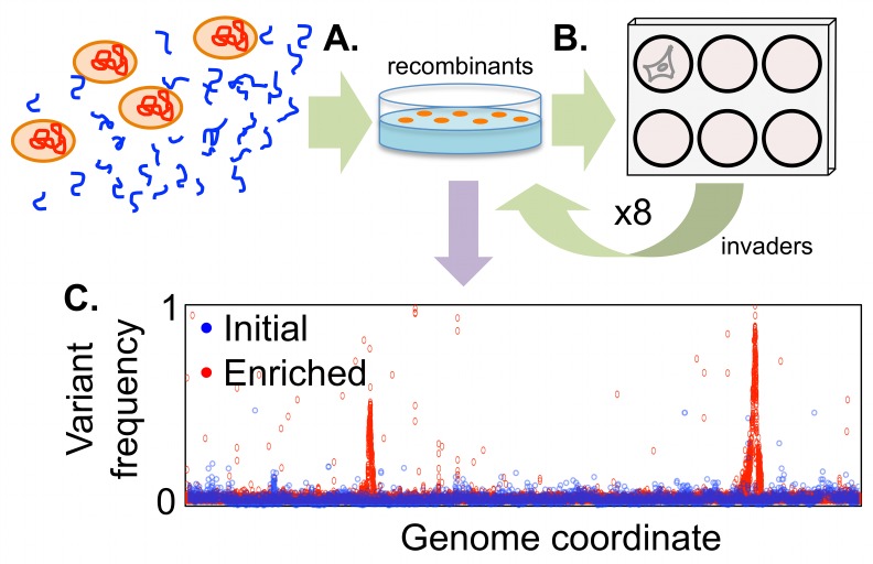
We chose the intracellular invasion phenotype as a model to test the genetic mapping strategy described above, which we have named “transformed recombinant enrichment profiling”, or TREP (summarized in Fig 1). We chose this phenotype because entry of H. influenzae into airways cells: (a) is easily selectable in lab culture, (b) displays wide phenotypic variation between clinical isolates, and (c) is likely to be an important factor in chronic infections. Application of TREP rapidly identified the hmw1 adhesin as a factor crucial for intracellular invasion.
Fig 1. Schematic of transformed recombinant enrichment profiling (TREP) to map H. influenzae intracellular invasion genes.
(A) Genomic DNA from a strain with high invasiveness is transformed into naturally competent cells of a strain with poor invasiveness. This produces complex pools of recombinants, in which short stretches of donor DNA replace homologous recipient DNA in individual recombinants. (B) Rare recombinants with increased invasiveness are enriched by serial passage of recombinant pools through A549 alveolar epithelial cells using gentamicin protection assays. Finally (C), the selected locus/loci are identified from donor allele frequencies measured by deep sequencing.
Results
Comparison of three diverse H. influenzae isolates
Three criteria were used to choose the donor and recipient strains: (a) substantially higher invasiveness of the donor over the recipients, (b) high natural transformability of the recipients, and (c) available genome references with many genetic markers distinguishing the donor from the recipients (S1 Text and S1 Fig). Three strains were assayed: the standard laboratory strain (Rd KW20, hereafter referred to as Rd) and two clinical isolates from pediatric patients with otitis media (Hi375 and 86-028NP). All three have complete genome sequences available [27–29]. Rd and Hi375 are highly transformable, while 86-028NP is not [30,31]. Rd is known to be a poor invader of several epithelial cell lines [32]; Hi375 has previously been used in studies of intracellular invasion [15,24]; and 86-028NP is known to be highly virulent in a chinchilla model of otitis media [33–36]. Antibiotic resistant derivatives of all three strains were produced to allow subsequent tracking of transformation events and genetic background, yielding strains Rd SpcR, Hi375 StrR, and 86-028NP NovR NalR, hereafter referred to as RdS, HiT, and NpNN, respectively (S1 Table).
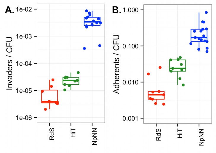
Intracellular invasion frequencies were evaluated by gentamicin protection assays with A549 airway epithelial cells, and quantified as gentamicin-protected bacterial colony forming units (CFU) relative to the original inoculated CFU (hereafter “Invaders/CFU”). Gentamicin protection assays found that NpNN is a highly efficient invader of A549 cells with ~10−2 invaders/CFU, whereas both HiT and RdS invade at ~100-fold and ~1,000-fold lower frequencies, respectively (Fig 2A; one-way ANOVA p<<0.01, and p<<0.01 for all three pairwise comparisons by post hoc testing using Tukey’s HSD).
Fig 2. Invasion and adhesion in the parent strains.
(A) “Invaders/CFU” was calculated as the total gentamicin-protected CFU / total input CFU. (B) “Adherents/CFU” was calculated as above with gentamicin treatment excluded. Triplicate experiments were conducted three times for RdS and HiT recipients and five times for the NpNN donor. Boxplots outline the first and third quartiles, with the thick horizontal line indicating the median, and the whiskers extend 1.5 times the interquartile distance.
Several controls were performed, both for assays of the three parental strains and during the serial passage experiments described below: (a) To ensure that spontaneous gentamicin resistance was not responsible for survival in gentamicin protection assays, equivalent bacterial suspensions were treated in the absence of A549 cells, yielding no viable CFUs (limit of detection <10−8), and gentamicin-protected CFUs recovered as colonies on plates remained gentamicin sensitive. (b) To ensure gentamicin treatment was complete, culture supernatants of infected A549 cells treated with gentamicin were plated, rendering no viable CFUs (limit of detection <10−8). (c) To ensure that introduction of selectable markers had no effect on intracellular invasion, these strains were compared to their progenitors, finding that they had comparable phenotypes (S2A Fig, one-way ANOVA p-values > 0.1 within each strain background).
Adhesiveness of the three parent strains to A549 cells was assayed similarly to invasiveness, except that the gentamicin treatment was omitted; “Adherents/CFU” was calculated as the CFU that remained associated with A549 cells after incubation and washing, relative to CFU of the input inoculum. The NpNN strain was the most adherent, with ~10% of the infecting cells remaining associated with A549 cells (Fig 2B). The HiT strain was intermediate (~10-fold lower than NpNN), whereas RdS had ~100-fold lower adherence (p<<0.01 by ANOVA and all for three pairwise comparisons). As with invasion, antibiotic resistant derivatives had adhesiveness comparable to that of the progenitors (S2B Fig).
Gentamicin protection strongly selects for intracellular invaders
A calibration experiment was used to test the strength of the experimental selection applied by the gentamicin protection assay. Mixtures of a high invasion strain (86-028NP NovR) and a low invasion strain (Rd StrR) were passaged twice: Bacterial cells were used to infect A549 cells and invaders were recovered after gentamicin treatment. The recovered colonies were then pooled and used for a second infection. This found that the highly invasive strain easily out-competed the poorly invasive one, even when starting at a 1 to 10,000 disadvantage, dominating the population after the second infection (S3 Fig). The high enrichment was not due to a growth rate advantage of 86-028NP, which has a slower doubling time than both recipients [30]. These data demonstrate that even rare recombinants could be highly enriched from complex pools using serial selection.
Summary of transformed recombinant enrichment profiling (TREP)
The experimental design for isolating bacterial intracellular invasion genes by TREP is depicted in Fig 1, summarized below, and described in detail in subsequent sections. (a) Donor genomic DNA from NpNN was used to transform naturally competent cells of two low invasion strains, RdS or HiT. (b) Pools of ~105 recombinant clones were enriched for those that conferred increased invasiveness by serial passages through A549 cells by gentamicin protection. Material from each cycle was stored to use for replicate quantitative assays and DNA extractions. (c) Genomic DNA from pools was sequenced to high coverage, and donor-specific allele frequencies were calculated at diagnostic SNPs. Sequencing and alignment statistics are summarized in S2, S3 and S4 Tables, and further details are presented in S1 Text and Materials and Methods.
By profiling genome-wide donor allele frequencies from independent experiments, a gene responsible for high invasion by NpNN was rapidly identified in both recipients, RdS or HiT. Individual clones were isolated after the fourth cycle of selection for genome sequencing and phenotypic analysis of recombinants and mutant derivatives. The results reveal a novel role for the adhesin-encoding hmw1A gene in intracellular invasion, beyond its previously described role in adhesion [37,38].
Generation of complex recombinant pools of natural transformants
Recombinant pools were made by incubating high molecular weight genomic DNA from NpNN with naturally competent cultures of either RdS or HiT (Fig 1A) [39]. Transformation by the two antibiotic resistance markers (NovR and NalR) indicated that ~15% of cells in each culture were competent and predicted that a given donor-specific SNP would be found in ~1% of NovR or NalR colonies, consistent with previously measured values (S5 Table) [8]. Selection for donor-specific antibiotic resistance alleles ensured elimination of untransformed recipient cells, and selection for recipient-specific resistances limited cross-contamination observed in preliminary experiments (Materials and Methods). This procedure generated four separate sets of recombinant clones (for the RdS recipient: SpcR NovR and SpcR NalR; for the HiT recipient: StrR NovR and StrR NalR). For each set, ~105 colonies were harvested into pools, thoroughly mixed, and stored prior to infections. We thus predicted that each pool would have millions of cells for which (nearly) any given donor-specific variant would be present. Sequencing of the initial recombinant pools provided several lines of evidence that the TREP approach would be practical (S1 Text): First, a single round of selection for the donor-specific antibiotic resistance alleles was sufficient to map the resistances to single nucleotide resolution (S4A and S4B Fig and S6 Table). Second, recombinant pools contained low frequency donor-specific alleles across the genome, both SNPs and and structural variants (S4C Fig and S7 Table).
Serial selection by gentamicin protection enriches for invasive recombinants
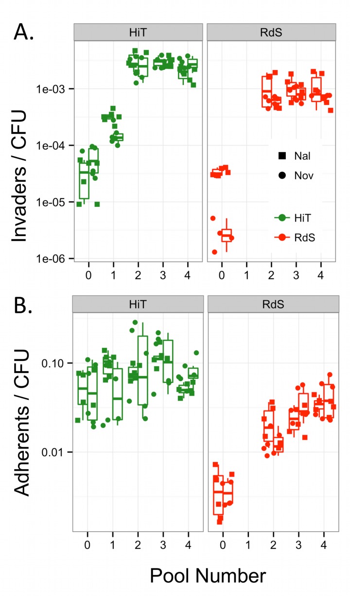
The four initial recombinant pools (Pool 0s) were used in separate infections of A549 cells (Fig 1B). Serial passages of these pools using gentamicin protection resulted in >100-fold increased invasion after only three cycles of enrichment (S5 Fig). Replicate invasion assays using material from the initial enrichments confirmed the dramatic increase in intracellular invasion by pools after serial enrichment (Fig 3A, one-way ANOVA p<<0.001, Tukey’s HSD for comparisons of Pool 0 to Pools 2–4 were p<<0.001, but p>0.2 among Pools 2–4). Comparison with donor invasiveness values measured in parallel showed that the HiT recombinants in the two Pool 4s were not significantly less invasive than the donor (p = 0.082); the two Pool 4s using the RdS recipient remained marginally less invasive (p = 0.025). These data suggest that after the third serial enrichment (Pool 4), donor-specific genetic variants conferring invasiveness were at or near fixation.
Fig 3. Serial enrichment for invasive recombinants by gentamicin protection.
(A) Boxplots of intracellular invasion frequencies from Pools 0–4 (2 experiments in triplicate). (B) Boxplots of adhesion frequencies (2 experiments in triplicate). The values show the combined ability of clones in Pool n to invade or adhere to airway epithelial cells, while the recovered colonies comprise Pool n+1. NalR and NovR pools are summarized by the left and right boxplots, with individual data points shown as circles and squares, respectively.
Increased invasion was not due to selection for de novo mutations that confer increased invasiveness, nor due to changes in bacterial gentamicin sensitivity (controls detailed above). Such events were unlikely given that the number of cell generations across the experiment was <100 in total. Furthermore, a control experiment using untransformed RdS or HiT cultures found no significant increases in invasiveness over 5 serial cycles of selection with either recipient strain (S6 Fig; p>0.1 by one-way ANOVA), strongly suggesting that de novo mutations conferring increased invasiveness were not captured in these experiments.
Since higher adhesion might concomitantly increase intracellular invasion, we tested how adhesion was affected by the serial selections (Fig 3B). This found that the RdS pools showed substantial progressive increases in adhesiveness (one-way ANOVA p<<0.001, Tukey’s HSD p<<0.001 for comparisons of Pool 0 to Pools 2–4, but p > 0.2 for comparisons among Pools 2–4). The HiT pools trended towards increasing adhesion over serial enrichments, but for this experiment, no significant change was observed (p>0.1). Both pairs of pools still had significantly lower adhesiveness than NpNN cultures run in parallel (p<0.05 for all comparisons against NpNN).
In sum, RdS and HiT recombinants acquired loci or alleles that enhanced intracellular invasion. Adhesion increased, though to a lesser extent. This suggests that, while adhesion might be a prerequisite for invasion, its increase may be insufficient to explain the elevated invasion displayed by invasiveness-enriched recombinants.
Serial enrichment for intracellular invaders selects for overlapping donor segments
Sequencing of genomic DNA across pools and serial passages (Fig 1C) showed that the four recombinant pools became progressively less complex, ultimately resulting in a total of only six recombinant clones dominating the four pools (out of ~4x10⁵ total). This suggests that the causative alleles transformed competent cells at lower rates than typical SNPs (~1,000-fold; S5 Table). The change in complexity was particularly apparent at the antibiotic-selected sites, where donor allele frequencies shifted from a smooth decline on either side of the resistance alleles to sharply demarcated donor segments (stretches of contiguous donor-specific variation) supporting the presence of only 1 or 2 antibiotic resistance-spanning segments dominating each pool (S7 Fig).
Coincident with decreasing complexity at the antibiotic-selected loci, serial enrichment also increased the frequency of donor segments in other intervals (Figs 4 and 5). At the end of selection, 1–2 recombinant clones dominated each pool, each carrying several donor segments (one segment in each clone spanning the antibiotic resistance allele).
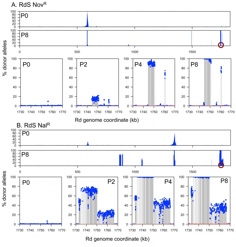
Fig 4. Genomic profile of the RdS recombinant pools.
(A) RdS NovR. (B) RdS NalR. The x-axes indicate recipient genome coordinate, and the y-axes indicate the percent donor alleles. Blue dots and grey lines indicate donor-specific SNPs. Salmon color shows the limits of detection, or 1/depth (alignments with mapping quality = 0 were ignored, which excludes multiply mapping reads). Pools are indicated as Pn. The two wide views at the top of A and B show the genome-wide donor allele frequencies for Pool 0 and Pool 8, whereas the lower four zoom around the nearly fixed locus selected for by serial gentamicin protection assay (indicated by purple circles) for Pools 0, 2, 4, and 8.
Fig 5. Genomic profile of the HiT recombinant pools.
(A) HiT NovR. (B) HiT NalR. Plotting as in Fig 4, with the green circles indicating a multi-mapping artifact. Low limits of detection (1/depth, salmon color) within the invasion locus are due to alignments with mapping quality = 0 (multiply mapping reads) caused by the same sequencing artifact.
Overlapping donor segment intervals between independent recombinants and pools are the best candidates for carrying invasion loci (i.e. the purple circles in Figs 4 and 5). In principle, donor-specific genetic variation found in only some clones (those seen with intermediate frequencies in the pools) could potentially modulate intracellular invasion, but “hitchhiking” segments that are not associated with invasion are expected to occur, since previous work has shown that competent cells typically take up and recombine multiple donor DNA molecules. Similarly, independent recombination tracts carrying the same invasion locus are expected to typically have independent recombination breakpoints [8,10].
All invasion-enriched RdS recombinants had acquired the hmw1 86-028NP operon
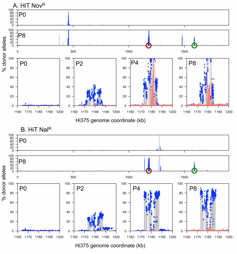
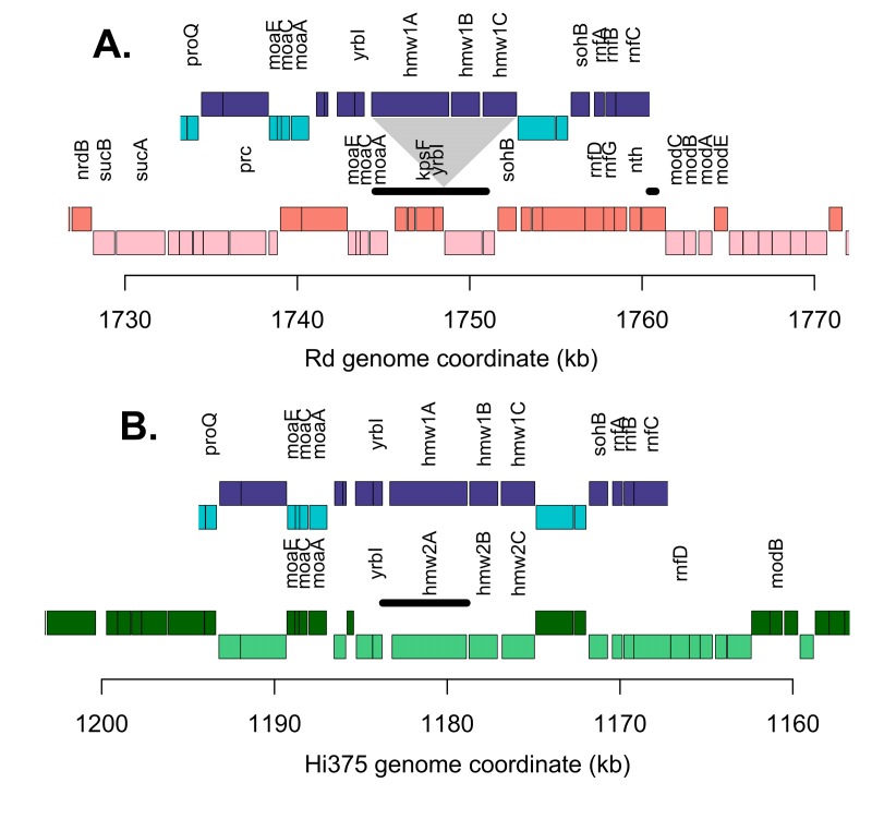
Sequencing of invader-enriched recombinant pools identified a single donor locus that was enriched to near-fixation in all four TREP experiments: hmw1 86-028NP (Fig 6). For the RdS recipient, a narrow interval reached near-fixation for both NovR and NalR-selected pools (Figs 4 and 6A; Rd coordinates 1,744,519–1,750,336 nt, accompanied by a short nearby interval at 1,760,431–1,760,794 nt with lower levels of enrichment). The Rd gene annotations in this interval have no obvious connection with host cell interactions, but the donor strain carries a large operon here that is absent from Rd: hmw1ABC, which is found in ~60% of H. influenzae strains [40–43] and located between yrbI and HI1680 (also known as NTHI1986 in the 86-028NP genome). Depth-of-coverage analysis and reciprocal mapping of sequence reads to the donor genome confirmed the insertion of hmw1ABC 86-028NP into the invasion-enriched RdS recombinant pools. Three independent recombination-mediated insertions of hmw1ABC 86-028NP dominated the two invasion-enriched pools: one recombinant clone in the NovR pool and two in the NalR pool, as indicated by the distinct recombination breakpoints flanking the insertion (Fig 4). Additional recombination tracts were detected distant from the putative invasion locus and the antibiotic resistance marker, but these were unique to one of the three Pool 4 clones, as expected for random “hitchhiking” recombination events.
Fig 6. Genomic map at the enriched invasion locus for the three parental strains.
For A and B, blue boxes indicate 86-028NP annotations; red is for Rd annotations, and green is for Hi375 annotations. Darker coloring indicates that sense is on the top strand, whereas lighter indicates bottom strand. Genes with names are indicated. The thick black horizontal lines indicate the minimum interval at or near fixation in both the NovR and NalR recombinant pools. (A) RdS recipient. Triangle indicates site of the hmw1 insertion. (B) HiT recipient.
Invader-enriched HiT recombinants had substituted their hmw2A Hi375 allele with the donor allele hmw1A 86-028NP
Selecting for invaders from the HiT recombinant pools likewise enriched for donor segments containing hmw1 86-028NP adjacent to the yrbI gene (Figs 5 and 6B, S9 Table). Only two donor-specific intervals reached fixation in HiT NovR: one spanning gyrB as expected, and another that completely spanned only the hmw1A 86-028NP gene carrying only short segments of the flanking genes (Hi375 genomic coordinates 1,178,771–1,183,728 nt). In HiT NalR, a long donor segment spanning this same interval reached ~75%.
In contrast to RdS, the HiT recipient already possesses the hmw1 operon and its paralogous operon hmw2, but the locations of the two adhesin genes (hmw1A and hmw2A) are swapped relative to their location in the donor NpNN. This is made evident through a comparison of the binding domains at the two hmw adhesin-encoding loci in HiT, NpNN, and the prototypic HMW-positive strain 12 (the strain that HMW adhesins were originally identified in and where they have been most extensively characterized, aka R2846) (Table 1). In strain 12 and the donor NpNN, hmw1 is adjacent to the yrbI gene (NTHI1982) and hmw2 is nearby the radA gene (NTHI1453). However, for Hi375, the radA-adjacent hmw adhesin has a binding domain with 100% amino acid identity to the yrbI-adjacent hmw1A gene from strain 12. Thus, whereas the hmw1 86-028NP operon was inserted into invasion-selected RdS recombinants, recombination events that increased HiT invasiveness were the result of an allelic substitution of hmw2 Hi375 for hmw1 86-028NP.
Table 1. HMW adhesin paralog assignment.
| Locus | Strain | HMW1strain12 | HMW2strain12 | Best Hit |
|---|---|---|---|---|
| YrbI- adjacent | Strain 12 | * | 33.8% | HMW1 |
| 86-028NP | 39.8% | 34.8% | HMW1 | |
| Hi375 | 35.8% | 50.7% | HMW2 | |
| RadA- proximal | Strain 12 | 33.8% | * | HMW2 |
| 86-028NP | 32.8% | 55.4% | HMW2 | |
| Hi375** | 100.0% | 33.8% | HMW1 |
Percent amino acid identity of six HMW binding domains to the prototype HMW1 and HMW2 paralogs in Strain 12, based on ClustalW2 alignments. Shared alignment gaps were excluded from pairwise comparisons. Self-alignments are indicated by *. The binding domains were defined by Strain 12 sequences as amino acids 555–914 in HMW1Astrain12 and 553–916 in HMW2Astrain12.
** The NCBI PGAAP annotation for hmw1A Hi375 ends with a serine instead of a stop codon and indicates a pseudogene. However, western analysis confirms its expression, and the presence of a stop codon 804 bp downstream accompanied by the expected pair of conserved C-terminal cysteine residues (codons 1624 and 1634) correctly defines the hmw1A Hi375 CDS as from coordinates 1,585,978 to 1,590,906 and encoding a 168 kDa protein.
The presence of the paralogous hmw locus nearby the radA gene caused an alignment artifact from multiply mapping sequence reads (Hi375 coordinates ~1,585–1,595 kb). Highly variable donor allele frequencies were seen across this interval ranging from ~0% to ~50% in Pool 8 for both HiT pools (S8 Fig). Furthermore, no donor-specific variation was detected flanking the radA-adjacent hmw1A Hi375 locus, in contrast to donor variation flanking the recombinant yrbI-adjacent hmw1A 86-028NP locus, which is expected for recA-mediated homologous recombination. Conclusive evidence for radA-proximal donor variation as a read alignment artifact is provided by allele-specific PCR assays on the isolated clones and mutants, as described below (S9 Fig).
Collectively, these data strongly support a role for hmw1 86-028NP in the increased intracellular invasion seen in enriched recombinants, though they do not strictly rule out a role for flanking donor-specific variation, particularly in the yrbI gene, since donor-specific variation in this gene was also near fixation in the invader-enriched recombinant pools. Comparison of sequence variants called between Pool 0s and Pool 8s identified that no novel mutations were fixed during serial selections, consistent with low per base mutation rates and the control experiment with untransformed recipients shown in S4 Fig.
Individual recombinant clones validate and disambiguate the pool sequencing
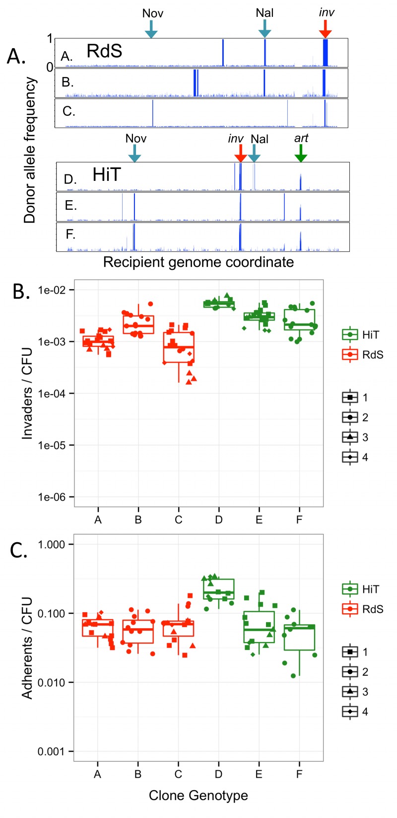
Four colonies from Pool 4 were isolated from each of the four TREP experiments and further analyzed. Each of the 16 clones was assayed in triplicate for its invasion and adhesion phenotypes (Fig 7). Subsequent genome sequencing (summarized in S3 Table) revealed a total of only six distinct genotypes: three clones from each recipient background (Fig 7A; S9 Table). This is consistent with predictions from the pool data and shows that the isolated clones represented all the high frequency recombinants observed in Pool 4. Intracellular invasion and adhesion were strongly enhanced for all 16 isolated clones compared to recipient controls (Fig 7B and 7C, Tukey’s HSD p<<0.001 for all six comparisons). Immunoblot analysis confirmed expression of HMW1A86-028NP protein in two recombinant clones, RdS genotype B (S10A Fig) and HiT genotype E (Fig 8A).
Fig 7. Genotype and phenotype of recombinant clones from Pool 4.
Sequencing grouped 16 clones into six genotypes, two RdS NalR (A and B), one RdS NovR (C), one HiT NalR (D) and two HiT NovR (E and F). Genome-wide donor allele frequencies of each clone are shown in (A). The x-axis indicates the recipient genome coordinate and the y-axis shows donor allele frequency from 0 to 1. Blue arrows mark the antibiotic resistance sites; red arrows mark the invasion locus; and the green arrow marks the hmw2 artifact. Clone assignments are in S8 Table and exact donor segment breakpoints are in S9 Table. All clones were phenotyped in triplicate and were aggregated by genotype for plots depicting their invasion (B) and adhesion (C) phenotypes. Different symbols within each genotype indicate the specific isolated colony tested (labeled 1–4, as in S8 Table).
Fig 8. The hmw1A 86-028NP gene confers increased adhesion and intracellular invasion.
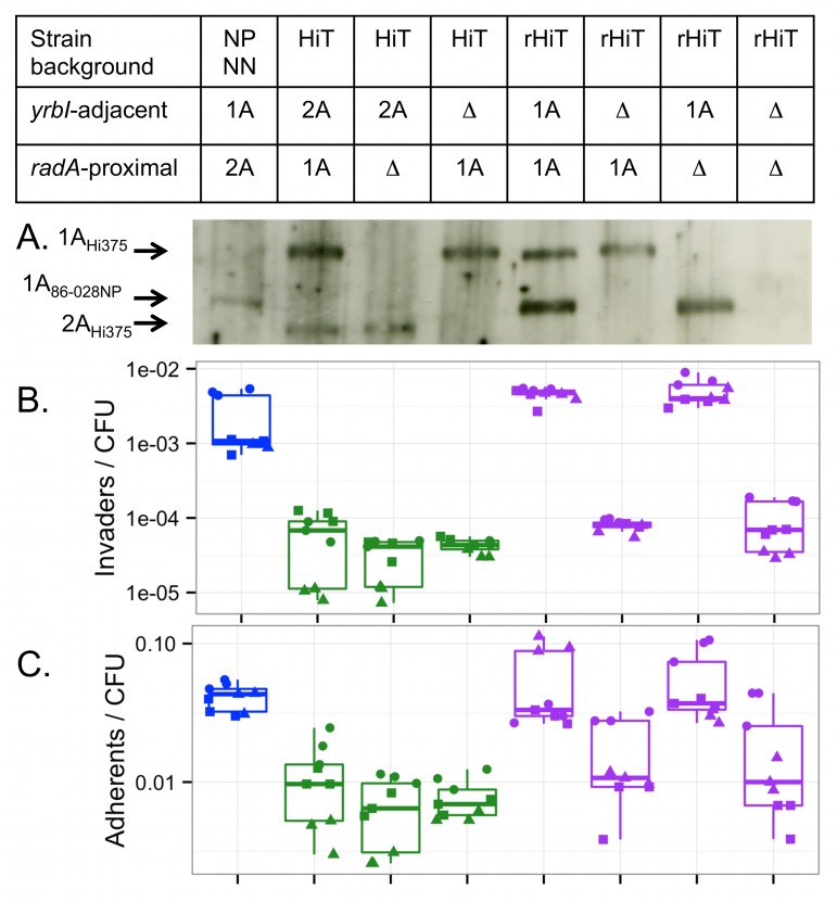
(A) Western blot detection of HMW adhesins with a guinea pig anti-HMW1A gp85 antibody. The 154 kDa protein corresponds to HMW1A86-028NP; the 168 kDa protein corresponds to HMW1AHi375, and the 150 kDa protein corresponds to HMW2AHi375. Expression of HMW2A86-028NP was not detectable with the antibody used. (B) Intracellular invasion and (C) adhesion by NpNN and HiT derivatives including mutant, recombinant and recombinant mutant strains. Blue indicates NpNN, green indicates the HiT recipient and mutant derivatives, and purple indicates the rHiT recombinant and mutant derivatives. Experiments were run in triplicate on three separate days, as indicated by the three distinct symbols.
Not all of the recovered genotypes had identical phenotypes. Genotype D colonies (HiT NalR from Pool 4) were significantly more invasive and adherent than genotype E and F colonies (one-way ANOVA p<<0.001, Tukey’s HSD gives p<0.001 for comparison of D against E or F, but p>0.1 for comparison of E and F). This indicates that donor-specific variation present in genotype D but absent from genotypes E and F slightly enhances adherence and invasion, albeit substantially less so than the hmw1ABC locus; the causative variation responsible remains unknown, but includes donor alleles of a QseBC-like two-component system (S9 Table, “D-specific” segments).
The sequencing of Pool 4 clones further supported a read alignment artifact at the radA-adjacent hmw1A Hi375 locus. Colonies had been collected after re-streaking to ensure they represented single clonal lineages. Despite this, all three HiT clone genotypes (D, E, and F) from Pool 4 showed a variable mixture of recipient- and donor-specific alleles across the radA-proximal hmw operon (Fig 6). In contrast, donor-specific allele frequencies observed at the yrbI-adjacent hmw1A 86-028NP were near fixation and accompanied by flanking donor-specific variation. As a final confirmation that this mixed signal was not due to merodiploidy or other complex genetic effect, allele-specific PCR assays were conducted that distinguished all four hmw adhesin genes (the yrbI-proximal hmw1A 86-028NP and hmw2A Hi375 genes and the radA-proximal hmw2A 86-028NP and hmw1A Hi375 genes), and these PCR assays unambiguously showed that HiT recombinants had replaced their yrbI-adjacent hmw2 Hi375 allele with the hmw1 86-028NP allele, whereas the radA-adjacent hmw1 Hi375 alleles were unchanged (S9 Fig).
Donor-specific variation in the hmw1ABC left flanking interval does not contribute to enhanced invasiveness
Despite the unambiguous acquisition of hmw1 86-028NP in all recombinant clones, other donor-specific variation in the recombinant clones could conceivably be responsible. Since recombination tracts that carried hmw1ABC 86-028NP also carried flanking donor-specific SNPs, we directly tested for a contribution by variation in the flanking interval. We cloned the 86-028NP alleles of the two genes upstream of hmw1ABC: kpsF and yrbI, encoding arabinose-5-phosphate isomerase and Kdo 8-phosphatase, respectively. Donor-specific variation in yrbI in particular might contribute to intracellular invasion, since the minimum interval that overlapped between all four experiments contained donor-specific variation in this gene (Fig 6). The resulting HA-tagged pSU20 plasmid (pSU20-kpsF-yrbI-HA) was then electroporated into Rd. Confirming expression from the plasmid, a ~19.3-kDa full-length YrbI86-028NP-HA protein was detected in whole cell extracts by immunoblot with an anti-HA antibody (S10B Fig). Strains Rd, Rd pSU20, and Rd pSU20-kpsF-yrbI-HA were tested for intracellular invasion into A549 cells. No significant difference was observed among strains (S10C Fig, one-way ANOVA p-value = 0.29), thereby excluding a significant role for these flanking loci in intracellular invasion.
Mutation of hmw1A 86-028NP confirms its role in intracellular invasion
Genetic confirmation of a role for hmw1A 86-028NP in intracellular invasion (rather than other donor-specific variation acquired by recombinants) was performed using the HiT recipient and one of the invasive HiT recombinant clones (strain P551 with genotype E, hereafter called rHiT). We generated a panel of knockouts of the genes encoding HMW adhesins in the HiT and rHiT strains, either the locus adjacent to yrbI (hmw2A Hi375 or hmw1A 86-028NP) or the locus nearby radA (hmw1A Hi375). In the case of rHiT, the double mutant was also produced (both hmw1A 86-028NP and hmw1A Hi375 deleted). Western blot analysis confirmed expression of the expected HMW adhesins in each strain (Fig 8A).
All mutant strains were assayed for invasion in parallel with NpNN, HiT, and rHiT controls (Fig 8B, one-way ANOVA p<<0.001). We hypothesized that knocking out hmw1A 86-028NP in the recombinant rHiT would show a strong defect in intracellular invasion, but that knocking out either hmw gene in the HiT recipient would have little or no effect. Indeed, deletion of hmw1A 86-028NP from rHiT reduced invasion frequencies 56-fold (Tukey’s HSD p<<0.01) down to HiT recipient levels. By contrast, deletion of the radA-proximal hmw1A Hi375 had no significant effect in either strain background, nor did deletion of either locus in the HiT recipient (Tukey’s HSD p>0.5 for all comparisons).
These results confirmed the role of hmw1A 86-028NP in the increased intracellular invasion of the HiT recombinant, and suggested that the HiT alleles of the HMW adhesins do not appreciably contribute to the HiT strain’s ability to invade A549 cells. Parallel adhesion assays of the knockout panel found qualitatively similar results (Fig 8C, one-way ANOVA p<<0.001). The recombinant rHiT was significantly more adherent than the HiT recipient (4.7-fold increase, Tukey’s HSD p<0.001)—comparable to adhesion by the donor NpNN (p = 0.99)—whereas deletion of hmw1A 86-028NP from rHiT brought adhesion down 3.3-fold to HiT recipient levels (Tukey’s HSD p = 0.0014 versus rHiT, p = 0.99 versus HiT).
The effect of hmw1A 86-028NP on adhesion is >10-fold lower than its effect on intracellular invasion, such that significant increases in adhesion had not been detected in the original pool experiments and were only marginally significant in adhesion assays with the isolated recombinant clones (see above). Nevertheless, these results indicate that hmw1A 86-028NP might contribute to intracellular invasion in part through an indirect effect of increasing adherence. While this is one contributing factor, the immunofluorescence data reported below indicate an unexpected intracellular invasion phenotype that cannot readily be explained by increased adherence alone.
Possession of hmw1ABC 86-028NP confers a self-aggregation phenotype to recombinants
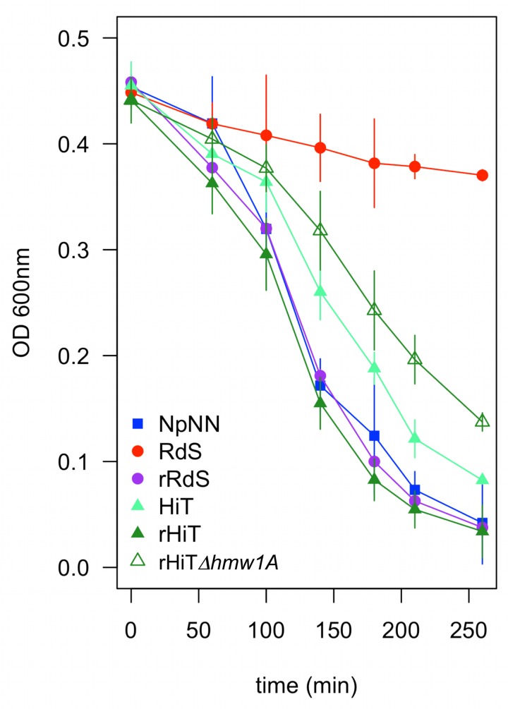
In the course of working with the parental strains, we noted that when cultures were left standing on the bench, NpNN (and 86-028NP) settled more quickly than RdS (and Rd), denoting a clumping or self-aggregation phenotype. Because self-aggregation could modulate bacterial-host cell interplay, we quantitatively tested this phenotype for a panel of strains: the three parents, an invasive recombinant RdS clone (P540, genotype B, hereafter rRdS), the rHiT recombinant, and the rHiTΔhmw1A 86-028NP mutant. This clearly demonstrated that hmw1A 86-028NP plays a major role in the high self-aggregation seen in the recombinants (Fig 9; one-way ANOVA p<<0.001 at the t = 140 min time point and higher). Both recombinants settled quicker than the recipient strains (Tukey’s HSD p<0.001) but were indistinguishable from NpNN (p>0.6). While the HiT recipient settled substantially faster than RdS, it and the rHiTΔhmw1A 86-028NP mutant settled slower than NpNN (p<0.01). These observations raised the question of how this clumping or self-aggregation phenotype modulates adhesion and intracellular invasion.
Fig 9. Self-aggregation is increased by hmw1A 86-028NP.
Bacteria scraped from chocolate agar plates were suspended into 35 ml of sBHI normalized to OD600 = 0.5 in a 50 ml conical tube and allowed to sit on the lab bench. The OD600 at the top of the cultures was followed over time as a proxy for clumping/self-aggregation. Error bars indicate the standard deviation from four replicate assays run on different days.
Immunofluorescence microscopy reveals that hmw1ABC 86-028NP confers a novel aggregated intracellular bacterial invasion phenotype
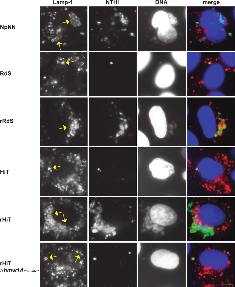
To directly assess intracellular location of bacterial cells, we used immunofluorescence microscopy (Fig 10). As expected, the donor (NpNN) and two recombinant (rRdS and rHiT) strains infected A549 cells at high rates, whereas the recipients (RdS and HiT) and the mutant recombinant rHiTΔhmw1A 86-028NP infected A549 cells at substantially lower rates (Table 2). Co-localization with the endosomal marker Lamp-1 was substantial for all strains, indicating bona fide intracellular invasion, rather than gentamicin resistance or occlusion by A549 cells. These results confirm: (a) that both recipients (including RdS) successfully enter A549 cells, albeit at low rates; (b) that donor and recombinants have substantially higher invasion rates than recipients; and (c) that hmw1A 86-028NP is responsible for elevated intracellular invasion rates in the rHiT recombinant.
Fig 10. Co-localization of intracellular bacteria and the Lamp-1 endosomal marker.
A549 cells were infected by NpNN, RdS, rRdS, HiT, rHiT, or rHiTΔhmw1A 86-028NP. In the merged images, reactivity with an anti-NTHi antibody is shown in green, Lamp-1 stain is shown in red, and DNA stained with Hoechst 33342 is shown in blue. Images were taken at 1 h post-gentamicin treatment. The scale bar in the lower right indicates 5 microns; individual bacterial cells are ~1 micron in diameter.
Table 2. Intracellular bacteria immunofluorescence microscopy.
| Strain | % infected (n = 250 cells) a | % bacteria:Lamp-1 co-localization (n = 150 infected cells) | % cells with # of infecting bacteria (n = 250 infected cells) | |
|---|---|---|---|---|
| <10 | >10 | |||
| RdS (P532) | 11.6 | 74.1 | 100 | 0 |
| HiT (P531) | 33.6 | 42.0 | 98.8 | 1.2 |
| NpNN (P351) | 100 | 88.0 | 0 | 100 |
| rRdS (P540) | 90.0 | 87.6 | 61.3 | 38.7 |
| rHiT (P551) | 80.8 | 93.9 | 76.2 | 23.8 |
| rHiTΔhmw1A 86-028NP (P834) | 44.7 | 65.3 | 92.1 | 7.9 |
aFrequencies are higher than from the plating assays above due to bacterial lysis during the saponin treatment that lyses A549 cells, as previously described [24].
The number of bacteria per infected cell was also distinct between strains. Cells infected by either recipient or the rHiTΔhmw1A 86-028NP mutant were infected with <10 bacteria/cell, whereas cells infected by the donor strain had >10 bacteria/cell. Although visually similar to the NpNN donor, scoring indicated that the rRdS and rHiT recombinants had an intermediate phenotype (Fig 10, Table 1). Whereas gentamicin protection assays with the recombinants gave invasion rates that approached donor levels, these data suggest that additional unidentified donor-specific factors besides hmw1A 86-028NP may augment the ability of bacteria to invade.
Unexpectedly, bacterial invaders had a distinct pattern of co-localization with Lamp-1-positive endosomes for the donor and two recombinants. When infected by bacteria of either recipient strain or the rHiTΔhmw1A 86-028NP mutant, Lamp-1-positive subcellular compartments typically enclosed single bacterial cells, as previously observed [24]. In contrast, cells of the donor or either recombinant had enlarged Lamp-1-positive subcellular compartments that surrounded groups of bacteria. The size of these groups varied, with ~5–50 bacteria per compartment (Fig 10).
These results indicate that increased intracellular invasion by the donor and recombinants relates—at least in part—to internalized bacteria localizing as groups in the same subcellular compartment, rather than as individual cells. Previous work showed that H. influenzae invaders do not replicate [24] and, in our assays, the time between infection and observation was sufficiently brief that the groups of intracellular bacteria seen are unlikely to reflect intracellular replication. Hi375 has previously been seen to clump at the surface of A549 cells prior to invasion, but with only single bacteria undergoing internalization [24]. Thus, our self-aggregation and immunofluorescence results strongly suggests that groups of hmw1ABC 86-028NP bacteria remain clumped during epithelial cell entry, thereby increasing overall invasion rates above and beyond the indirect effect of elevated adhesion, and also explaining the observed Lamp-1 reorganization around groups of bacteria.
Addition of hmw1ABC strain12 to Rd increases intracellular invasion
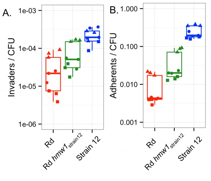
Previous studies of hmw1 focused on its role in adhesion, including detailed characterization of an Rd derivative carrying the hmw1 operon from strain 12 with only minimal flanking variation from strain 12 [44]. We used the Rd hmw1 strain12 strain to independently test for the role of hmw1 in intracellular invasion. We evaluated invasiveness and adhesiveness of Rd, Rd hmw1 strain12, and strain 12 using A549 cells, as described above. Rd hmw1 strain12 had intermediate levels of both invasion and adhesion between both parents (Fig 11, p-values<0.01 for one-way ANOVA and all three comparisons by Tukey’s HSD).
Fig 11. The hmw1 strain12 allele confers increased intracellular invasion to Rd KW20.
(A) Invasion and (B) adherence phenotypes of Rd, Rd hmw1 strain12, and strain 12 were evaluated in A549 cells in triplicate on three separate days (shown by distinct symbols).
To test how these results depended on the specific protocol or cell type used, we evaluated invasion and adhesion following an alternative protocol in both A549 and Chang cells [18]. Rd hmw1 strain12 had significantly higher adhesion and invasion than Rd for both cell types, though the effect was much stronger with Chang cells (S11 Fig, p-value < 0.01 for all three comparisons, except p = 0.054 for adherence to Chang cells by Rd hmw1 strain12 and Strain 12). Rd hmw1 strain12 invaded Chang cells nearly as well as strain 12. In contrast, when infecting A549 cells, Rd hmw1 strain12 had an intermediate phenotype. These results indicate that hmw1’s contribution to invasion depends on the host cell type and confirm a more general role for hmw1 in intracellular invasion beyond that seen for the 86-028NP allele.
Discussion
Is hmw1 directly involved in H. influenzae intracellular invasion?
In contrast to bacterial pathogens with well-characterized intracellular life styles like Salmonella, Listeria, Legionella or Brucella [45–47], the mechanism and functional role of intracellular invasion in H. influenzae has been less well understood. It has been suggested that intracellular invasion of airway epithelial cells by non-typeable H. influenzae (NTHi) allows the bacterium to evade the immune system (antibodies, surfactant, antimicrobial peptides, galectins, professional phagocytes) and therapeutic interventions (antibiotics, anti-inflammatory agents), and to facilitate access to essential nutrients [14,48–50]. Thus, entry into airways cells may better equip bacterial cells for survival during long-term infections, particularly in the context of chronic infections that are often treated with intense antibiotic regimes.
Potential factors that contribute to invasion include those known to facilitate H. influenzae’s interactions with host cell surfaces. Among these are bacterial surface proteins that participate in H. influenzae binding to extracellular matrix proteins, mucin, or epithelial cells (including P5, OapA, PE, Hap, Hia, the HMW1 and HMW2 adhesins, IgA1 proteases A and B, and type IV pili [26,33,38,51–54]). Some factors, such as IgaA1 protease, appear to be more directly involved in H. influenzae entry into epithelial cells [26]. The PE and Hap adhesins have also been implicated in H. influenzae entry into epithelial cells [55,56]. While adherence to host cells may be a prerequisite for invasion, we lacked information on the specific involvement of adhesins or other factors that modulate intracellular invasion by H. influenzae. TREP was designed to identify invasion-promoting genes in an unbiased manner, but we were nonetheless surprised to isolate the well-characterized adhesin-encoding hmw1 operon.
Gain of hmw1 86-028NP by a poorly invading strain naturally lacking both hmw1 and hmw2 (RdS) dramatically enhanced both adhesion and invasion, showing that hmw1 86-028NP is sufficient to confer high adhesion and invasion levels. When a recipient strain already possessing both hmw1 and its paralog hmw2 (HiT) was transformed, allelic substitution of the hmw2A Hi375 allele with hmw1A 86-028NP also strongly enhanced invasion. Characterization of recombinant clones and mutants lacking functional hmw1 86-028NP confirmed these results. Important roles for other donor-specific variation carried by the transformed recombinants can be excluded, except that one of the HiT recombinants (genotype D) showed significantly higher adhesion and invasion than the others, suggesting at least one additional invasion-promoting factor, albeit a smaller contributor; further analysis will be of interest in future studies.
The striking intracellular phenotype we observed by immunofluorescence for hmw1 86-028NP strains—in which groups of bacteria occupy engorged intracellular vesicles—suggests that increased adhesion alone is insufficient to fully explain the role of hmw1 in intracellular invasion. We instead suggest that elevated invasion is an emergent property of HMW1-mediated self-aggregation; whereas adhesion is increased as an indirect result, we speculate that invasion by bacterial groups directly enhances overall invasion rates. Alternatively, possession of hmw1 86-028NP may increase independent entry by bacteria into cells, followed by subsequent aggregation of bacterium-containing vesicles. Ruling out self-aggregation per se, recent results show that deletion of the Hap autotransporter from Hi375 (naturally present in both 86-028NP and Hi375 but absent from Rd) eliminates self-aggregation, but epithelial cell adhesion and invasion were not significantly affected [57]. Altogether, we propose that, whether or not it can truly be called an “invasin”, allelic variation in HMW1A affects invasion by increasing adhesion and also directly through a novel mechanism that allows for entry by groups of aggregated bacterial cells (model summary in Fig 12).
Fig 12. Model of enhanced intracellular invasion by HMW186-028NP.
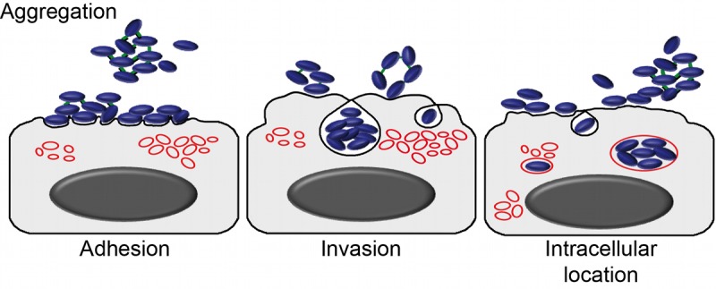
Bacteria self-aggregate and adhere to the epithelial cell surface in clumps, and HMW1-mediated attachments are maintained during uptake by cells into subcellular compartments with endosomal features.
The HMW1 and HMW2 adhesins
The hmw1 and hmw2 operons are found in ~60% of H. influenzae isolates, and they always co-occur, despite being at different chromosomal loci (one adjacent to HI1679 in Rd and the other to HI1598) [40,58,59]. Co-occurrence of the hmw1 and hmw2 loci in all tested clinical isolates suggests that the laboratory-created hmw1-only strains studied here must be at some unknown fitness disadvantage in nature.
The hmw1 and hmw2 operons are phase-variable, and expression is inversely correlated with the number of 7-bp tandem repeats found within their promoter regions [60–63]. Though subtle expression variation was not ruled out, western blot analysis indicated that HMW1 adhesin levels were mostly unchanged in recombinants (Fig 8A; 16 repeats upstream of hmw1 86-028NP but 17 upstream of hmw2 Hi375). This indicates that allelic variation in the hmw coding sequences is likely responsible for differences in adhesion and invasion.
HMW1A and HMW2A display wide amino acid diversity both within and between isolates, with the region of lowest sequence identity in the host cell binding domain, which has been predicted to affect tissue tropism and immune evasion [42,59,63–67]. Phylogenetic analyses of the HMW adhesin binding domain has revealed four distinct sequence clusters, and the majority of sequences belonging to one of two dominant sequence clusters [41]. Of note, 86-028NP and strain 12 hmw1A binding domains belong to clusters 4 and 2, respectively, which might contribute to their strong and intermediate phenotypes; future studies using mosaic proteins with binding domains from distinct clusters and using multiple human cell lines could identify any clade-specific functions for HMW proteins.
The two hmw operons encode high molecular weight non-pilus adhesins (HMW1/HMW2) [37,64], along with two co-factor proteins encoded by the downstream genes. The co-factors are required for proper surface localization of HMW adhesins, and the paralogs of the co-factors are functionally interchangeable [37,58]. The hmw1B/hmw2B genes encode outer membrane pore-forming translocators that export HMW1 and HMW2 to the cell surface [68]. The hmw1C/hmw2C genes encode glycosyltransferases responsible for adding mono-hexose or di-hexose residues at asparagines in conserved NX(S/T) motifs of HMW1 and HMW2 [69], likely involved in stabilizing the adhesins during or after their synthesis [70]. Due to the high diversity in HMW adhesin sequences, differential glycosylation patterns might in part be responsible for distinct activities of different alleles.
Another key distinction between the HMW1 and HMW2 adhesins is that the former recognizes sialylated glycoprotein receptors on cultured human epithelial cells [71]. HMW1 confers high adherence to Chang, Hep-2, HaCaT and NCI-H292 cells mediated by interactions with α-2,3 N-linked sialic acids. By contrast, HMW2 confers adherence to HaCaT and NCI-H292 cells via a sialic acid-independent mechanism [59,67,71]. This suggests that the role of hmw1A 86-028NP in intracellular invasion may involve specific interactions between H. influenzae cells and sialylated glycoprotein receptors, both on the bacterial cell surface to mediate self-aggregation and possibly also specific host sialylated glycoproteins on epithelial cells. Glycoproteins play important roles in many cellular activities, and new methods for investigating their expression and sialylation states are being developed and applied to multiple cell types including A549 [72], opening new avenues to identify host glycoproteins hijacked by bacterial proteins such as HMW adhesins during the infection process.
To our knowledge, this is the first report of an involvement for hmw1A in H. influenzae intracellular invasion and, more strikingly, we further found that intracellular invasion is modulated by allelic diversity at hmw1A. Finding that hmw1 86-028NP results in clumps of intracellular bacteria was unexpected and indicates that increased self-aggregation and adhesion per se are not sufficient to explain its effects, offering new avenues of investigation. Bacterial factors contributing to adhesion are already potential targets for antimicrobial therapies, and the additional role of HMW1 in intracellular invasion further increases its attractiveness as a target. Understanding the relationship between hmw1 allelic variation and within-host adaptive evolution poses interesting challenges for future studies.
Transformed recombinant enrichment profiling
To better understand intracellular invasion by H. influenzae, we have successfully employed a gain-of-function genetic mapping strategy, TREP, which takes advantage of within-species phenotypic variation, natural competence, and deep sequencing. In total, our experiment isolated six highly invasive recombinants from a total of ~400,000 independent recombinants. Thus, while transformation rates of SNPs was much higher (e.g. ~0.4% for the antibiotic resistance alleles), this approach was able to isolate even very rare recombinants, in the case of the Rd strain requiring the insertion of a particularly long operon (>9kb). TREP proved to be a rapid method to map genes. Once the donor and recipient were assayed and the effectiveness of the selections was determined, the total hands-on time was only about six weeks, from generating the recombinant pools, performing the serial selections, extracting DNA, making libraries and sequencing, with serial selections comprising the most time-consuming step. Owing to the strong selection used, we found that strains that have slight advantages in invasion were able to overtake the pools after serial selection. Thus, it was crucial to add selectable markers to our recipient background and to ensure that the serial selections were performed without the donor strain or any other strains assayed in parallel.
The TREP method holds great promise for studying a wide range of traits that show natural phenotypic variation in other naturally competent species, which includes many virulence traits and pathogens important to human health. In contrast to screening/selecting clones transformed by plasmids [73], TREP does not depend on dominance, a suitable vector, nor is it restricted to monogenetic traits. The approach should be readily applicable to any selectable trait in any bacterial species for which a natural competence protocol has been developed, and the number of such species continues to grow. Similar approaches have recently been reported in other organisms, for example to identify conjugation genes in Mycobacterium and causative alleles responsible for antibiotic resistance in Streptococcus [7,74]. Importantly, TREP is a general genetic mapping strategy agnostic to the type of variation (i.e. SNPs or whole loci can be identified), and we expand the utility of the transformation-based genetic mapping to include quantitative differences that go beyond absolute phenotypic differences (i.e. resistance versus sensitivity) by incorporating serial selection.
Traditionally, microbial experimental evolution studies rely on “hard” selective sweeps, in which newly arising beneficial mutations fix in a laboratory population [75]; more recently this has also included experimental evolution of pathogenic traits [76,77]. But “soft” sweeps, in which pre-existing genomic variation recombines within/into a population [78], may also play an important role in the adaptation of naturally competent species to new environments [79]. Allowing for introgression of natural variation has been used in experimental evolution in sexual eukaryotes (e.g. [80,81]), but its role in bacterial adaptation has been explored only recently and only in the context of standard experimental evolution studies that start with clonal populations [82,83]. Here, we found that rare recombinants generated in a single round of natural transformation could reach fixation after a small number of serial selections, illustrating the powerful contribution of natural competence to adaptation.
Finally, applying TREP to understand bacterial pathogenesis could use large “zoos” of donor genomic DNAs, rather than single donor-recipient combinations. This would better mimic the situation in chronic infections, where diverse polyclonal infections are common, and it would more fully sample the genomic diversity of these organisms in single experiments. However, caution must be exercised with such an approach: depending on the organism and trait under study, this could inadvertently generate novel hyper-virulent strains by combining multiple pathogenicity factors from different genetic backgrounds; a similar ethical concern has already been raised for studying pathogens using gain-of-function mutagenesis [84,85].
Materials and Methods
Bacterial strains
Bacterial strains and plasmids used are listed in S1 Table, and all PCR primers used are in S10 Table. General culturing and manipulation of Haemophilus influenzae followed standard methods [39]. Strains were grown at 37°C with 5% CO2, on chocolate agar or brain heart infusion (BHI) supplemented with 10 μg/ml hemin and 10 μg/ml β-nicotinamide (sBHI). Antibiotics were added as required: novobiocin (Nov) at 2.5 μg/ml, nalidixic acid (Nal) at 3 μg/ml, spectinomycin (Spc) at 25 μg/ml, streptomycin (Str) at 100 μg/ml, chloramphenicol (Cm) at 2 μg/ml, and erythromycin (Erm) at 11 μg/ml. Escherichia coli strains were grown at 37°C on Luria Bertani (LB), and Cm at 30 μg/ml was added as required.
Donor and recipient strains
Donor genomic DNA was obtained from P351, a derivative of the non-typeable otitis media isolate 86-028NP that also carries NovR and NalR alleles of gyrB and gyrA from the multi-antibiotic resistant strain MAP7 [10,28,39] (hereafter NpNN). Two recipient strains were used for natural transformation by this donor DNA: P532, a SpcR derivative of the laboratory strain Rd KW20 [27] (hereafter RdS), and P531, a StrR derivative of the non-typeable otitis media isolate Hi375 [29,86] (hereafter HiT). RdS was produced by transformation of Rd with MAP7 genomic DNA, selection for SpcR, followed by screening against other MAP7 resistance alleles. HiT was produced by transformation of Hi375 with a PCR amplicon spanning the StrR allele of the rpsL gene from an Rd KW20 StrR strain (P193) produced with “RdS” primers 1497+1798, and selection of transformants on sBHI-agar containing Str 100 μg/ml.
Cloning of the HMW1 flanking interval
To test whether the region flanking hmw1 86-028NP played a role in invasion, an interval encompassing NTHI1981 (kpsF) and NTHI1982 (yrbI), and 675 bp upstream, was PCR amplified using 86-028NP genomic DNA as template and “cloning” primers 1219 and 1218. Primer 1218 included an HA tag to add at the 3´end of the yrbI gene. This 2,241bp blunt PCR product was phosphorylated with T4 kinase, and cloned into pSU20 [87] pre-digested with HincII and dephosphorylated with Antarctic phosphatase, generating pSU20-kpsF-yrbI-HA. pSU20 and pSU20-Pr::kpsF-yrbI-HA were transformed into electrocompetent Rd. Tranformants were selected by plating to sBHI-agar containing Cm.
HMW adhesin knockouts
Deletion mutations of hmw adhesins were generated using one of two approaches. In the first recombineering approach, the first 1kb of the hmw1A 86-028NP gene (7,000bp) was replaced with a SpcR cassette. This was used to generate an hmw1A 86-028NP knockout in the rHiT recombinant clone (P551). Briefly, a ~3kb interval was amplified from NpNN using “interval” primers 13CAN+1273, which spanned the 1kb region targeted for deletion ±1kb, and the purified amplicon was cloned into the pGEMT-easy vector. Separately, a SpcR selectable marker was amplified from pRSM2832 [88] using primers carrying 50 bp overhangs flanking the deletion target (“Deletion” primers 1199 and 1274). The plasmid and amplicon were co-electroporated into DY380/SW102, which carries a heat-shock inducible λ Red recombinase. After recombinase induction, recovery, and selection for AmpR SpcR resistant E. coli colonies [88], disruption cassettes were confirmed by PCR. Finally, the “interval” primers (13CAN and 1273) were used to amplify the complete disruption cassette from the plasmid prior to natural transformation into rHiT and selection on SpcR. Mutants were selected on sBHI-agar containing Spc, and correct targeting was confirmed by PCR and western blot. This procedure generated strain P834.
In the second approach, PCR was used to amplify two ~1kb intervals flanking the region targeted for deletion (Flanks A and B), and an erythromycin resistance cassette (ErmR) was added between them. This was used to generate knockouts in HiT (P531), rHiT (P551), and rHiT Δhmw1A 86-028NP (P834) in two genes: hmw1A Hi375 (Flank A primers 1463 and 1464; Flank B primers 1465 and 1466) and hmw2A Hi375 (Flank A primers 1467 and 1468; Flank B primers 1469 and 1470). Flanks A and B were amplified with SmaI sites included in the R primer of Flank A and the F primer of Flank B, digested with SmaI, and blunt-end cloned into pJET1.2 by tri-molecular ligation. The ErmR cassette was excised from pBSLerm [89] using SmaI (1,188 bp fragment) and added to the pJET1.2 clone by blunt-ended ligation into the SmaI site joining Flanks A and B. Finally, the whole disruption cassette was amplified and transformed into MIV-competent cells of the appropriate strain using the standard protocol [39]. Mutants were selected on sBHI-agar containing Erm, and correct targeting was confirmed by PCR and western blot. This procedure generated strains P836-P839.
TREP design
The spectrum and distribution of recombinants in transformed pools that carry invasion alleles/loci depends upon the number of loci involved, their genetic interactions, the rates of recombination at those loci, and the experimental environment. To maximize the chance of enriching invasive recombinants from transformed pools: (a) We selected for a donor-specific marker (either NovR or NalR) after transformations to ensure that recombinant clones were not derived from non-competent cells in the original culture [8,10]. (b) We selected for a recipient-specific marker (SpcR or StrR) to limit cross-contamination. (c) Colonies were pooled, so that each independent recombinant in the pools was represented by many (>106) cells. (d) We maximized the complexity of the recombinant pools emerging from the first round of selection. (e) We progressively increased the frequency of invasive recombinants by serial selection, using a pool of the total CFU output from the gentamicin protection assay as the infecting material for the next cycle of selection.
Natural transformations
Recipient strains were made naturally competent using the standard protocol [39], except scaled up to 10 ml (~1010 CFU). Briefly, exponentially dividing cells growing in rich medium (sBHI) were transferred to starvation medium (MIV) for 100 min. Purified genomic DNA from the donor was incubated with naturally competent cultures at a concentration of ~1 genome per cell, or ~2 μg / 109 CFU / ml, for 30 min on a roller drum at 37°C, followed by a 1:5 dilution into sBHI and further incubation for 80 min to allow for expression of resistance alleles. Cultures were diluted and plated on sBHI-agar ± antibiotics to measure transformation and co-transformation frequencies (as NovR or NalR resistant colonies / CFU). Percent competence was calculated as (NovR NalR / CFU) / (NovR/CFU * NalR/CFU), as previously described [8,10,90]. To generate high complexity pools of recombinants, we plated 0.75 ml of a 10−2 dilution to 20 large petri dishes (20 cm diameter): 10 containing Nov and 10 with Nal (plus Spc or Str, depending on the recipient). This yielded ~104 resistant colonies per plate. Colonies from each set of 10 plates were scraped into a single 10 ml sBHI pool, titrated by dilution and plating to sBHI+antibiotics, and immediately stored as 1.25 ml aliquots in 15% glycerol at -80°C. This generated a total of four pools with an initial complexity of ~105 independent recombinants each, two for each recipient (Rd KW20 SpcR and Hi375 StrR), selected for either the NovR or the NalR donor allele, as well as a second antibiotic to select for the appropriate recipient background.
Infection of cultured epithelial cells and measurements of adhesion and intracellular invasion frequencies
The carcinomic human alveolar basal epithelial cell line A549 (ATCC CCL-185) was maintained in RPMI 1640 medium supplemented with 10 mM Hepes, 10% heat-inactivated fetal calf serum (FCS) and antibiotics (penicillin 100 units/ml and Str 0.1 mg/ml) in 25 cm2 tissue culture flasks at 37°C in a humidified 5% CO2 atmosphere. Chang cells (ATCC CCL-13) were cultured under the same atmospheric conditions in Minimum Essential Medium Eagle supplemented with 10% FCS and 1x MEM non-essential amino acid mixture (Sigma). Cells were seeded to 6×10⁴ or to 1.2×10⁵ cells / well in 24- or in 6-well tissue culture plates, respectively, for 32 h, and then serum starved for 16 h before infection. A ~90% confluence was reached by the time of infection. Adhesion and intracellular invasion assays in 24-well plates were conducted as previously described [15,24], starting with H. influenzae cells scraped from chocolate-agar plates (freshly grown for 16 h at 37°C with 5% CO2) into PBS and adjusted to OD600 = 1. A small aliquot of this adjusted suspension was diluted and plated on sBHI-agar to titrate the input CFU.
For invasion assays, A549 cells were incubated with 0.2 ml of each adjusted bacterial suspension for 2 h, washed 3 times with PBS and incubated for 1 h with RPMI 1640 containing 10% FCS, Hepes 10 mM and gentamicin 200 μg/ml to kill extracellular bacteria (the bacterial isolates used all had minimum inhibitory concentrations of < 5 μg/mL), washed 3 times with PBS, and human cells were lysed with 300 μl of PBS-saponin 0.025% for 10 min at room temperature. To quantify intracellular invasion frequencies, lysates were serial diluted and plated onto sBHI-agar with appropriate antibiotics. Recovered CFU was divided by the input CFU to calculate “Invaders/CFU”. Unless otherwise indicated, all infections were carried out in triplicate on three separate occasions. Adhesion assays were carried out similarly, excluding gentamicin treatment to calculate “Adherents/CFU”. For adhesion assays, cells were incubated with 0.1 ml of each adjusted bacterial suspension for 30 min. Wells were then washed 5 times with PBS and lysed as above. An alternative method was also used for both invasion and adhesion for comparisons of Rd, Strain 12, and Rd/HMW1Strain12 following a previously published protocol [18]. The primary differences were the lack of a serum starvation step, a low-speed centrifugation step to quickly bring bacteria into contact with the monolayer, and a lower MOI.
Serial enrichment for invasive recombinants
To enrich for recombinants carrying donor-specific invasion alleles, we performed eight serial selections for invasive clones for each recombinant pool. To maximize the complexity of the initial recombinant pools (Pool 0), one frozen aliquot per pool (~1010 CFU of ~105 independent recombinants per aliquot) was used to infect three wells of A549 cells seeded onto 6-well plates. Pool 0 aliquots were first recovered by thawing, pelleting, resuspending in 5 ml sBHI, and incubating on a roller drum for 60 min at 37°C under 5% CO2. Cultures were pelleted prior to proceeding with the invasion protocol, performed as described above, scaled up to cells seeded on 6-well plates (in 4 ml EBSS, with 0.8 ml of bacterial adjusted suspension / well), starting with resuspension of pellets in PBS, and ending with the total lysate plated on sBHI-agar (+appropriate antibiotics) at varying dilutions. This allowed measurement of intracellular invasion frequency and provided material for the next cycle.
For each subsequent serial invasion cycle, all CFU were scraped off plates into PBS and thoroughly mixed before normalizing to OD600 = 1 and proceeding with the infection. For all cycles, unused material was stored as (i) 15% glycerol stocks at -80°C for repeat assays and isolation of individual clones, and (ii) as a pellet at -20°C for DNA extractions (except for the RdS Pool 1 material, for which none was left over). In practical terms, this serial infection procedure was repeated for four enrichment cycles, at which point recovered pools were frozen as 15% glycerol stocks to allow for a new set of confluent A549 cells to grow up; frozen stocks were then restarted to carry out four additional cycles of selection. Untransformed recipient controls were run in parallel to exclude potential issues related to cell seeding. At Pool 4, several single gentamicin-protected clones per enrichment were isolated on sBHI-agar plates and stored at -80°C in 15% glycerol for adhesion and invasion assays, as well as clone sequencing.
Western blot
To monitor YrbI-HA expression, whole cell extracts from strain Rd alone, Rd carrying pSU20, and Rd carrying pSU20-Pr::kpsF-yrbi-HA were prepared from bacterial cultures grown to OD600 = 0.9 in sBHI containing Cm, when required. YrbI-HA expression was analyzed by western blot with a primary rabbit anti-HA antibody (Sigma) diluted 1:4000, and a secondary goat anti-rabbit IgG (whole molecule, Sigma) antibody conjugated to horseradish peroxidase, diluted 1:1000.
To investigate HMW adhesin protein expression in strains NpNN, RdS, rRdS, HiT, HiTΔhmw1A Hi375, HiTΔhmw2A Hi375, rHiT, rHiTΔhmw1A 86-028NP, rHiTΔhmw1A Hi375, rHiTΔhmw1A 86-028NPΔhmw1A Hi375, whole cell extracts were prepared from bacterial suspensions recovered from overnight grown chocolate-agar plates and adjusted to OD600 = 1 in PBS. HMW1A expression was analyzed by western blot with a primary guinea pig anti-HMW1A (gp85) antibody diluted 1:2000 [91], and a secondary goat anti-guinea pig IgG (Santa Cruz) antibody conjugated to horseradish peroxidase, diluted 1:5000.
Bacterial self-aggregation
H. influenzae cells were scraped from chocolate-agar plates freshly grown for 16 h at 37°C with 5% CO2 into PBS solution, and adjusted to OD600 = 0.45 in a 35 ml volume, and left standing at room temperature for at least 260 min. OD600 readings were performed at regular time intervals on 500 μl aliquots gently collected from the top of each bacterial suspension. Four independent experiments were performed for each strain.
Immunofluorescence microscopy
A549 cells were seeded on 13 mm circular coverslips in 24-well tissue culture plates. Cells were infected at an MOI ~1:8 (5 μl) of each adjusted bacterial suspension for 2 h, and infected cells were incubated in RPMI 1640 containing 10% FCS, Hepes 10mM and gentamicin 200 μg/ml for 1 h. Cells were washed three times with PBS and fixed with 3.7% paraformaldehyde (PFA) in PBS pH 7.4 for 15 min at room temperature. Immunofluorescence staining was carried out as previously described [24]. H. influenzae cells were stained with a rabbit anti-NTHi serum (raised against a pool of strains Hi375, 2019, and 398 [24]) diluted 1:600. Late endosomes were stained with mouse monoclonal anti-human Lamp-1 H4A3 antibody (Developmental Studies Hybridoma Bank) diluted 1:100. DNA was stained with Hoechst 33342 (Invitrogen) diluted 1:2500. Donkey anti-rabbit conjugated to Cy2 and donkey anti-goat or donkey anti-mouse conjugated to Rhodamine secondary antibodies (Jackson) were diluted 1:100.
Samples were analyzed with a Carl Zeiss Axioskop 2 plus fluorescence microscope and a Carl Zeiss Axio Cam MRm monochrome camera. We quantified: (a) the percentage of infected cells, counting at least 250 cells per sample; (b) the number of bacteria per infected cell in at least 250 cells per sample type, scoring <10 bacteria/cell or >10 bacteria/cell; (c) co-localization of bacteria and Lamp-1—an NTHi-containing vacuole (NTHi-CV) was considered positive for Lamp-1 when the marker was detected throughout the area occupied by the bacterium, or around/enclosing the bacterium. To determine the percentage of bacteria that co-localized with Lamp-1, all bacteria located inside a minimum of 150 infected cells were scored in each experiment. Results were calculated from two independent experiments.
DNA sequencing
Genomic DNA was extracted from the donor and recipients, stored pools, and isolated clones by phenol/chloroform extraction as described [8]. Purity and quality were evaluated by Nanodrop spectrophotometry (Thermo Scientific) and agarose gel electrophoresis, and quantification was performed with Qbit fluorometry prior to sequencing library construction. Multiplexed sequencing libraries were produced using the Nextera XT kit following manufacturer recommendations (Illumina). Paired-end sequencing (2x101nt) was conducted on an Illumina HiSeq in RapidRun mode over several independent runs/lanes. Raw base call data (bcl) was converted into FastQ format (Illumina version 1.8) using the bcl2fastq conversion software from Illumina (version 1.8.3, setting—no-eamss). For recombinant clones, paired-end sequencing (2x151nt) was conducted on an Illumina MiSeq, which automates demultiplexing to provide raw FastQ files. Properties of the genomic DNA samples and sequencing statistics (including donor and recipient controls) are in S2 and S3 Tables.
Read alignments and variant calling
The genome sequence references for the donor and recipients were: 86-028NP (NC_007146.2) [28], Rd_KW20 (NC_000907.1) [27], and Hi375 (CP009610.1) [29]. For the Rd genome, all non-ACGT bases were first converted to Ns (some non-N ambiguous IUPAC nucleotide characters lead to errors running samtools mpileup). Reads from control strains were used to identify variation between the derivative strains’ genomes and their deposited parental reference sequences (as described below). For all raw Illumina sequence processing, paired-end reads were trimmed of adapter sequences with Trimmomatic (v0.32) [92] and overlapping pairs were merged with COPE (v1.1.3; simple-connect mode) [93]. Next, reads were mapped using bwa mem (v0.7.8) with default settings [94], duplicates were marked with SamBlaster (v0.1.14) [95], and aligned reads were sorted and compressed using SamBamba (v0.4.6) [96]. Subsequent steps filtered out reads with a mapping quality = 0, which excludes multiply mapping reads that align equally well to different reference genome coordinates.
For donor and recipient controls, as well as recombinant clones, single-nucleotide polymorphism (SNP) and small indel variant calling used samtools mpileup and bcftools view (v0.1.19) [97]. Variant frequency calling from recombinant pools used a python script (available at https://github.com/photonchang/allelecount/) to count reads supporting each of the 4 bases at each reference position directly from samtools mpileup output (for base calls with quality score >10), and subsequent parsing used linux commands (mostly awk). BedTools (v2.19.1) [98] was used for subsetting (using the intersect tool) with the variants detected between donor and recipient genomes. Variant tables were first corrected for “self” variants identified between reads and their own reference (with the exception of resistance-associated markers). This allowed calculation of recipient-specific, donor-specific, and erroneous base frequencies (i.e. bases with neither donor nor recipient identity). Manual validation of recombination breakpoints and clone assignments used the Integrative Genomics Viewer (v2.3.1) [99]. Identification of novel alleles that had approached fixation compared the variants called from Pool 8 reads to those from Pool 0 reads (using samtools mpileup and bcftools view). Due to systematic alignment artifacts that arise when mapping donor reads to recipient genomes in regions of high divergence, putative novel variation that was also identified only in reciprocal alignments of control reads (“unreliable” SNP positions) was excluded, leaving no observed fixed new mutations.
PCR validation
To distinguish between the four possible hmw genotypes, allele- and locus-specific PCR was used, with allele-specific primer pairs listed under “Allele ID” in S10 Table. Each pair is specific for one of the four possibilities and generates a distinct PCR product size as determined by standard agarose gel electrophoresis. Primers 1456+1458 were used for hmw1A Hi375 (product size 1,364 bp), primers 1456+1457 for hmw2A Hi375 (product size 1,076 bp); primers 1459+1460 for hmw1A 86-028NP (product size 744 bp); and primers 1461+1462 for hmw2A 86-028NP (product size 582 bp).
Statistics and plotting
Significant differences in invasion, adhesion, and self-aggregation phenotypes among strains and pools were evaluated using one-way ANOVA with post hoc hypothesis testing using Tukey’s HSD (“honest significant differences”). Invasion and adhesion frequencies were first log-transformed prior to testing to account for the highly unequal variances observed between strains/pools that were quantified at distinct plating dilutions. Pairwise student’s t-tests with untransformed data and Bonferroni correction gave qualitatively similar results. Plotting used the R statistical programming language including add-on packages seqinr, genoplotr, ggplot2, and Rcolorbrewer.
Data deposition
All sequence data were deposited at NCBI under BioProject PRJNA308311. BioSample accessions are included in S2 and S3 Tables. Parental strains were submitted to the SRA as BAM files aligned to their own reference sequence. Recombinant pool and clone data were submitted to the SRA as BAM files aligned to the appropriate recipient reference sequence (Hi375 or Rd KW20).
Supporting Information
(DOCX)
Turquoise lines above the x-axis indicate the position of SNPs distinguishing donor from recipient, while grey lines below the x-axis indicate positions in the recipient genome missing from the donor genome (at indels). (A) Hi375 recipient. (B) Rd KW20 recipient. Note that SNPs between Hi375 and 86-028NP are punctate, with stretches of very low SNP density punctuated by stretches of high SNP density. Genomic positions exclusive to the recipient strains are shown in grey; these coincide with areas that appear as regions of low SNP density, but these artifacts are insufficient to explain the pattern seen in Hi375. Conversely Rd-specific positions do largely explain low SNP density regions in Rd KW20.
(TIF)
(A) Invasion of and (B) adhesion to A549 cells is shown for H. influenzae strains Rd KW20, Hi375, 86-028NP, and antibiotic resistant derivatives, including the parental strains.
(TIF)
Two serial cycles of selection for intracellular invaders were conducted using three mixtures of 86-028NP NovR and Rd StrR cells, at 1:100, 1:1,000, or 1:10,000 ratios. Prior to the first infection (input.A), the bacterial cell suspension was titrated for the total NovR and StrR CFU used per well, and this closely matched the expected frequencies. After the first round of selection (output.A), dramatically fewer CFU were recovered, but NovR were proportionally much more abundant. Total unselected CFUs were pooled and titrated (input.B), showing that the proportion of NovR remained relatively the same in between cycles of selection for invasion. Finally, the second cycle of selection resulted in a higher yield with an even higher proportion of NovR colonies, representing a strong enrichment of 86-028NP over Rd, even when at a low relative abundance in the starting mixture.
(TIF)
(A) and (B) NpNN-specific SNP frequencies as a function of chromosome coordinate for the RdS and HiT recipients, respectively, at Pool 0, prior to enrichment for invasive recombinants. Left panels: NovR-selected pools. Right panels: NalR-selected pools. Top panels: chromosome-wide view. Bottom panels: zoom on 60 kb windows around the antibiotic resistance markers. The peak SNP is the one conferring antibiotic resistance. (C) “Bean plots” summarizing 16 histograms of non-recipient allele frequencies for untransformed controls and the initial transformed recombinant pools. The left side (salmon-colored) of each bean shows a histogram for allele frequencies with donor allele identities, whereas the right side (light blue) shows a histogram for “novel” alleles (neither recipient nor donor). The latter are sequencing errors, while the former are sequencing errors for the control strains and a combination of sequencing errors and transformants for the transformed pools.
(TIF)
Invaders/CFU for pools during the initial eight serial selections for invasive recombinants. Recovered CFU that survived gentamicin treatment (Pool 1) served as input for the next cycle (which generated Pool 2). The values show the combined ability of clones in Pool n to invade airway epithelial cells, while the recovered colonies comprise Pool n+1. This procedure was carried out eight times. The apparent decline in invasiveness seen at Pool 4 appears to be an artifact, since no such decline was seen in the replicate assays (Fig 3A). Instead, this drop likely reflects that Pool 4 bacteria had been frozen and re-inoculated prior to the next cycle, combined with batch-to-batch variation of the confluent A549 cells used.
(TIF)
Control experiment using untransformed recipients cultures in triplicate found no increase in invasiveness over 5 serial selections. This experiment was conducted independently for each of the recipients and separately from the experimental enrichments to minimize enrichment of cross-contaminants.
(TIF)
Genomic profiling at antibiotic-selected sites for both the (A) RdS and (B) HiT recipients at NovR (top) and NalR (bottom) sites (gyrB and gyrA respectively, see S6 Table) for Pools 0, 2, 4, and 8. Axes are as in other figures with x-axes indicating recipient genome coordinate (in kb) and the y-axis indicating donor allele frequency. RdS NovR contains a single clone at ~95% by Pool 8, while RdS NalR contains two dominant clones, one at ~70% and the other ~30%. HiT NovR contains two dominant clones (at ~30% and 70%), whereas HiT NalR appears to contain two clones at ~80% and ~20%. For this pool, only a single genotype (the one at ~80%) was recovered in the four individual clones collected from Pool 4. No other donor segments appeared at ~20%, so this is likely due to incomplete fixation of the invasive genotype after several rounds of selection.
(TIF)
(A) Pools 0, 2, 4, and 8 for HiT NovR and HiT NalR as in other figures (x-axis is HiT recipient coordinate in kb, and y-axis is donor allele frequency). (B) Genomic map around the same interval. The thick black horizontal line shows the entire range of positions containing donor frequencies >5%. The affected interval spans only the hmw1 locus; no flanking variation was detected, unlike the situation at the yrbI-adjacent hmw2 Hi375, which was replaced by the hmw1 86-028NP allele. Donor allele frequencies are highly variable in this region. They are also highly consistent between the two pools, which was unexpected, as all other overlapping donor segments detected had distinct recombination breakpoints. Allele-specific PCR assays confirm this as read alignment artifact and confirm that the radA-proximal adhesin remain hmw1 Hi375 across strains (S9 Fig).
(TIF)
Strains are listed as it follows: (1) NpNN, (2) RdS, (3) rRdS, (4) HiT, (5) HiTΔhmw1A Hi375, (6) HiTΔhmw2A Hi375, (7) rHiT, (8) rHiTΔhmw1A 86-028NP, (9) rHiTΔhmw1A Hi375, (10) rHiTΔhmw1A 86-028NPΔhmw1A Hi375, and primers are in S10 Table. (A) Primers 1456+1458 identify hmw1A Hi375 (1,364 bp product); (B) primers 1456+1457 identify hmw2A Hi375 (1,076 bp product); (C) primers 1459+1460 identify hmw1A 86-028NP (744 bp product); and (D) primers 1461+1462 for hmw2A 86-028NP (582 bp product). Lanes 8 and 10 rendered a correct size band upon PCR with primers 1459+1460 because mutant strains lacking hmw1A 86-028NP were generated by partial deletion that maintains the annealing sites for the primers and product size.
(TIF)
(A) Western blot showing expression of HMW1A86-028NP (154 KDa) adhesin. Whole cell extracts of NpNN, RdS and rRdS (P540, genotype B) were prepared and used to detect HMW by immunoblot with the guinea pig anti-HMW1A gp85 antibody. (B and C) Addition of the kpsF and yrbI alleles from 86-028NP on a plasmid does not increase intracellular invasion frequencies. (B) Western blot showing expression from plasmid carrying an interval carrying kpsF-yrbI from 86-028NP. Whole cell extracts of cultures (Rd, Rd pSU20, and Rd pSU20-kpsF-yrbI-HA) were prepared and used to detect Hap-HA by immunoblot with a rabbit anti-HA antibody, finding expression of the expected ~19.3-kDa protein in the expected strain. (C) The same strains were used to infect A549 cells and measure bacterial intracellular invasion. Experiments were performed three times in triplicate (different symbols denote independent experiments).
(TIF)
Invasion (A) and adhesion (B) by Rd, Rd hmw1 strain12, and Strain 12 bacterial strains into Chang and A549 epithelial cell lines. An alternative protocol that includes centrifugation to quickly bring bacteria into contact with the cell monolayer was used for these experiments, showing that both cell type and details of the infection procedure give qualitatively similar results.
(TIF)
(DOCX)
(DOCX)
(DOCX)
(DOCX)
(DOCX)
(DOCX)
(DOCX)
(DOCX)
(DOCX)
(DOCX)
Acknowledgments
We thank Nathaniel Lin for work on pileup parsing script and Begoña Euba for help with DNA preparations and cloning.
Data Availability
All sequence data were deposited at NCBI under BioProject PRJNA308311. BioSample accessions are included in S2 and S3 Tables. Parental strains were submitted to the SRA as BAM files aligned to their own reference sequence. Recombinant pool and clone data were submitted to the SRA as BAM files aligned to the appropriate recipient reference sequence (Hi375 or Rd KW20).
Funding Statement
This work was supported by National Institutes of Health Ruth Kirschstein postdoctoral fellowship F32AI084427 (to JCM); a Canadian Institute of Health Research operating grant (to RJR); Genome British Columbia grant SOF122 (to RJR and JCM); the Faculty of Pharmaceutical Sciences, Canadian Foundation for Innovation (to CN); National Institutes of Heath R01 grant DC002873 (to JWSG); and (to JG) grants from Ministerio Economía y Competitividad-MINECO SAF2012-31166 and SAF2015-66520-R, Dpto. Salud Gobierno Navarra 359/2012 and Ministerio de Educación PRX12/00191. The funders had no role in study design, data collection and analysis, decision to publish, or preparation of the manuscript.
References
- 1. Wong SM, Bernui M, Shen H, Akerley BJ (2013) Genome-wide fitness profiling reveals adaptations required by Haemophilus in coinfection with influenza A virus in the murine lung. Proc Natl Acad Sci U S A 110: 15413–15418. 10.1073/pnas.1311217110 [DOI] [PMC free article] [PubMed] [Google Scholar]
- 2. Akerley BJ, Rubin EJ, Novick VL, Amaya K, Judson N, et al. (2002) A genome-scale analysis for identification of genes required for growth or survival of Haemophilus influenzae . Proc Natl Acad Sci U S A 99: 966–971. [DOI] [PMC free article] [PubMed] [Google Scholar]
- 3. Johnston C, Martin B, Fichant G, Polard P, Claverys JP (2014) Bacterial transformation: distribution, shared mechanisms and divergent control. Nat Rev Microbiol 12: 181–196. 10.1038/nrmicro3199 [DOI] [PubMed] [Google Scholar]
- 4. Mell JC, Redfield RJ (2014) Natural competence and the evolution of DNA uptake specificity. J Bacteriol 196: 1471–1483. 10.1128/JB.01293-13 [DOI] [PMC free article] [PubMed] [Google Scholar]
- 5. Avery OT, Macleod CM, McCarty M (1944) Studies on the Chemical Nature of the Substance Inducing Transformation of Pneumococcal Types: Induction of Transformation by a Desoxyribonucleic Acid Fraction Isolated from Pneumococcus Type Iii. J Exp Med 79: 137–158. [DOI] [PMC free article] [PubMed] [Google Scholar]
- 6. Dalia AB, McDonough E, Camilli A (2014) Multiplex genome editing by natural transformation. Proc Natl Acad Sci U S A 111: 8937–8942. 10.1073/pnas.1406478111 [DOI] [PMC free article] [PubMed] [Google Scholar]
- 7. Gray TA, Krywy JA, Harold J, Palumbo MJ, Derbyshire KM (2013) Distributive conjugal transfer in mycobacteria generates progeny with meiotic-like genome-wide mosaicism, allowing mapping of a mating identity locus. PLoS Biol 11: e1001602 10.1371/journal.pbio.1001602 [DOI] [PMC free article] [PubMed] [Google Scholar]
- 8. Mell JC, Lee JY, Firme M, Sinha S, Redfield RJ (2014) Extensive cotransformation of natural variation into chromosomes of naturally competent Haemophilus influenzae . G3 (Bethesda) 4: 717–731. [DOI] [PMC free article] [PubMed] [Google Scholar]
- 9. Freddolino PL, Goodarzi H, Tavazoie S (2014) Revealing the genetic basis of natural bacterial phenotypic divergence. J Bacteriol 196: 825–839. 10.1128/JB.01039-13 [DOI] [PMC free article] [PubMed] [Google Scholar]
- 10. Mell JC, Shumilina S, Hall IM, Redfield RJ (2011) Transformation of natural genetic variation into Haemophilus influenzae genomes. PLoS Pathog 7: e1002151 10.1371/journal.ppat.1002151 [DOI] [PMC free article] [PubMed] [Google Scholar]
- 11. Agrawal A, Murphy TF (2011) Haemophilus influenzae infections in the H. influenzae type b conjugate vaccine era. J Clin Microbiol 49: 3728–3732. 10.1128/JCM.05476-11 [DOI] [PMC free article] [PubMed] [Google Scholar]
- 12. Garmendia J, Viadas C, Calatayud L, Mell JC, Marti-Lliteras P, et al. (2014) Characterization of nontypable Haemophilus influenzae isolates recovered from adult patients with underlying chronic lung disease reveals genotypic and phenotypic traits associated with persistent infection. PLoS One 9: e97020 10.1371/journal.pone.0097020 [DOI] [PMC free article] [PubMed] [Google Scholar]
- 13. Murphy TF, Brauer AL, Schiffmacher AT, Sethi S (2004) Persistent colonization by Haemophilus influenzae in chronic obstructive pulmonary disease. Am J Respir Crit Care Med 170: 266–272. [DOI] [PubMed] [Google Scholar]
- 14. Clementi CF, Murphy TF (2011) Non-typeable Haemophilus influenzae invasion and persistence in the human respiratory tract. Front Cell Infect Microbiol 1: 1 10.3389/fcimb.2011.00001 [DOI] [PMC free article] [PubMed] [Google Scholar]
- 15. Lopez-Gomez A, Cano V, Moranta D, Morey P, Garcia del Portillo F, et al. (2012) Host cell kinases, alpha5 and beta1 integrins, and Rac1 signalling on the microtubule cytoskeleton are important for non-typable Haemophilus influenzae invasion of respiratory epithelial cells. Microbiology 158: 2384–2398. 10.1099/mic.0.059972-0 [DOI] [PubMed] [Google Scholar]
- 16. Ahren IL, Williams DL, Rice PJ, Forsgren A, Riesbeck K (2001) The importance of a beta-glucan receptor in the nonopsonic entry of nontypeable Haemophilus influenzae into human monocytic and epithelial cells. J Infect Dis 184: 150–158. [DOI] [PubMed] [Google Scholar]
- 17. Ketterer MR, Shao JQ, Hornick DB, Buscher B, Bandi VK, et al. (1999) Infection of primary human bronchial epithelial cells by Haemophilus influenzae: macropinocytosis as a mechanism of airway epithelial cell entry. Infect Immun 67: 4161–4170. [DOI] [PMC free article] [PubMed] [Google Scholar]
- 18. St Geme JW 3rd, Falkow S (1990) Haemophilus influenzae adheres to and enters cultured human epithelial cells. Infect Immun 58: 4036–4044. [DOI] [PMC free article] [PubMed] [Google Scholar]
- 19. Swords WE, Buscher BA, Ver Steeg Ii K, Preston A, Nichols WA, et al. (2000) Non-typeable Haemophilus influenzae adhere to and invade human bronchial epithelial cells via an interaction of lipooligosaccharide with the PAF receptor. Mol Microbiol 37: 13–27. [DOI] [PubMed] [Google Scholar]
- 20. Virji M, Kayhty H, Ferguson DJ, Alexandrescu C, Moxon ER (1991) Interactions of Haemophilus influenzae with cultured human endothelial cells. Microb Pathog 10: 231–245. [DOI] [PubMed] [Google Scholar]
- 21. Bandi V, Apicella MA, Mason E, Murphy TF, Siddiqi A, et al. (2001) Nontypeable Haemophilus influenzae in the lower respiratory tract of patients with chronic bronchitis. Am J Respir Crit Care Med 164: 2114–2119. [DOI] [PubMed] [Google Scholar]
- 22. Forsgren J, Samuelson A, Ahlin A, Jonasson J, Rynnel-Dagoo B, et al. (1994) Haemophilus influenzae resides and multiplies intracellularly in human adenoid tissue as demonstrated by in situ hybridization and bacterial viability assay. Infect Immun 62: 673–679. [DOI] [PMC free article] [PubMed] [Google Scholar]
- 23. St Geme JW 3rd (2002) Molecular and cellular determinants of non-typeable Haemophilus influenzae adherence and invasion. Cell Microbiol 4: 191–200. [DOI] [PubMed] [Google Scholar]
- 24. Morey P, Cano V, Marti-Lliteras P, Lopez-Gomez A, Regueiro V, et al. (2011) Evidence for a non-replicative intracellular stage of nontypable Haemophilus influenzae in epithelial cells. Microbiology 157: 234–250. 10.1099/mic.0.040451-0 [DOI] [PubMed] [Google Scholar]
- 25. Woo JI, Oh S, Webster P, Lee YJ, Lim DJ, et al. (2014) NOD2/RICK-dependent beta-defensin 2 regulation is protective for nontypeable Haemophilus influenzae-induced middle ear infection. PLoS One 9: e90933 10.1371/journal.pone.0090933 [DOI] [PMC free article] [PubMed] [Google Scholar]
- 26. Clementi CF, Hakansson AP, Murphy TF (2014) Internalization and trafficking of nontypeable Haemophilus influenzae in human respiratory epithelial cells and roles of IgA1 proteases for optimal invasion and persistence. Infect Immun 82: 433–444. 10.1128/IAI.00864-13 [DOI] [PMC free article] [PubMed] [Google Scholar]
- 27. Fleischmann RD, Adams MD, White O, Clayton RA, Kirkness EF, et al. (1995) Whole-genome random sequencing and assembly of Haemophilus influenzae Rd. Science 269: 496–512. [DOI] [PubMed] [Google Scholar]
- 28. Harrison A, Dyer DW, Gillaspy A, Ray WC, Mungur R, et al. (2005) Genomic sequence of an otitis media isolate of nontypeable Haemophilus influenzae: comparative study with H. influenzae serotype d, strain KW20. J Bacteriol 187: 4627–4636. [DOI] [PMC free article] [PubMed] [Google Scholar]
- 29. Mell JC, Sinha S, Balashov S, Viadas C, Grassa CJ, et al. (2014) Complete Genome Sequence of Haemophilus influenzae Strain 375 from the Middle Ear of a Pediatric Patient with Otitis Media. Genome Announc 2. [DOI] [PMC free article] [PubMed] [Google Scholar]
- 30. Maughan H, Redfield RJ (2009) Tracing the evolution of competence in Haemophilus influenzae . PLoS One 4: e5854 10.1371/journal.pone.0005854 [DOI] [PMC free article] [PubMed] [Google Scholar]
- 31. Maughan H, Redfield RJ (2009) Extensive variation in natural competence in Haemophilus influenzae . Evolution 63: 1852–1866. 10.1111/j.1558-5646.2009.00658.x [DOI] [PubMed] [Google Scholar]
- 32. Daines DA, Cohn LA, Coleman HN, Kim KS, Smith AL (2003) Haemophilus influenzae Rd KW20 has virulence properties. J Med Microbiol 52: 277–282. [DOI] [PubMed] [Google Scholar]
- 33. Hong W, Mason K, Jurcisek J, Novotny L, Bakaletz LO, et al. (2007) Phosphorylcholine decreases early inflammation and promotes the establishment of stable biofilm communities of nontypeable Haemophilus influenzae strain 86-028NP in a chinchilla model of otitis media. Infect Immun 75: 958–965. [DOI] [PMC free article] [PubMed] [Google Scholar]
- 34. Mason KM, Munson RS Jr., Bakaletz LO (2003) Nontypeable Haemophilus influenzae gene expression induced in vivo in a chinchilla model of otitis media. Infect Immun 71: 3454–3462. [DOI] [PMC free article] [PubMed] [Google Scholar]
- 35. Mason KM, Munson RS Jr., Bakaletz LO (2005) A mutation in the sap operon attenuates survival of nontypeable Haemophilus influenzae in a chinchilla model of otitis media. Infect Immun 73: 599–608. [DOI] [PMC free article] [PubMed] [Google Scholar]
- 36. Novotny LA, Mason KM, Bakaletz LO (2005) Development of a chinchilla model to allow direct, continuous, biophotonic imaging of bioluminescent nontypeable Haemophilus influenzae during experimental otitis media. Infect Immun 73: 609–611. [DOI] [PMC free article] [PubMed] [Google Scholar]
- 37. St Geme JW 3rd, Falkow S, Barenkamp SJ (1993) High-molecular-weight proteins of nontypable Haemophilus influenzae mediate attachment to human epithelial cells. Proc Natl Acad Sci U S A 90: 2875–2879. [DOI] [PMC free article] [PubMed] [Google Scholar]
- 38. St Geme JW 3rd, Yeo HJ (2009) A prototype two-partner secretion pathway: the Haemophilus influenzae HMW1 and HMW2 adhesin systems. Trends Microbiol 17: 355–360. 10.1016/j.tim.2009.06.002 [DOI] [PubMed] [Google Scholar]
- 39. Poje G, Redfield RJ (2003) Transformation of Haemophilus influenzae . Methods Mol Med 71: 57–70. [DOI] [PubMed] [Google Scholar]
- 40. De Chiara M, Hood D, Muzzi A, Pickard DJ, Perkins T, et al. (2014) Genome sequencing of disease and carriage isolates of nontypeable Haemophilus influenzae identifies discrete population structure. Proc Natl Acad Sci U S A 111: 5439–5444. 10.1073/pnas.1403353111 [DOI] [PMC free article] [PubMed] [Google Scholar]
- 41. Davis GS, Patel M, Hammond J, Zhang L, Dawid S, et al. (2014) Prevalence, distribution, and sequence diversity of hmwA among commensal and otitis media non-typeable Haemophilus influenzae . Infect Genet Evol 28: 223–232. 10.1016/j.meegid.2014.09.035 [DOI] [PMC free article] [PubMed] [Google Scholar]
- 42. Ecevit IZ, McCrea KW, Pettigrew MM, Sen A, Marrs CF, et al. (2004) Prevalence of the hifBC, hmw1A, hmw2A, hmwC, and hia Genes in Haemophilus influenzae Isolates. J Clin Microbiol 42: 3065–3072. [DOI] [PMC free article] [PubMed] [Google Scholar]
- 43. Vuong J, Wang X, Theodore JM, Whitmon J, Gomez de Leon P, et al. (2013) Absence of high molecular weight proteins 1 and/or 2 is associated with decreased adherence among non-typeable Haemophilus influenzae clinical isolates. J Med Microbiol 62: 1649–1656. 10.1099/jmm.0.058222-0 [DOI] [PubMed] [Google Scholar]
- 44. Grass S, Buscher AZ, Swords WE, Apicella MA, Barenkamp SJ, et al. (2003) The Haemophilus influenzae HMW1 adhesin is glycosylated in a process that requires HMW1C and phosphoglucomutase, an enzyme involved in lipooligosaccharide biosynthesis. Mol Microbiol 48: 737–751. [DOI] [PubMed] [Google Scholar]
- 45. Fonseca MV, Swanson MS (2014) Nutrient salvaging and metabolism by the intracellular pathogen Legionella pneumophila . Front Cell Infect Microbiol 4: 12 10.3389/fcimb.2014.00012 [DOI] [PMC free article] [PubMed] [Google Scholar]
- 46. Liss V, Hensel M (2015) Take the tube: remodelling of the endosomal system by intracellular Salmonella enterica . Cell Microbiol 17: 639–647. 10.1111/cmi.12441 [DOI] [PubMed] [Google Scholar]
- 47. Winchell CG, Steele S, Kawula T, Voth DE (2015) Dining in: intracellular bacterial pathogen interplay with autophagy. Curr Opin Microbiol 29: 9–14. 10.1016/j.mib.2015.09.004 [DOI] [PMC free article] [PubMed] [Google Scholar]
- 48. Raffel FK, Szelestey BR, Beatty WL, Mason KM (2013) The Haemophilus influenzae Sap transporter mediates bacterium-epithelial cell homeostasis. Infect Immun 81: 43–54. 10.1128/IAI.00942-12 [DOI] [PMC free article] [PubMed] [Google Scholar]
- 49. Garmendia J, Marti-Lliteras P, Moleres J, Puig C, Bengoechea JA (2012) Genotypic and phenotypic diversity of the noncapsulated Haemophilus influenzae: adaptation and pathogenesis in the human airways. Int Microbiol 15: 159–172. [DOI] [PubMed] [Google Scholar]
- 50. Euba B, Moleres J, Segura V, Viadas C, Morey P, et al. (2015) Genome Expression Profiling-Based Identification and Administration Efficacy of Host-Directed Antimicrobial Drugs against Respiratory Infection by Nontypeable Haemophilus influenzae . Antimicrob Agents Chemother 59: 7581–7592. 10.1128/AAC.01278-15 [DOI] [PMC free article] [PubMed] [Google Scholar]
- 51. Prasadarao NV, Lysenko E, Wass CA, Kim KS, Weiser JN (1999) Opacity-associated protein A contributes to the binding of Haemophilus influenzae to chang epithelial cells. Infect Immun 67: 4153–4160. [DOI] [PMC free article] [PubMed] [Google Scholar]
- 52. Rosadini CV, Ram S, Akerley BJ (2014) Outer membrane protein P5 is required for resistance of nontypeable Haemophilus influenzae to both the classical and alternative complement pathways. Infect Immun 82: 640–649. 10.1128/IAI.01224-13 [DOI] [PMC free article] [PubMed] [Google Scholar]
- 53. Singh B, Brant M, Kilian M, Hallstrom B, Riesbeck K (2010) Protein E of Haemophilus influenzae is a ubiquitous highly conserved adhesin. J Infect Dis 201: 414–419. 10.1086/649782 [DOI] [PubMed] [Google Scholar]
- 54. Spahich NA, St Geme JW 3rd (2011) Structure and function of the Haemophilus influenzae autotransporters. Front Cell Infect Microbiol 1: 5 10.3389/fcimb.2011.00005 [DOI] [PMC free article] [PubMed] [Google Scholar]
- 55. Ronander E, Brant M, Eriksson E, Morgelin M, Hallgren O, et al. (2009) Nontypeable Haemophilus influenzae adhesin protein E: characterization and biological activity. J Infect Dis 199: 522–531. 10.1086/596211 [DOI] [PubMed] [Google Scholar]
- 56. St Geme JW 3rd, de la Morena ML, Falkow S (1994) A Haemophilus influenzae IgA protease-like protein promotes intimate interaction with human epithelial cells. Mol Microbiol 14: 217–233. [DOI] [PubMed] [Google Scholar]
- 57. Euba B, Moleres J, Viadas C, Ruiz de los Mozos I, Valle J, et al. (2015) Relative Contribution of P5 and Hap Surface Proteins to Nontypable Haemophilus influenzae Interplay with the Host Upper and Lower Airways. PLoS One 10: e0123154 10.1371/journal.pone.0123154 [DOI] [PMC free article] [PubMed] [Google Scholar]
- 58. St Geme JW 3rd, Grass S (1998) Secretion of the Haemophilus influenzae HMW1 and HMW2 adhesins involves a periplasmic intermediate and requires the HMWB and HMWC proteins. Mol Microbiol 27: 617–630. [DOI] [PubMed] [Google Scholar]
- 59. Buscher AZ, Burmeister K, Barenkamp SJ, St Geme JW 3rd (2004) Evolutionary and functional relationships among the nontypeable Haemophilus influenzae HMW family of adhesins. J Bacteriol 186: 4209–4217. [DOI] [PMC free article] [PubMed] [Google Scholar]
- 60. Dawid S, Barenkamp SJ, St Geme JW 3rd (1999) Variation in expression of the Haemophilus influenzae HMW adhesins: a prokaryotic system reminiscent of eukaryotes. Proc Natl Acad Sci U S A 96: 1077–1082. [DOI] [PMC free article] [PubMed] [Google Scholar]
- 61. Cholon DM, Cutter D, Richardson SK, Sethi S, Murphy TF, et al. (2008) Serial isolates of persistent Haemophilus influenzae in patients with chronic obstructive pulmonary disease express diminishing quantities of the HMW1 and HMW2 adhesins. Infect Immun 76: 4463–4468. 10.1128/IAI.00499-08 [DOI] [PMC free article] [PubMed] [Google Scholar]
- 62. Davis GS, Marino S, Marrs CF, Gilsdorf JR, Dawid S, et al. (2014) Phase variation and host immunity against high molecular weight (HMW) adhesins shape population dynamics of nontypeable Haemophilus influenzae within human hosts. J Theor Biol 355: 208–218. 10.1016/j.jtbi.2014.04.010 [DOI] [PMC free article] [PubMed] [Google Scholar]
- 63. Giufre M, Carattoli A, Cardines R, Mastrantonio P, Cerquetti M (2008) Variation in expression of HMW1 and HMW2 adhesins in invasive nontypeable Haemophilus influenzae isolates. BMC Microbiol 8: 83 10.1186/1471-2180-8-83 [DOI] [PMC free article] [PubMed] [Google Scholar]
- 64. Barenkamp SJ, Leininger E (1992) Cloning, expression, and DNA sequence analysis of genes encoding nontypeable Haemophilus influenzae high-molecular-weight surface-exposed proteins related to filamentous hemagglutinin of Bordetella pertussis . Infect Immun 60: 1302–1313. [DOI] [PMC free article] [PubMed] [Google Scholar]
- 65. Dawid S, Grass S, St Geme JW 3rd (2001) Mapping of binding domains of nontypeable Haemophilus influenzae HMW1 and HMW2 adhesins. Infect Immun 69: 307–314. [DOI] [PMC free article] [PubMed] [Google Scholar]
- 66. Giufre M, Muscillo M, Spigaglia P, Cardines R, Mastrantonio P, et al. (2006) Conservation and diversity of HMW1 and HMW2 adhesin binding domains among invasive nontypeable Haemophilus influenzae isolates. Infect Immun 74: 1161–1170. [DOI] [PMC free article] [PubMed] [Google Scholar]
- 67. van Schilfgaarde M, van Ulsen P, Eijk P, Brand M, Stam M, et al. (2000) Characterization of adherence of nontypeable Haemophilus influenzae to human epithelial cells. Infect Immun 68: 4658–4665. [DOI] [PMC free article] [PubMed] [Google Scholar]
- 68. Li H, Grass S, Wang T, Liu T, St Geme JW 3rd (2007) Structure of the Haemophilus influenzae HMW1B translocator protein: evidence for a twin pore. J Bacteriol 189: 7497–7502. [DOI] [PMC free article] [PubMed] [Google Scholar]
- 69. Grass S, Lichti CF, Townsend RR, Gross J, St Geme JW 3rd (2010) The Haemophilus influenzae HMW1C protein is a glycosyltransferase that transfers hexose residues to asparagine sites in the HMW1 adhesin. PLoS Pathog 6: e1000919 10.1371/journal.ppat.1000919 [DOI] [PMC free article] [PubMed] [Google Scholar]
- 70. McCann JR, St Geme JW 3rd (2014) The HMW1C-like glycosyltransferases—an enzyme family with a sweet tooth for simple sugars. PLoS Pathog 10: e1003977 10.1371/journal.ppat.1003977 [DOI] [PMC free article] [PubMed] [Google Scholar]
- 71. St Geme JW 3rd (1994) The HMW1 adhesin of nontypeable Haemophilus influenzae recognizes sialylated glycoprotein receptors on cultured human epithelial cells. Infect Immun 62: 3881–3889. [DOI] [PMC free article] [PubMed] [Google Scholar]
- 72. Liang Y, Hua Q, Pan P, Yang J, Zhang Q (2015) Development of a novel method to evaluate sialylation of glycoproteins and analysis of gp96 sialylation in Hela, SW1990 and A549 cell lines. Biol Res 48: 52 10.1186/s40659-015-0041-8 [DOI] [PMC free article] [PubMed] [Google Scholar]
- 73. Sanders JD, Cope LD, Hansen EJ (1994) Identification of a locus involved in the utilization of iron by Haemophilus influenzae . Infect Immun 62: 4515–4525. [DOI] [PMC free article] [PubMed] [Google Scholar]
- 74. Todorova K, Maurer P, Rieger M, Becker T, Bui NK, et al. (2015) Transfer of penicillin resistance from Streptococcus oralis to Streptococcus pneumoniae identifies murE as resistance determinant. Mol Microbiol 97: 866–880. 10.1111/mmi.13070 [DOI] [PubMed] [Google Scholar]
- 75. Barrick JE, Lenski RE (2013) Genome dynamics during experimental evolution. Nat Rev Genet 14: 827–839. 10.1038/nrg3564 [DOI] [PMC free article] [PubMed] [Google Scholar]
- 76. Traverse CC, Mayo-Smith LM, Poltak SR, Cooper VS (2013) Tangled bank of experimentally evolved Burkholderia biofilms reflects selection during chronic infections. Proc Natl Acad Sci U S A 110: E250–259. 10.1073/pnas.1207025110 [DOI] [PMC free article] [PubMed] [Google Scholar]
- 77. van Ditmarsch D, Boyle KE, Sakhtah H, Oyler JE, Nadell CD, et al. (2013) Convergent evolution of hyperswarming leads to impaired biofilm formation in pathogenic bacteria. Cell Rep 4: 697–708. 10.1016/j.celrep.2013.07.026 [DOI] [PMC free article] [PubMed] [Google Scholar]
- 78. Messer PW, Petrov DA (2013) Population genomics of rapid adaptation by soft selective sweeps. Trends Ecol Evol 28: 659–669. 10.1016/j.tree.2013.08.003 [DOI] [PMC free article] [PubMed] [Google Scholar]
- 79. Moradigaravand D, Engelstadter J (2013) The evolution of natural competence: disentangling costs and benefits of sex in bacteria. Am Nat 182: E112–126. 10.1086/671909 [DOI] [PubMed] [Google Scholar]
- 80. Burke MK, Dunham JP, Shahrestani P, Thornton KR, Rose MR, et al. (2010) Genome-wide analysis of a long-term evolution experiment with Drosophila . Nature 467: 587–590. 10.1038/nature09352 [DOI] [PubMed] [Google Scholar]
- 81. Zhou D, Udpa N, Gersten M, Visk DW, Bashir A, et al. (2011) Experimental selection of hypoxia-tolerant Drosophila melanogaster . Proc Natl Acad Sci U S A 108: 2349–2354. 10.1073/pnas.1010643108 [DOI] [PMC free article] [PubMed] [Google Scholar]
- 82. Engelmoer DJ, Donaldson I, Rozen DE (2013) Conservative sex and the benefits of transformation in Streptococcus pneumoniae . PLoS Pathog 9: e1003758 10.1371/journal.ppat.1003758 [DOI] [PMC free article] [PubMed] [Google Scholar]
- 83. Cooper TF (2007) Recombination speeds adaptation by reducing competition between beneficial mutations in populations of Escherichia coli . PLoS Biol 5: e225 [DOI] [PMC free article] [PubMed] [Google Scholar]
- 84. Duprex WP, Fouchier RA, Imperiale MJ, Lipsitch M, Relman DA (2015) Gain-of-function experiments: time for a real debate. Nat Rev Microbiol 13: 58–64. 10.1038/nrmicro3405 [DOI] [PMC free article] [PubMed] [Google Scholar]
- 85. Casadevall A, Howard D, Imperiale MJ (2014) An epistemological perspective on the value of gain-of-function experiments involving pathogens with pandemic potential. MBio 5: e01875–01814. 10.1128/mBio.01875-14 [DOI] [PMC free article] [PubMed] [Google Scholar]
- 86. Hood DW, Makepeace K, Deadman ME, Rest RF, Thibault P, et al. (1999) Sialic acid in the lipopolysaccharide of Haemophilus influenzae: strain distribution, influence on serum resistance and structural characterization. Mol Microbiol 33: 679–692. [DOI] [PubMed] [Google Scholar]
- 87. Sanchez R (1998) A medium-copy-number plasmid for insertional mutagenesis of Streptococcus mutans . Plasmid 40: 247–251. [DOI] [PubMed] [Google Scholar]
- 88. Tracy E, Ye F, Baker BD, Munson RS Jr. (2008) Construction of non-polar mutants in Haemophilus influenzae using FLP recombinase technology. BMC Mol Biol 9: 101 10.1186/1471-2199-9-101 [DOI] [PMC free article] [PubMed] [Google Scholar]
- 89. Allen S, Zaleski A, Johnston JW, Gibson BW, Apicella MA (2005) Novel sialic acid transporter of Haemophilus influenzae . Infect Immun 73: 5291–5300. [DOI] [PMC free article] [PubMed] [Google Scholar]
- 90. Goodgal SH, Herriott RM (1961) Studies on transformations of Hemophilus influenzae. I. Competence. J Gen Physiol 44: 1201–1227. [DOI] [PMC free article] [PubMed] [Google Scholar]
- 91. Buscher AZ, Grass S, Heuser J, Roth R, St Geme JW 3rd (2006) Surface anchoring of a bacterial adhesin secreted by the two-partner secretion pathway. Mol Microbiol 61: 470–483. [DOI] [PubMed] [Google Scholar]
- 92. Bolger AM, Lohse M, Usadel B (2014) Trimmomatic: a flexible trimmer for Illumina sequence data. Bioinformatics 30: 2114–2120. 10.1093/bioinformatics/btu170 [DOI] [PMC free article] [PubMed] [Google Scholar]
- 93. Liu B, Yuan J, Yiu SM, Li Z, Xie Y, et al. (2012) COPE: an accurate k-mer-based pair-end reads connection tool to facilitate genome assembly. Bioinformatics 28: 2870–2874. 10.1093/bioinformatics/bts563 [DOI] [PubMed] [Google Scholar]
- 94.Li H (2013) Aligning sequence reads, clone sequences and assembly contigs with BWA-MEM. arXiv:13033997v2.
- 95. Faust GG, Hall IM (2014) SAMBLASTER: fast duplicate marking and structural variant read extraction. Bioinformatics 30: 2503–2505. 10.1093/bioinformatics/btu314 [DOI] [PMC free article] [PubMed] [Google Scholar]
- 96. Tarasov A, Vilella AJ, Cuppen E, Nijman IJ, Prins P (2015) Sambamba: fast processing of NGS alignment formats. Bioinformatics. [DOI] [PMC free article] [PubMed] [Google Scholar]
- 97. Li H (2011) A statistical framework for SNP calling, mutation discovery, association mapping and population genetical parameter estimation from sequencing data. Bioinformatics 27: 2987–2993. 10.1093/bioinformatics/btr509 [DOI] [PMC free article] [PubMed] [Google Scholar]
- 98. Quinlan AR, Hall IM (2010) BEDTools: a flexible suite of utilities for comparing genomic features. Bioinformatics 26: 841–842. 10.1093/bioinformatics/btq033 [DOI] [PMC free article] [PubMed] [Google Scholar]
- 99. Robinson JT, Thorvaldsdottir H, Winckler W, Guttman M, Lander ES, et al. (2011) Integrative genomics viewer. Nat Biotechnol 29: 24–26. 10.1038/nbt.1754 [DOI] [PMC free article] [PubMed] [Google Scholar]
Associated Data
This section collects any data citations, data availability statements, or supplementary materials included in this article.
Supplementary Materials
(DOCX)
Turquoise lines above the x-axis indicate the position of SNPs distinguishing donor from recipient, while grey lines below the x-axis indicate positions in the recipient genome missing from the donor genome (at indels). (A) Hi375 recipient. (B) Rd KW20 recipient. Note that SNPs between Hi375 and 86-028NP are punctate, with stretches of very low SNP density punctuated by stretches of high SNP density. Genomic positions exclusive to the recipient strains are shown in grey; these coincide with areas that appear as regions of low SNP density, but these artifacts are insufficient to explain the pattern seen in Hi375. Conversely Rd-specific positions do largely explain low SNP density regions in Rd KW20.
(TIF)
(A) Invasion of and (B) adhesion to A549 cells is shown for H. influenzae strains Rd KW20, Hi375, 86-028NP, and antibiotic resistant derivatives, including the parental strains.
(TIF)
Two serial cycles of selection for intracellular invaders were conducted using three mixtures of 86-028NP NovR and Rd StrR cells, at 1:100, 1:1,000, or 1:10,000 ratios. Prior to the first infection (input.A), the bacterial cell suspension was titrated for the total NovR and StrR CFU used per well, and this closely matched the expected frequencies. After the first round of selection (output.A), dramatically fewer CFU were recovered, but NovR were proportionally much more abundant. Total unselected CFUs were pooled and titrated (input.B), showing that the proportion of NovR remained relatively the same in between cycles of selection for invasion. Finally, the second cycle of selection resulted in a higher yield with an even higher proportion of NovR colonies, representing a strong enrichment of 86-028NP over Rd, even when at a low relative abundance in the starting mixture.
(TIF)
(A) and (B) NpNN-specific SNP frequencies as a function of chromosome coordinate for the RdS and HiT recipients, respectively, at Pool 0, prior to enrichment for invasive recombinants. Left panels: NovR-selected pools. Right panels: NalR-selected pools. Top panels: chromosome-wide view. Bottom panels: zoom on 60 kb windows around the antibiotic resistance markers. The peak SNP is the one conferring antibiotic resistance. (C) “Bean plots” summarizing 16 histograms of non-recipient allele frequencies for untransformed controls and the initial transformed recombinant pools. The left side (salmon-colored) of each bean shows a histogram for allele frequencies with donor allele identities, whereas the right side (light blue) shows a histogram for “novel” alleles (neither recipient nor donor). The latter are sequencing errors, while the former are sequencing errors for the control strains and a combination of sequencing errors and transformants for the transformed pools.
(TIF)
Invaders/CFU for pools during the initial eight serial selections for invasive recombinants. Recovered CFU that survived gentamicin treatment (Pool 1) served as input for the next cycle (which generated Pool 2). The values show the combined ability of clones in Pool n to invade airway epithelial cells, while the recovered colonies comprise Pool n+1. This procedure was carried out eight times. The apparent decline in invasiveness seen at Pool 4 appears to be an artifact, since no such decline was seen in the replicate assays (Fig 3A). Instead, this drop likely reflects that Pool 4 bacteria had been frozen and re-inoculated prior to the next cycle, combined with batch-to-batch variation of the confluent A549 cells used.
(TIF)
Control experiment using untransformed recipients cultures in triplicate found no increase in invasiveness over 5 serial selections. This experiment was conducted independently for each of the recipients and separately from the experimental enrichments to minimize enrichment of cross-contaminants.
(TIF)
Genomic profiling at antibiotic-selected sites for both the (A) RdS and (B) HiT recipients at NovR (top) and NalR (bottom) sites (gyrB and gyrA respectively, see S6 Table) for Pools 0, 2, 4, and 8. Axes are as in other figures with x-axes indicating recipient genome coordinate (in kb) and the y-axis indicating donor allele frequency. RdS NovR contains a single clone at ~95% by Pool 8, while RdS NalR contains two dominant clones, one at ~70% and the other ~30%. HiT NovR contains two dominant clones (at ~30% and 70%), whereas HiT NalR appears to contain two clones at ~80% and ~20%. For this pool, only a single genotype (the one at ~80%) was recovered in the four individual clones collected from Pool 4. No other donor segments appeared at ~20%, so this is likely due to incomplete fixation of the invasive genotype after several rounds of selection.
(TIF)
(A) Pools 0, 2, 4, and 8 for HiT NovR and HiT NalR as in other figures (x-axis is HiT recipient coordinate in kb, and y-axis is donor allele frequency). (B) Genomic map around the same interval. The thick black horizontal line shows the entire range of positions containing donor frequencies >5%. The affected interval spans only the hmw1 locus; no flanking variation was detected, unlike the situation at the yrbI-adjacent hmw2 Hi375, which was replaced by the hmw1 86-028NP allele. Donor allele frequencies are highly variable in this region. They are also highly consistent between the two pools, which was unexpected, as all other overlapping donor segments detected had distinct recombination breakpoints. Allele-specific PCR assays confirm this as read alignment artifact and confirm that the radA-proximal adhesin remain hmw1 Hi375 across strains (S9 Fig).
(TIF)
Strains are listed as it follows: (1) NpNN, (2) RdS, (3) rRdS, (4) HiT, (5) HiTΔhmw1A Hi375, (6) HiTΔhmw2A Hi375, (7) rHiT, (8) rHiTΔhmw1A 86-028NP, (9) rHiTΔhmw1A Hi375, (10) rHiTΔhmw1A 86-028NPΔhmw1A Hi375, and primers are in S10 Table. (A) Primers 1456+1458 identify hmw1A Hi375 (1,364 bp product); (B) primers 1456+1457 identify hmw2A Hi375 (1,076 bp product); (C) primers 1459+1460 identify hmw1A 86-028NP (744 bp product); and (D) primers 1461+1462 for hmw2A 86-028NP (582 bp product). Lanes 8 and 10 rendered a correct size band upon PCR with primers 1459+1460 because mutant strains lacking hmw1A 86-028NP were generated by partial deletion that maintains the annealing sites for the primers and product size.
(TIF)
(A) Western blot showing expression of HMW1A86-028NP (154 KDa) adhesin. Whole cell extracts of NpNN, RdS and rRdS (P540, genotype B) were prepared and used to detect HMW by immunoblot with the guinea pig anti-HMW1A gp85 antibody. (B and C) Addition of the kpsF and yrbI alleles from 86-028NP on a plasmid does not increase intracellular invasion frequencies. (B) Western blot showing expression from plasmid carrying an interval carrying kpsF-yrbI from 86-028NP. Whole cell extracts of cultures (Rd, Rd pSU20, and Rd pSU20-kpsF-yrbI-HA) were prepared and used to detect Hap-HA by immunoblot with a rabbit anti-HA antibody, finding expression of the expected ~19.3-kDa protein in the expected strain. (C) The same strains were used to infect A549 cells and measure bacterial intracellular invasion. Experiments were performed three times in triplicate (different symbols denote independent experiments).
(TIF)
Invasion (A) and adhesion (B) by Rd, Rd hmw1 strain12, and Strain 12 bacterial strains into Chang and A549 epithelial cell lines. An alternative protocol that includes centrifugation to quickly bring bacteria into contact with the cell monolayer was used for these experiments, showing that both cell type and details of the infection procedure give qualitatively similar results.
(TIF)
(DOCX)
(DOCX)
(DOCX)
(DOCX)
(DOCX)
(DOCX)
(DOCX)
(DOCX)
(DOCX)
(DOCX)
Data Availability Statement
All sequence data were deposited at NCBI under BioProject PRJNA308311. BioSample accessions are included in S2 and S3 Tables. Parental strains were submitted to the SRA as BAM files aligned to their own reference sequence. Recombinant pool and clone data were submitted to the SRA as BAM files aligned to the appropriate recipient reference sequence (Hi375 or Rd KW20).


