

Case Report
A 40-year-old male was struck by lightning resulting in loss of consciousness. Ten months later, he presented with complaints of metamorphopsia in the left eye. The best-corrected visual acuity was 6/6, N6 in the right eye and 6/7.5, N6 in the left eye. Anterior segment examination was normal. Fundus examination of the left eye revealed a lamellar hole at macula and a group of pigment clumps arranged in a wedge-shaped pattern in the temporal periphery of retina [Fig. 1]. Fundus fluorescein angiogram of the left eye revealed retinal pigment epithelium (RPE) window defects at fovea and blocked fluorescence in the temporal periphery due to pigments, better seen on red-free photograph [Figs. 2 and 3]. Spectral domain optical coherence tomography (SD-OCT) analysis of the left eye demonstrated a lamellar hole with intraretinal cystic spaces and a defect in the inner segment/outer segment (IS-OS) junction [Fig. 4]. Fundus examination and imaging studies in the right eye were normal.
Figure 1.
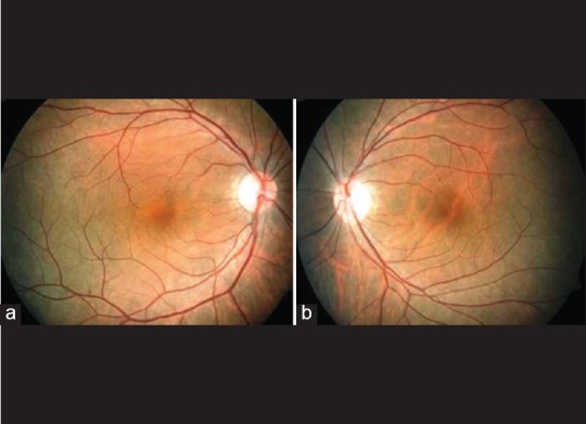
(a) Color fundus picture of the right eye, (b) color fundus picture of the left eye of the same patient with retinal pigment epithelial alterations at fovea after lightening injury
Figure 2.
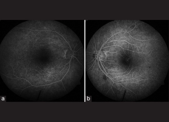
(a) Fundus fluorescein angiogram of the right eye, (b) fundus fluorescein angiography picture of the left eye shows retinal pigment epithelial window defects at macula following lightening injury
Figure 3.
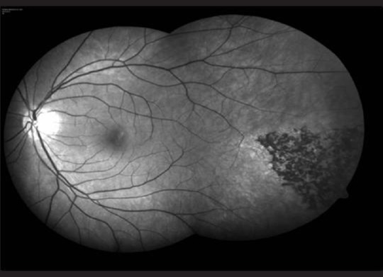
Red-free fundus image of the left eye showing pigment clumps in temporal periphery after lightening injury
Figure 4.

(a) Optical coherence tomography images of macula of the right eye, (b) optical coherence tomography image of the left eye of the same patient with a lamellar macular hole following lightening injury
Discussion
Lightning-induced maculopathy is caused by the heat generated at the level of RPE due to resistance by melanin.[1] It often manifests as cystoid macular edema and macular hole.[2,3]
SD-OCT analysis shows loss of foveal photoreceptors and IS-OS junction disruption.[4] Peripheral pigmentary changes following lightning injury as seen in our patient have also been described in the literature.[4] Visual prognosis in patients with lightning-induced ocular injury depends on the extent of irreversible retinal and macular damage. Therefore, long-term follow-up of these patients is recommended.
Financial support and sponsorship
Nil.
Conflicts of interest
There are no conflicts of interest.
Acknowledgment
We would like to acknowledge photography and OCT services at Sankara Nethralya, Chennai.
References
- 1.Lagrèze WD, Bömer TG, Aiello LP. Lightning-induced ocular injury. Arch Ophthalmol. 1995;113:1076–7. doi: 10.1001/archopht.1995.01100080128041. [DOI] [PubMed] [Google Scholar]
- 2.Handa JT, Jaffe GJ. Lightning maculopathy. A case report. Retina. 1994;14:169–72. [PubMed] [Google Scholar]
- 3.Rao KA, Rao LG, Kamath AN, Jain V. Bilateral macular hole secondary to remote lightning strike. Indian J Ophthalmol. 2009;57:470–2. doi: 10.4103/0301-4738.57156. [DOI] [PMC free article] [PubMed] [Google Scholar]
- 4.Armstrong B, Fecarotta C, Ho AC, Baskin DE. Evolution of severe lightning maculopathy visualized with spectral domain optical coherence tomography. Ophthalmic Surg Lasers Imaging. 2010;41(Suppl):S70–3. doi: 10.3928/15428877-20101031-02. [DOI] [PubMed] [Google Scholar]