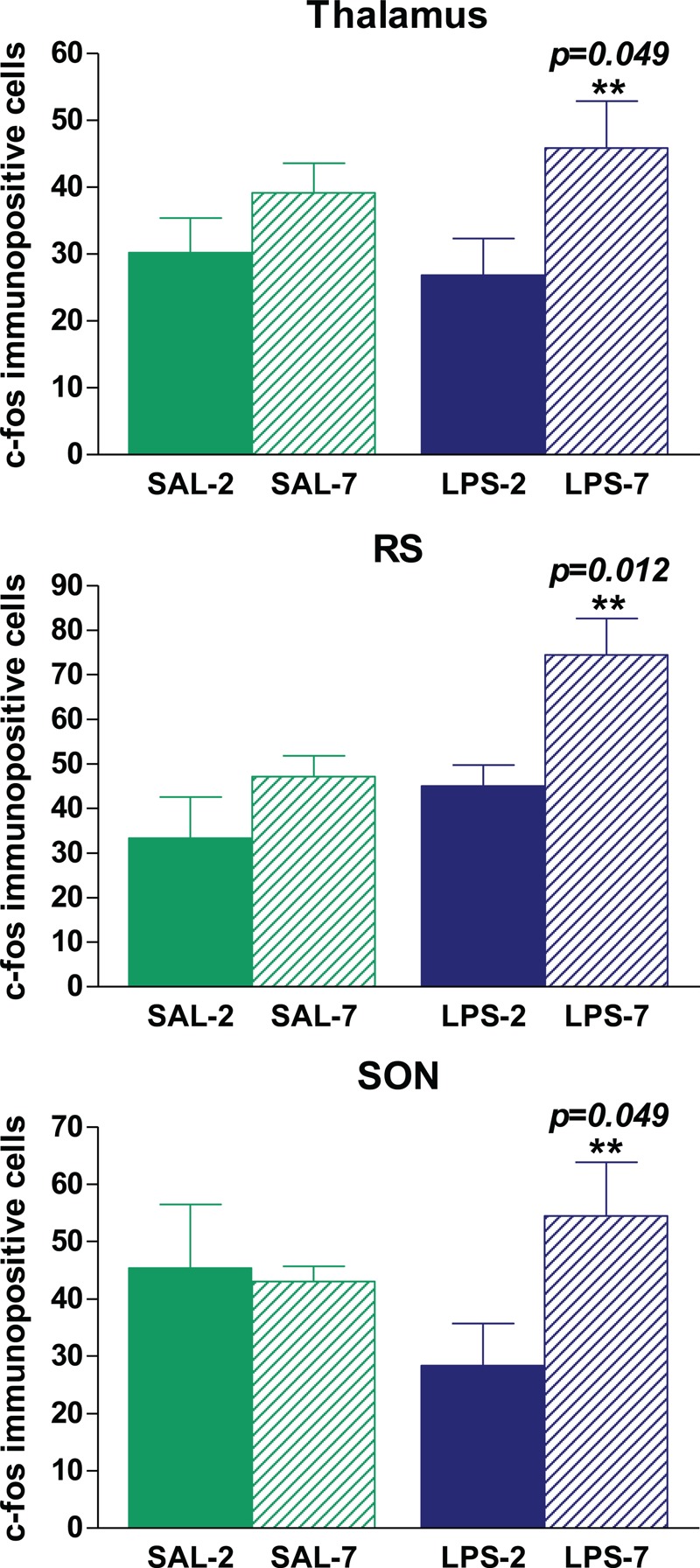
Fig. 6.
Quantification of c-Fos-immunopositive cells per field in different areas of the brain.

No immunopositive cells were found in nonventilated BAS rats. The administration of LPS did not modify the number of immunopositive cells in the thalamus, RS, or SON. LPS-instilled rats receiving 7 cmH2O PEEP had more immunopositive cells in the thalamus, RS, and SON than those receiving 2 cmH2O PEEP (∗∗ P < 0.05). Bars represent means ± SEM.
