Abstract
Objective:
Assess the effectiveness of selected yoga postures in raising bone mineral density (BMD).
Methods:
Ten-year study of 741 Internet-recruited volunteers comparing preyoga BMD changes with postyoga BMD changes.
Outcome Measures:
Dual-energy x-ray absorptiometric scans. Optional radiographs of hips and spine and bone quality study (7 Tesla).
Results:
Bone mineral density improved in spine, hips, and femur of the 227 moderately and fully compliant patients. Monthly gain in BMD was significant in spine (0.0029 g/cm2, P = .005) and femur (0.00022 g/cm2, P = .053), but in 1 cohort, although mean gain in hip BMD was 50%, large individual differences raised the confidence interval and the gain was not significant for total hip (0.000357 g/cm2). No yoga-related serious injuries were imaged or reported. Bone quality appeared qualitatively improved in yoga practitioners.
Conclusion:
Yoga appears to raise BMD in the spine and the femur safely.
Keywords: osteoporosis, yoga
Osteoporosis and osteopenia affect up to 200 000 000 people worldwide today, with numbers likely to grow with our aging population. Many people are without access to medications or professional help after the fractures that are more likely without them. A low-cost, low-risk alternative is desirable.
Annual spinal fractures in the United States exceed 700 000, with more than 300 000 hip fractures. After hip fracture, 25% of Americans will succumb, and another 25% will never leave the nursing institution to which they are admitted following hospitalization.1–4 The United States currently spends an estimated $19 billion on the more than 2 million annual fragility fractures and the 500 000 hospitalizations these entail.1–2
Some have termed hip fracture a “sentinel event,” indicating a general and irreversible decline in many aspects of health, but since the medications themselves are associated with spontaneous fracture, atrial fibrillation, slowed healing, gastric distress, osteonecrosis, and, in the newer intravenous forms, scleritis and episcleritis, some of this general decline actually may be due to pharmaceutical treatment rather than the patient herself or himself.5–7
Yoga classes are a dramatically low-cost and less dangerous alternative to medications and the elaborate health care their absence is alleged to engender. The “side effects” of yoga include better posture, improved balance, enhanced coordination, greater range of motion, higher strength, reduced levels of anxiety, and better gait.8–17 Improved posture directly addresses spinal fractures, while all of these documented benefits of yoga reduce the risk of falling, which is the main cause of all other osteoporotic fractures.8–17 The current study examines the proposition that yoga is a safe and effective means of preventing osteoporosis-related fracture.
Contemporary medicine accounts for bone loss or gain by genetics, nutrition, hormones, medications, and activity. The main preventive measures are based on nutrition, such as vitamin D and calcium supplementation;18–20 medications, such as bisphosphonates and serum estrogen receptor manipulation; and activities such as gym workouts, running, and sports.20
We held other variables as constant as possible to isolate the effect of one activity: yoga, to determine the response of 3 common sites of fracture—the spine, hip, and femur, associated with daily use of a DVD of 12 common yoga poses. We selected the poses specifically for their safety and their calculated effects on those 3 sites, among the most commonly fractured. Qualitative bone quality measurements were performed on the left hips of 18 subjects.
By pitting one group of muscles against another, yoga exposes bones to greater forces and, therefore, might enhance bone mineral density (BMD) more than other means. The advantages of such a program include universal applicability, virtual absence of side effects, and minimal cost.
MATERIALS AND METHODS
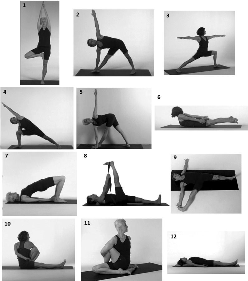
Encouraged by a successful pilot study, 21 and approved by Sound Shore Medical Center Institutional Review Board, currently part of Montefiore Medical Center, we devised a 12-minute DVD of 12 yoga poses that we believed would stimulate increasing BMD in the lumbar vertebrae, the hip, and the femoral neck (see Figure 1). We then announced through the Web site “sciatica.org” and at talks and workshops that we would give the DVD without cost to yoga practitioners and nonpractitioners that satisfied the criteria for study entry.
Figure 1.
Poses of the DVD from top left: (1) Vriksasana—tree, (2) Trikonasana—triangle, (3) Virabhadrasana II—warrior II, (4) Parsvakonasana—side-angle pose, (5) Parivrtta Trikonasana—twisted triangle, (6) Salabhasana—Locust, (7) Setu Bandhasana—bridge, (8) Supta Padangusthasana I—supine hand-to-foot I, (9) Supta Padangusthasana II—supine hand-to-foot II, (10) Marichyasana II—straight-legged twist, (11) Matsyendrasana—bent-knee twist, (12) Savasana—corpse pose.
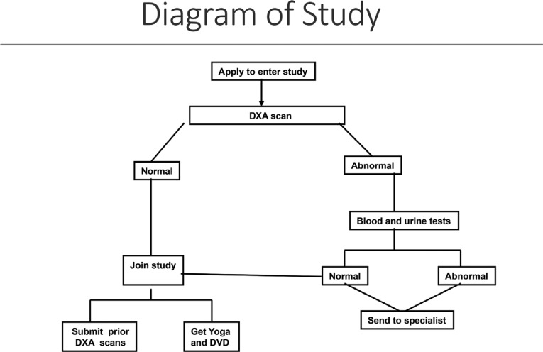
Using the Internet, we collected baseline data, urine, blood chemistries, BMD, and spinal and hip radiographs of volunteers worldwide who intended to perform the 12-minute yoga regimen daily and distributed to them a disk of 12 poses that we had studied in a pilot study21 accompanied by simultaneous verbal descriptions with each pose (see Figure 1 and 2). We began our pilot study in June 2005, employing an online scoresheet for subjects to report their levels of compliance. Subjects received a periodic newsletter and were encouraged to communicate with each other via a bulletin board. Instructional webinars were held in 2006, 2008, 2011, and 2013, with seminars in 2007 and 2012 in Westchester County and Lenox, Massachusetts, stressing the proper way to do the poses. Changes in BMD over 1 or more years leading up to study inclusion were compared with change in BMD 1 or more years following study entry. Bone quality measurements were taken on 18 patients with long-term yoga practices.
Figure 2.
Study flow diagram. DXA indicates dual-energy x-ray absorptiometry
Inclusion criteria:
Dual-energy x-ray absorptiometric (DXA) scan within the 6 months preceding study entry.
- If the current DXA scan was normal, no laboratory tests were required. If the DXA scan contained osteopenic or osteoporotic values on the T-scale for spine, hip, or femur, then normal values on the following tests were necessary within 6 months of joining the study:
- Comprehensive Metabolic Panel (SMA-18)
- Parathyroid hormone
- Thyroid stimulating hormone
- Erythrocyte sedimentation rate
- Vitamin D, 25 hydroxy
- Vitamin D, 1, 25 dihydroxy
- Urine collagen cross-linkage (NTX).
Completion of a questionnaire including information, such as medical and surgical history, medication usage, and diet, that can be seen on the Web site “sciatica.org.”
Agreement to continue the current medication, nutritional and exercise regimen for the following 2 years, and to repeat the DXA scan within or just after that time period. Study subjects were encouraged but not required to submit spinal and hip radiographs obtained within the same time frame, submit previous DXA scans, and further encouraged to enter one of the authors' (GC) bone quality study.
Exclusionary criteria:
Abnormal values on the above tests.
Metabolic or bone diseases such as osteogenesis imperfecta, or Paget disease.
Individuals with abnormal blood tests, for example, with thyroid stimulating hormone (TSH), were referred to appropriate specialists, an endocrinologist in the case of elevated TSH. Once the value of the test was normal, they naturally became eligible for the study (see Figure 1).
The DVD consisted of 3 versions of each pose, the classical pose, an elementary pose that contained most elements of the classical pose but stressed safety and simplicity, and an intermediate or transitional version. Entry-level poses often used a wall for stabilization and a prop such as a bolster, belt, or chair to reduce the challenges and risks of the posture. All entrants were asked to begin by practicing the elementary poses for 1 week and to progress thereafter as far as they safely could from each elementary pose to the intermediate version of the pose and thereafter to the classical pose. Entrants were asked to perform each pose displayed and described in the DVD for 30 seconds daily and to log their yoga activities on a Health Insurance Portability and Accountability Act–compliant electronic scorecard available on the Internet. Subjects without yoga experience were encouraged to have at least 1 private lesson from a yoga teacher or yoga therapist, preferably one knowledgeable in the Iyengar type of yoga.
The primary measure was mean BMD change during the years before the study began versus mean BMD change following 2 years of daily yoga. Spine, right or left hip, and right or left femur (g/cm2) constituted the metrics of choice. Because the timing of the DXA scans varied from patient to patient, all comparisons were in BMD changes per month. Secondary measures were based on compliance and self-reported and radiological measures of injury. Bone quality measures were qualitative. All DXA studies and bone quality studies were blinded in the sense that no radiologist was aware of the status of study subjects.
RESULTS
A total of 741 patients joined the study between 2005 and 2015, of whom 227 were identified as compliant with more than every-other-day yoga. Women comprised 202 of these patients. Mean age at joining the study was 68.2 years. Entry DXA scans showed that 174 (83%) of the compliant patients had osteoporosis or osteopenia.
Dropout rate was lower than that in a previous pilot study,21 in which recruitment was not Internet-based. Many people failed to submit DXA scans from years prior to study entry. Others did not submit subsequent DXA scans; still others offered inadequate information about their in-study yoga practice. Because of the variable information that patients submitted subsequent to their enrollment in the study, data analysis was done in 3 tiers, as a function of the adequacy and relative certainty of the data supplied.
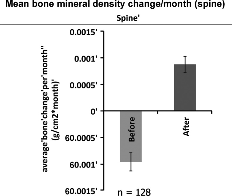
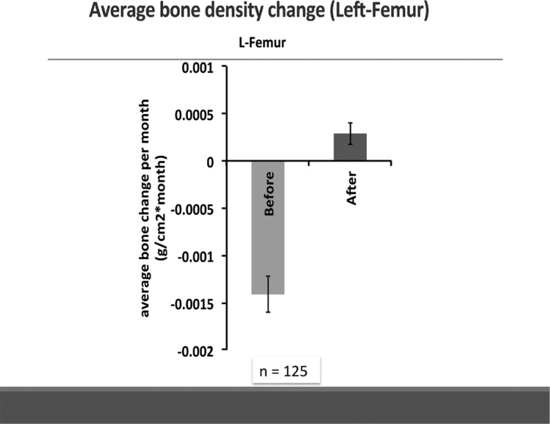
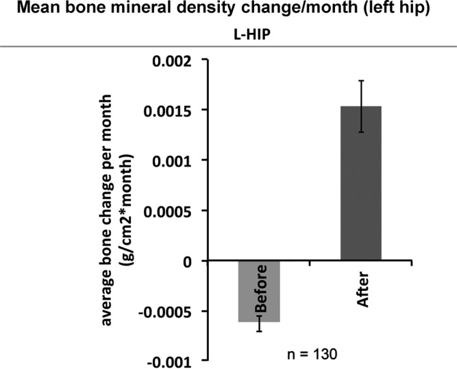
In approximately 4 years preceding study entry, 128, 130, and 125 patients presented prestudy DXA scans that revealed a mean monthly decline in BMD of −0.0036 g/cm2 for the spine, −0.00008 g/cm2 for the hips, and −0.009 for the femora, over a mean 47, 52, and 48 months, respectively; standard deviations/95% confidence intervals = 0.125/0.118, 0.117/0.183, and 0.317/0.192, for spine, hips, and femora, respectively. After practicing the yoga of the DVD over 22, 22, and 24 months, respectively, 72, 81, and 83 of these subjects reported mean gains of 0.048, 0.088, and 0.0003 g/cm2 per month, for spine, hips, and femora; standard deviations/95% confidence intervals = 0.551/0.44, 0.103/0.159, and 0.129/0.133, respectively (see Table 1 and Figures 3–5).
TABLE 1. Most Recent Changes in BMD Before Yoga vs Those Following Two Years of Yogaa.
| Location | Preyogaa | Number | Timingb | SD/CI | Postyoga | Timing | Number | SD |
|---|---|---|---|---|---|---|---|---|
| Spine | −0.036 | 128 | 47 | 0.125/0.118 | 0.048 | 21.7 | 71 | 0.551/0.44 |
| Left hip | −0.017 | 130 | 52 | 0.117/0.183 | 0.088 | 22 | 81 | 0.103/0.159 |
| Left femur | −0.03 | 125 | 48 | 0.317/0.192 | 0.003 | 24 | 83 | 0.129/0.133 |
Abbreviations: BMD, bone mineral density; SD/CI, standard deviation/confidence interval.
aAll pre-yoga values are negative while all post-yoga values are positive.
bLoss of bone mineral density in g/cm2/mo.
cMonths between dual-energy x-ray absorptiometry scans.
Figure 3.
Change (mg/cm2/mo) in spine dual-energy x-ray absorptiometry for all patients.
Figure 5.
Change (mg/cm2/mo) in femur dual-energy x-ray absorptiometry for all patients.
Figure 4.
Change (mg/cm2/mo) in total hip dual-energy x-ray absorptiometry for all patients
Compliant patients were classified as having done more than half the poses at the highest level (high level, grade 3), more than half at the lowest level (low level, grade 1), or neither (moderate level, grade 2). We identified 57 patients of moderate to full compliance (grades 2 or 3).
For lumbar spine L1-L4, 29 participants showed average −0.00080 ± 0.00025 g/cm2 change per month over a mean 35 ± 7 months before yoga practice and showed significant improvement with 0.0010 ± 0.00043 g/cm2 change per month over a mean 21 ± 1 month after yoga practice (P = .002). For total hip, 25 (16 from left hip, 5 from right hip, 2 bilateral) measurements showed mean 0.000082 ± 0.00038 g/cm2 change per month over a mean 38 ± 8 months before and mean 0.00012 ± 0.00031 g/cm2 change per month over 23 ± 2 months after yoga practice, which was nearly a 50% improvement but not significant. For femoral neck, 18 measurements were analyzed (13 from left side, 3 from right side, and both right and left of 1 participant). Bone mineral density fell a mean −0.00090 ± 0.00027 g/cm2 change per month over average 46 ± 12 months before yoga and showed significant improvement with mean 0.00030 ± 0.00048 g/cm2 change per month over average 24 ± 2 months after yoga (P = .05) (see Table 2).
TABLE 2. Mean Monthly Change Before Yoga vs. After Two Years of Yoga in Moderately and Completely Compliant Patients.
| Location | Before Yoga or After Yoga | Number | Time Span in Months/Confidence Interval | Change Per Month/Confidence Interval | P |
|---|---|---|---|---|---|
| Spine | Before | 29 | 35 ± 7 | −0.00080/0.00025 | |
| After | 21 ± 1 | 0.0010/0.000043 | .002 | ||
| Total hip | Before | 25 | 38 ± 8 | 0.000082/0.00038 | |
| After | 23 ± 2 | 0.00012/0.00031 | ... | ||
| Femur | Before | 18 | 46 ± 12 | −0.00090/0.00027 | |
| After | 24 ± 2 | 0.00030/0.00048 | .05 |
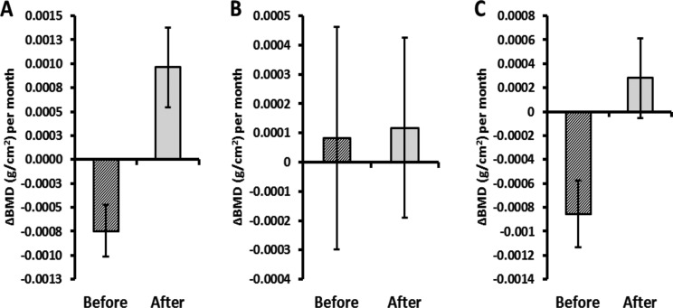
Selecting only those individuals with complete pre-and poststudy entry data (who sent in the actual DXA reports rather than just sending us the numerical results) and moderate to full compliance, we found that 43 patients—spine (n = 27), hip (n = 16), and femur (n = 14), within a span of 11 to 59, 23 to 59, and 11 to 59 months, respectively—showed monthly BMD gains of 0.001 (±0.0007), 0.0003 (±0.0007), and 0.0009 (±0.0003), g/cm2, respectively. The P values for these changes are .02, .01, and .0002, respectively (see Table 3 and Figure 6).
TABLE 3. Improvements in Fully Documented Patients Who Followed the Daily Regimen for Two Years.
| Location | Number | Range of Time Between DXA Scans in Months | Difference in Change Per Month (Before Yoga—After Yoga)/Confidence Interval | P |
|---|---|---|---|---|
| Lumbar spine | 27 | 11-59 | 0.001/0.0007 | .02 |
| Total hip | 16 | 23-59 | 0.0003/0.0007 | .01 |
| Femur | 14 | 11-59 | 0.0009/0.0003 | .0002 |
Abbreviation: DXA, dual-energy x-ray absorptiometry.
Figure 6.
Change (mg/cm2/mo) in spine dual-energy x-ray absorptiometry for all fully documented patients: A: Spine; B: Hip; and C: Femur.
In all, 109 fractures were reported on prestudy radiographs and in the induction forms, and 19 in subjects postentry. As of this writing, with more than 90 000 hours of yoga practiced largely by people with osteoporosis or osteopenia, there have been no reported or X-ray detected fractures or serious injuries of any kind related to the practice of yoga in any of the 741 participants. Bone quality of the 18 patients completing the study was judged as superior to those without yoga experience.
CONCLUSION
The 12 yoga poses studied here appear to be a safe and effective means to reverse bone loss in the spine and the femur and have weaker indications of positive effects on the total hip measurement of the DXA scan. There is qualitative evidence suggesting improved bone quality as a result of the practice of yoga.
DISCUSSION
Globally, there are 200 000 000 people with osteopenia or osteoporosis.1–2,4 A safe, effective, and stunningly low-cost means of reversing this condition would be welcome for the many people who have neither the means to acquire the recommended medications nor access to the treatment of the fractures that may occur without them.
Mean rate of improvement in total hip BMD increased from baseline by 50% after a mean 23 ± 2 months of yoga, but wide variation in individual improvement caused the confidence intervals to overlap, thus precluding statistical significance. The root cause of this increased variation is possibly due to the wide differences in yoga skills within the study population that was more important in the more difficult twisting, extension, and standing poses in which the hips bear the most pressure. There are also several different formulae used for calculating total hip BMD which might add to the variability in this number.
Some studies suggest that BMD is not as closely associated with fracture as falling itself.8 By improving posture, balance, range of motion, strength, and coordination, decreasing anxiety and improving gait,8–17,19–21 yoga opposes falls in ways no medicine can provide. Although some medications do reduce anxiety, they also impair balance.
However, this study suffers from many drawbacks.
Recruitment
People signing up for the study were largely those with already weakened or weakening bones and, very often, people already doing some yoga. Therefore, the study may have selected the very people for whom yoga does not give maximum benefit. The bone-building effects of yoga in younger and in healthier people, without the genetic predisposition that is possibly overrepresented in our sample, might well be greater and at any rate are unknown.
Study focus
Yoga poses were selected specifically to produce torque and bending of the proximal femur, compression of the pelvis, and twisting of the lumbar vertebral bodies. The choice was determined because these are the most common sites of osteoporotic fractures and the anatomical regions measured by the DXA scan. However, osteoporotic fractures frequently occur in the thoracic spine, the forearm, and the ribs. These sites were not studied and might not respond to yoga directed toward them in the same way.
Data
Some subjects used the electronic scorecard incorrectly and/or inadequately, limiting the data on actual compliance. This vitiated the calculation of dose-response curves enough to preclude their utility in this study.
Bone quality
The qualitative measure of bone quality can be supplemented and improved with finite element analysis, which is yet to be performed on this sample.
Study design
This was perforce a crossover study since none of the subjects were doing yoga for osteoporosis before joining the study. However, many were doing yoga for other reasons, reducing the calculated effect of the 12 poses studied here.
SUMMARY
The current study supports the efficacy and safety of yoga as a treatment of osteopenia and osteoporosis. However, its use in prevention, on a wider selection of possible fracture sites, with more precise data for dose-response relationships, improved bone quality measures and with standard randomized, controlled, double-blinded inquiries, requires further investigation. Many of these goals could be accomplished with a younger osteopenia-free population. What is suggested by this study's results is that yoga can reverse bone loss that has reached the stages of osteopenia and osteoporosis.
Footnotes
Dr Fishman funded the study.
The authors declare no conflicts of interest.
References
- 1.National Osteoporosis Foundation. org http://nof.org/live/treating. Accessed May 17, 2015.
- 2.www.ncbi.nlm.nih.gov/books/NBK45502/. Accessed November 26, 2014. http://www.aaos.org/news/aaosnow/jan12/advocacy6.asp. Accessed November 26, 2014.
- 3.Leland NE, Gozalo P, Bynum J, Mor V, Christian TJ, Teno JM. What happens to patients when they fracture their hip during a skilled nursing facility stay? J Am Med Dir Assoc. 2015;16(9):767–774. [DOI] [PMC free article] [PubMed] [Google Scholar]
- 4.National Osteoporosis Foundation. http://www.nof.org/osteoporosis/facts.htm. Accessed July 14, 2006.
- 5.Sorensen HT, Christensen S, Mehnert FF, et al. Use of bisphosphonates among women and risk of atrial fibrillation and flutter: population based case-control study. BMJ. 2008;336:813–826. [DOI] [PMC free article] [PubMed] [Google Scholar]
- 6.MacLean C, Newberry S, Maglione M, et al. Systematic review: comparative effectiveness of treatments to prevent fractures in men and women with low bone density or osteoporosis. Ann Intern Med. 2008;148:197–213. [DOI] [PubMed] [Google Scholar]
- 7.Reid IR. Short-term and long-term effects of osteoporosis therapies [published online ahead of print May 12, 2015]. Nat Rev Endocrinol. 2015; 11(7):418–428. 10.1038/nrendo.2015.71. [DOI] [PubMed] [Google Scholar]
- 8.Järvinen TLN, Sievänen H, Khan KM, Heinonen A, Kannus P. Shifting the focus in fracture prevention from osteoporosis to falls. BMJ. 2008;336:124. [DOI] [PMC free article] [PubMed] [Google Scholar]
- 9.Smith EN, Boser A. Yoga, vertebral fractures and osteoporosis: research and recommendations. Int J Yoga Therap. 2013;23(1):17–23. [PubMed] [Google Scholar]
- 10.Prado ET, Raso V, Scharlach RC, Kasse CA. Hatha yoga on body balance. Int J Yoga. 2014;7(2):133–137. [DOI] [PMC free article] [PubMed] [Google Scholar]
- 11.Ni M, Mooney K, Balachandran A, Richards L, Harriell K, Signorile JF. Muscle utilization patterns vary by skill levels of the practitioners across specific yoga poses (asanas). Complement Ther Med. 2014;22(4):662–669. [DOI] [PubMed] [Google Scholar]
- 12.Ni M, Mooney K, Harriell K, Balachandran A, Signorile J. Core muscle function during specific yoga poses. Complement Ther Med. 2014;22(2):235–243. [DOI] [PubMed] [Google Scholar]
- 13.Telles S, Hanumanthaiah BH, Nagarathna R, Nagendra HR. Plasticity of motor control systems demonstrated by yoga training. Indian J Physiol Pharmacol. 1994;38(2):143–144. [PubMed] [Google Scholar]
- 14.Salem GJ, Yu SS, Wang MY, et al. Physical demand profiles of hatha yoga postures performed by older adults. Evid Based Complement Alternat Med. 2013;2013:165763. [DOI] [PMC free article] [PubMed] [Google Scholar]
- 15.Payne P, Crane-Godreau MA. Meditative movement for depression and anxiety. Front Psychiatry. 2013;4:71. 10.3389/fpsyt.2013.00071. eCollection 2013. [DOI] [PMC free article] [PubMed] [Google Scholar]
- 16.DiBenedetto M, Innes KE, Taylor AG, et al. Effect of a gentle Iyengar yoga program on gait in the elderly: an exploratory study. Arch Phys Med Rehabil. 2005;86:1830–1837. [DOI] [PubMed] [Google Scholar]
- 17.Bruno AG, Anderson DE, D'Agostino J, Bouxsein ML. The effect of thoracic kyphosis and sagittal plane alignment on vertebral compressive loading. J Bone Miner Res. 2012;27(10):2144–2151. [DOI] [PMC free article] [PubMed] [Google Scholar]
- 18.Ward E. Addressing nutritional gaps with multivitamin and mineral supplements. Nutr J. 2014;13:72. [DOI] [PMC free article] [PubMed] [Google Scholar]
- 19.Beck TJ, Fuerst T, Gaither KW, et al. The effects of bazedoxifene on bone structural strength evaluated by hip structure analysis. Bone. 2015;77:115–119. [DOI] [PubMed] [Google Scholar]
- 20.Heidari B, Hosseini R, Javadian Y, Bijani A, Sateri MH, Nouroddini HG. Factors affecting bone mineral density in postmenopausal women [published online ahead of print May 14, 2015]. Arch Osteoporos. 2015;10(1):217. 10.1007/s11657-015-0217-4. [DOI] [PubMed] [Google Scholar]
- 21.Fishman LM. Yoga for osteoporosis—a pilot study. Topics Geriatr Rehabil. 2009;25(3):244–250. [Google Scholar]


