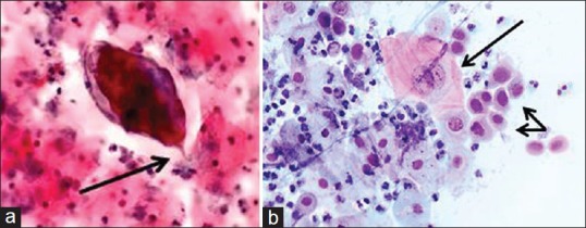
Figure 1.

(a) Cervical smear, (Papanicolaou stain, ×40). Schistosoma haematobium ovum with an embryo miracidium. Note the diagnostic terminal spine(arrow). (b) Cervical smear, (Papanicolaou stain, ×40). Cytological changes consistent with high grade squamous intraepithelial lesion including human papillomavirus infection. Long arrow: Human papillomavirus cytopathic changes including nuclear and cellular enlargement, and koilocytosis. Double arrow: Cells with increased nuclear to cytoplasmic ratios, nuclear membrane irregularities and marked hyperchromasia consistent with high-grade squamous intraepithelial lesion. The background contains neutrophil granulocytes denoting inflammation
