Abstract
Background
Pulmonary veins (PV) and the atria undergo electrical and structural remodeling in atrial fibrillation (AF). This study aimed to determine PV and left atrial (LA) reverse remodeling after catheter ablation for AF assessed by chest computed tomography (CT).
Methods
PV electrophysiologic studies and catheter ablation were performed in 63 patients (68% male; mean ± SD age: 56 ± 10 years) with symptomatic AF (49% paroxysmal, 51% persistent). Chest CT was performed before and 3 months after catheter ablation.
Results
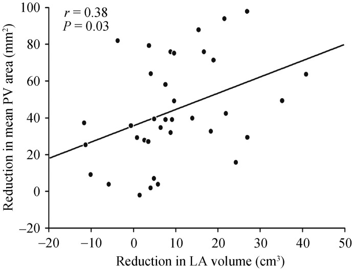
At baseline, patients with persistent AF had a greater LA volume (91 ± 29 cm3 vs. 66 ± 27 cm3; P = 0.003) and mean PV ostial area (241 ± 43 mm2 vs. 212 ± 47 mm2; P = 0.03) than patients with paroxysmal AF. There was no significant correlation between the effective refractory period and the area of the left superior PV ostium. At 3 months of follow-up after ablation, 48 patients (76%) were AF free on or off antiarrhythmic drugs. There was a significant reduction in LA volume (77 ± 31 cm3 to 70 ± 28 cm3; P < 0.001) and mean PV ostial area (224 ± 48 mm2 to 182 ± 43 mm2; P < 0.001). Patients with persistent AF had more reduction in LA volume (11.8 ± 12.8 cm3 vs. 4.0 ± 11.2 cm3; P = 0.04) and PV ostial area (62 mm2 vs. 34 mm2; P = 0.04) than those who have paroxysmal AF. The reduction of the averaged PV ostial area was significantly correlated with the reduction of LA volume (r = 0.38, P = 0.03).
Conclusions
Catheter ablation of AF improves structural remodeling of PV ostia and left atrium. This finding is more apparent in patients with persistent AF treated by catheter ablation.
Keywords: Ablation, Atrial fibrillation, Computed tomography, Left atrium, Pulmonary vein isolation, Pulmonary vein ostial area
1. Introduction
Catheter ablation to interrupt the conduction from the left atrium to the pulmonary vein (PV) to treat atrial fibrillation (AF) has emerged as an important intervening therapy for restoration of sinus rhythm in patients with symptomatic AF since Haïssaguerre, et al.[1],[2] and others found that PV musculature is a potential arrhythmogenic source initiating AF.[3],[4] Atrial electrical and structural remodeling subsequent to ablation for AF have been well recognized.[5]–[8] Numerous investigators have reported the benefit of PV isolation on reversal of atrial remodeling, including reduced left atrial (LA) chamber size and improved atrial contractility after restoration of sinus rhythm.[9]–[14] However, despite the important role of PVs in the pathophysiology of AF, the PV remodeling is poorly understood. Computed tomography (CT) is considered a reliable imaging modality for assessing the PV ostia and LA anatomy.[15],[16] In the present study, we aimed to characterize the PV anatomic remodeling in AF ablation and reverse remodeling after ablative therapy using CT.
2. Methods
2.1. Study patients
Consecutive AF patients who were referred to the Mayo Clinic for catheter-based PV isolation were screened, and 63 subjects [43 (68%) male; mean ± SD age, 56 ± 10 years] who were willing to participate in this prospective study were enrolled. The inclusion criteria were symptomatic paroxysmal or persistent AF (duration < 1 year) refractory to at least one antiarrhythmic drug. Patients with a left ventricular (LV) ejection fraction of less than 50%, structural heart disease, cardiomyopathy, valvular heart disease, congenital heart disease, or long last persistent AF of more than 1 year's duration were excluded. The study was approved by the Institutional Review Board, and all patients provided informed consent.
2.2. Clinical evaluation
All patients underwent clinical evaluation before the ablation, including a detailed history and physical examination, 12-lead ECG, and a 24-h Holter study to assess the presence, frequency, and duration of AF. Transthoracic and transesophageal echocardiography were performed to assess LV function, LA size and volume, PV anatomy, and presence of intracardiac thrombi.
2.3. Computed tomography
All patients underwent chest CT to determine LA and PV anatomy before and 3 months after catheter ablation. Non-ionic intravenous contrast material (150 mL Omnipaque 350) was administered through an antecubital vein using a power injector at a rate of 4 mL/s. CT images were taken using 64 detector scanners (120 kV, 400 to 850 mA, 0.3 to 0.5 s per rotation, slice thickness of 1.5 mm and reconstructed slice thickness of 0.6 mm). The images were analyzed with visualization and analysis software for noninvasive diagnostic evaluation of multimodality imaging data (Vitrea 2; Vital Images, Inc., Minneapolis, Minnesota) using the 3-dimensional maximum intensity projection mode. Multiplanar LA views were projected in the sagittal, coronal, and axial planes. The left atrium was first centered using three orthogonal views. Then, the LA dimensions were recorded as anterior to posterior in the axial plane (AP); superior to inferior, from the roof of the left atrium to the mitral annulus in the sagittal plane (SI); and medial to lateral, from the inferior limbus of the fossa ovalis to the lateral mitral annulus in the coronal plane (ML). The LA volume was calculated using the biplane dimension-length formula: LA volume = 4/3 π × (anteroposterior diameter/2) × (longitudinal diameter/2) × (transversal diameter/2).[17]
Pulmonary vein ostial plane was defined by the intersection of tangents extending from the surface of the pulmonary vein and the wall of the adjacent left atrium, as previously described.[18] Pulmonary vein ostial area was measured using commercially available software (Volume Viewer, GE Healthcare, Milwaukee, WI, USA). Multiplanar reformat images were used to select a plane perpendicular through each pulmonary vein ostia. The wall of the pulmonary vein ostium was manually traced and the area automatically calculated. All measurements were made by a single observer.
2.4. Electrophysiology study and AF ablation procedure
Antiarrhythmic drugs were discontinued at least five half-lives prior to the ablation procedure (amiodarone was discontinued at least 1 month before ablation). Catheters were introduced percutaneously through the left and right femoral veins and the right jugular vein and positioned in the high right atrium, the right ventricle, and the coronary sinus. Dual transseptal punctures were performed to deliver ablation and circular PV mapping catheters. Heparin was administered to maintain an activated clotting time of 300 to 400 s during the entire time of LA mapping and ablation.
The effective refractory period (ERP) of the right atrium, left atrium, and proximal and distal left superior PV ostia was measured by using programmed single atrial extrastimulus. The ERP was defined as the longest S1-S2 interval that failed to induce tissue response. Electrical impulses of 2.0-ms duration at twice the pacing threshold were delivered at driving cycle lengths (S1) of 600 ms, with premature (S2) stimulation in 10-ms decrements. A 3.5-mm open, saline-irrigated ablation catheter (ThermoCool; Biosense Webster Inc, Diamond Bar, California) was placed at the left superior PV proximal and distal ostium for ERP measurement. If patient was in AF at the time of procedure, sinus rhythm was restored by direct-current cardioversion.
Three-dimensional mapping of the left atrium and the PV ostia was performed using a CARTO navigation system (Biosense Webster Inc). Ablation was accomplished by creating a wide-area circumferential ring of lesions that were 5 to 15 mm away from the venoatrial junctions of the left and right PVs. The radiofrequency energy was delivered at 25 to 40 W. A “drag and burn” technique was used to deliver the radiofrequency energy and to create linear lesions. Additional (“touch-up”) ablation of residual PV potentials was undertaken to achieve an end point of complete entrance block from the left atrium to each PV. Additional ablation of LA linear lesions and non-PV arrhythmogenic foci was performed at the operator's discretion.
2.5. Follow-up
All patients were seen at the outpatient clinic for follow-up at 3 months after the ablation procedure. Rhythm status was determined using the patient's history, ECG, and 24-h Holter recording. Chest CT scanning was repeated using the same preablation protocol to reevaluate LA and PV anatomy. Thereafter, patients were asked to complete a survey about clinical symptoms and recurrent AF. Follow-up data were also obtained by direct telephone interview of patients. Repeat Holter or event monitoring was provided to document arrhythmia when symptoms recurred. Because early recurrences of AF after catheter ablation may be transient, a blanking period of 2 months after the ablation procedure was applied.
2.6. Statistical analysis
Statistical analysis was performed using SPSS 11.5. Continuous variables, presented as mean ± SD, were compared with either the Student t-test or a paired t test. Univariate analysis was performed to identify variables that may impact on LA size and PV ostial area change after ablation. A χ2 test was used to compare discrete variables. Correlations between continuous variables were examined with Pearson correlation. A P-value less than 0.05 was considered statistically significant.
3. Results
3.1. Baseline patient characteristics
Patient baseline characteristics are shown in the Table 1. Paroxysmal AF was present in 31 patients (49%), and 32 patients (51%) had persistent AF. Presence of hypertension was common, occurring in 36 patients (57%).
Table 1. Baseline patient characteristics (n = 63).
| Characteristic | Value |
| Age, yrs | 56 ± 10 |
| Male | 43 (68) |
| Body mass index, kg/m2 | 31.4 ± 6.2 |
| Type of atrial fibrillation | |
| Paroxysmal | 31 (49) |
| Persistent | 32 (51) |
| Duration of AF, yrs | 6.2 ± 5.7 |
| Coronary heart disease | 9 (14) |
| Hypertension | 36 (57) |
| Medications | |
| β-blocker | 42 (67) |
| ACE inhibitor/ARB | 14 (22) |
| Calcium channel blocker | 20 (32) |
| Antiarrhythmic drugs used | 1.46 ± 0.95 |
Data are expressed as n (%) or mean ± SD. ACE: angiotensin-converting enzyme; AF: atrial fibrillation; ARB: angiotensin II receptor blocker.
3.2. LA volume and PV ostial area
At baseline, the mean LA volume of the whole study group was 77 ± 31 cm3 by CT Patients with persistent AF had a greater LA volume than patients with paroxysmal AF (91 ± 29 cm3 vs. 66 ± 27 cm3; P = 0.003). Patients with AF recurrence had a larger LA volume before ablation than patients who remained in sinus rhythm (105 ± 35 cm3 vs. 69 ± 26 cm3; P = 0.002).
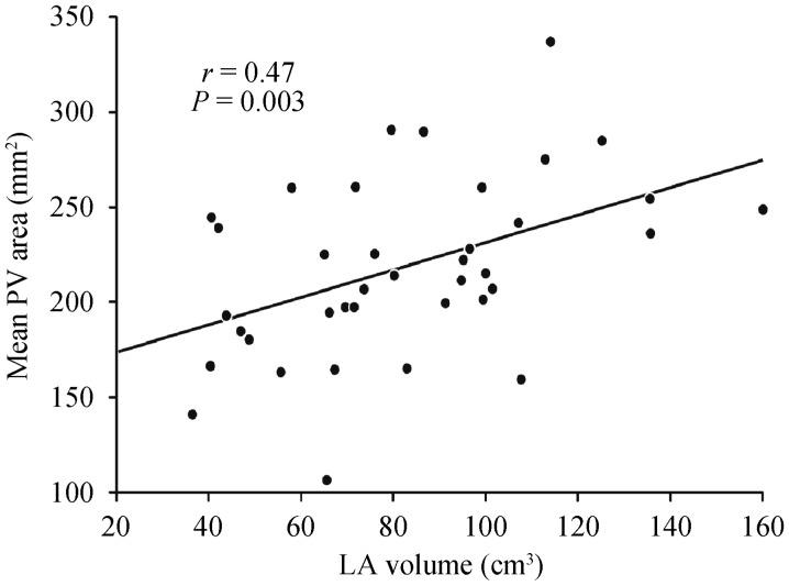
Forty-eight patients had baseline and repeat chest CT satisfied for imaging analysis at 3 months follow-up. A total of 185 PV areas before and after ablation were compared: 41 of left superior PVs (LSPVs); 41 of left inferior PVs (LIPVs); 48 of right superior PVs (RSPVs); 48 of right inferior PVs (RIPVs); and seven of the left common antrum. A significant difference was seen among the four PV ostial areas (P < 0.001), in descending order: RSPV (263 ± 89 mm2), LSPV (244 ± 94 mm2), RIPV (242 ± 84 mm2), and LIPV (168 ± 50 mm2). At baseline, patients with persistent AF had a greater mean PV ostial area than those with paroxysmal AF (241 ± 43 mm2 vs. 212 ± 47 mm2; P = 0.03). At baseline, the mean PV ostial area was significantly correlated with the LA volume (r = 0.47, P = 0.003; Figure 1).
Figure 1. Correlation of LA volume and mean PV area.
LA: left atrial; PV: pulmonary vein.
3.3. Electrophysiology findings
At a pacing cycle length of 600 ms, the proximal LSPVs (217.3 ± 37.4 ms) and distal LSPVs (205.1 ± 40.8 ms) had significantly shorter ERPs than the right atria (270.5 ± 47.0 ms; P < 0.001) and left atria (248.7 ± 36.7 ms; P < 0.001). No significant difference was seen in the proximal (210 ± 37 ms vs. 233 ± 37 ms; P = 0.21) and distal (196 ± 37 ms vs. 215 ± 43 ms; P = 0.09) ERPs between persistent AF and paroxysmal AF patients. There was no significant correlation between the ERP of LSPV and the area of the LSPV ostium (P = 0.77, proximal; P = 0.97, distal).
3.4. Changes in LA and PV size after AF ablation
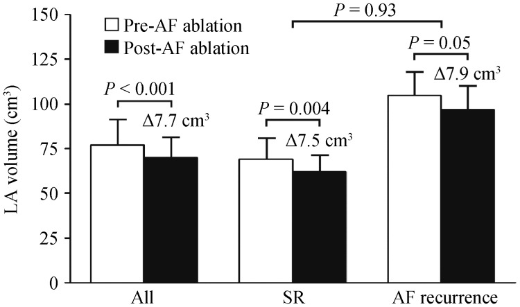
After AF ablation, there was a significant decrease in LA volume, on average from 77 ± 31 cm3 to 70 ± 28 cm3 (P < 0.001). The reduction of LA volume was seen in patients with paroxysmal AF (65 ± 27 cm3 to 61 ± 21 cm3; P = 0.05) and those with persistent AF (93 ± 30 cm3 to 82 ± 32 cm3; P = 0.001). The extent of reduction in LA volume was greater in patients with persistent AF than that in those with paroxysmal AF (11.8 ± 12.8 cm3 vs. 4.0 ± 11.2 cm3; P = 0.04). At 3-month follow-up, 48 patients (76%) were AF free on or off antiarrhythmic drugs. The reduction in LA volume was seen regardless of whether they remained in sinus rhythm (from 69 ± 26 cm3 to 62 ± 20 cm3; P = 0.004) or had AF recurrence (from 105 ± 35 cm3 to 97 ± 36 cm3; P = 0.05). The extent of reduction in LA volume was not significantly different between patients who remained in sinus rhythm and those who had AF recurrence (7.5 ± 13.8 cm3 vs. 7.9 ± 10.9cm3; P = 0.93) (Figure 2).
Figure 2. LA volume before and after ablation of AF.
Delta (Δ) represents the mean difference before and after ablation. The error bars indicate LA volume + standard deviation. AF: atrial fibrillation; LA: left atrial; PV: pulmonary vein; SR: sinus rhythm.
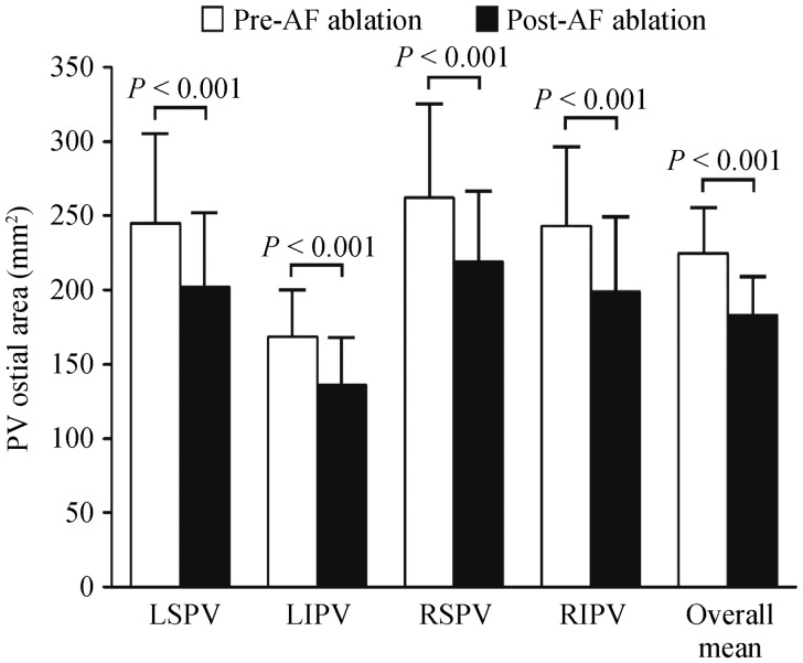
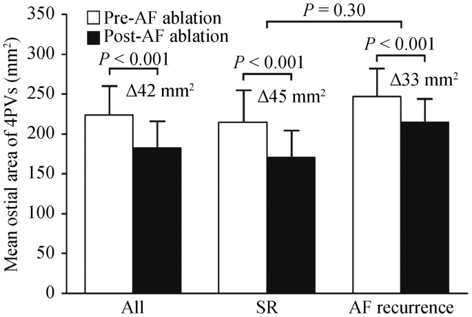
After AF ablation, the ostial area was reduced in all 4 PVs (LSPV, 14%, P < 0.001; LIPV, 19%, P < 0.001; RSPV, 14%, P < 0.001; RIPV, 16%, P < 0.001) (Figure 3). The average PV ostial area was reduced from 224 ± 48 mm2 to 182.2 ± 43 mm2 (P < 0.001). None of the study patients had more than 50% reduction in any of the four PV areas. The reduction in the mean area of the four PVs was observed in patients either remaining in sinus rhythm (from 215 ± 46 mm2 to 169.2 ± 39 mm2; P < 0.001) or having AF recurrences (from 247 ± 47 mm2 to 214.2 ± 38 mm2; P < 0.001), although patients with AF recurrence had a larger baseline mean PV area than those without AF recurrence (P = 0.02). The mean change in the ostial area of all the four PVs in patients without AF recurrence was not significantly different from the change in those with AF recurrence (45 mm2 vs. 33 mm2; P = 0.30) (Figure 4). However, the PV area reduction after ablation in patients with persistent AF was significantly greater than that in patients with paroxysmal AF (62 mm2 vs. 34 mm2; P = 0.04). The reduction of the mean PV ostial area was significantly correlated with the reduction of LA volume (r = 0.38, P = 0.03) (Figure 5).
Figure 3. PV ostial areas are shown before and after ablation of AF.
The error bars indicate PV ostial area + standard deviation. AF: atrial fibrillation; LIPV: indicates left inferior pulmonary vein; LSPV: left superior pulmonary vein; PV: pulmonary vein; RIPV: right inferior pulmonary vein; RSPV: right superior pulmonary vein.
Figure 4. Mean ostial area for all the four PVs before and after ablation of AF.
AF: atrial fibrillation; LA: left atrial; PV: pulmonary vein; SR: sinus rhythm.
Figure 5. Correlation in reduction of LA volume and mean PV area.
LA: left atrial; PV: pulmonary vein.
4. Discussion
4.1. Main findings
The study presented here had four main findings. (1) At baseline, the proximal and distal pulmonary veins had significantly shorter ERPs than those in the right atrium and the left atrium. There was no correlation between the ERP and geometric area of the pulmonary vein ostia. (2) Patients with persistent AF had greater LA volume and PV ostial areas than patients with paroxysmal AF. (3) After ablation, the reduction of LA volume and PV ostial areas was observed in both patients with sinus rhythm and with AF recurrence. (4) The reduction in PV ostial area was greater in those with persistent AF than in patients with paroxysmal AF, suggesting a greater structural reverse remodeling in this group.
4.2. ERP and geometry of PV
Since Haïssaguerre, et al.[1] demonstrated that the PV musculature is a potential arrhythmogenic source initiating AF, catheter ablation to eliminate AF has rapidly become an effective alternative therapy for patients with symptomatic AF. In this study, we observed that proximal PV and distal PV had significantly shorter ERPs than the right atrium and left atrium. This finding is consistent with the results of others.[19]–[21] Jais, et al.[21] reported that the ERP of the PV was shorter than the ERP of the left atrium in patients with paroxysmal AF but longer in the controls. We did not observe a difference in ERP of PV ostia between the patients with paroxysmal AF and persistent AF, which agrees with a recent report by Teh, et al.[19] The PV ostial size is individualized in each patient; usually, the superior veins are larger than the inferior veins, and the right veins are larger than the left veins.[6] The greater size of the left atrium and PV ostia seen more often in persistent AF than in paroxysmal AF conveys the evidence of concurrent atrial and PV ostial eccentric remodeling, more apparent in persistent AF. Our study showed no correlation between the ERP and the area of the PV, suggesting that the extent of PV structural and electrical remodeling may not coincide.
4.3. Changes in LA size after AF ablation
Beneficial reduction of the LA dimension or volume after catheter ablation for AF has been well documented.[22], [23] Reduced or eliminated AF burden, could lead to the reduction of LA volume after catheter ablation.[24],[25] In our study, the reduction in LA volume was seen regardless of whether patients remained in sinus rhythm or had AF recurrence, suggesting that a reduced AF burden may occur in recurrent AF group after ablation. It is well known that LA size is a risk factor for AF recurrence, and patients with persistent AF had greater LA volume than patients with paroxysmal AF. After AF ablation, structural reverse remodeling was more prevalent in persistent AF patients in the present study. These patients may be subject to structural remodeling to a greater extent.
4.4. Changes in PV area after PV isolation
Clinical information describing PV reverse remodeling after catheter ablation is limited. In our study, the PV ostial area was reduced after ablation regardless of whether patients were in sinus rhythm or had recurrent AF. A 14% to 20% reduction in PV ostial area could be explained by mild PV ostial narrowing caused by ablation-induced scarring. Alternatively, restoration of sinus rhythm has improved PV ostial dilation. The baseline PV ostia area was correlated with the LA volume shown in our study, suggesting that in AF, the PV structure remodeling was accompanied by LA enlargement. The reduction of PV area that was moderately correlated with the reduction of LA volume suggests that the reduction of PV ostia area represents a concomitant reverse remodeling of PV ostial geometry parallel to the reduction of LA volume.
4.5. Study limitations
The present study has a few limitations. It is well known that the success rate of catheter ablation of AF is dependent on the screening protocol for AF recurrences. Asymptomatic AF recurrences, which were not recorded by ECG or Holter monitoring, may underestimate AF recurrence. The moderate sample size of study patients may have limited the power of our results.
4.6. Conclusions
Catheter ablation of AF resulted in significant reverse structural remodeling, reducing the PV ostial area and LA volume. These findings occur concomitantly and are more apparent in patients with persistent AF treated with ablation.
References
- 1.Haïssaguerre M, Jais P, Shah DC, et al. Spontaneous initiation of atrial fibrillation by ectopic beats originating in the pulmonary veins. N Engl J Med. 1998;339:659–666. doi: 10.1056/NEJM199809033391003. [DOI] [PubMed] [Google Scholar]
- 2.Haïssaguerre M, Shah DC, Jaïs P, et al. Electrophysiological breakthroughs from the left atrium to the pulmonary veins. Circulation. 2000;102:2463–2465. doi: 10.1161/01.cir.102.20.2463. [DOI] [PubMed] [Google Scholar]
- 3.Chen SA, Hsieh MH, Tai CT, et al. Initiation of atrial fibrillation by ectopic beats originating from the pulmonary veins: electrophysiological characteristics, pharmacological responses, and effects of radiofrequency ablation. Circulation. 1999;100:1879–1886. doi: 10.1161/01.cir.100.18.1879. [DOI] [PubMed] [Google Scholar]
- 4.Packer DL, Asirvatham S, Munger TM. Progress in nonpharmacologic therapy of atrial fibrillation. J Cardiovasc Electrophysiol. 2003;14:S296–S309. doi: 10.1046/j.1540-8167.2003.90403.x. [DOI] [PubMed] [Google Scholar]
- 5.Pappone C, Oreto G, Rosanio S Vicedomini G, et al. Atrial electroanatomic remodeling after circumferential radiofrequency pulmonary vein ablation: efficacy of an anatomic approach in a large cohort of patients with atrial fibrillation. Circulation. 2001;104:2539–2544. doi: 10.1161/hc4601.098517. [DOI] [PubMed] [Google Scholar]
- 6.Tsao HM, Yu WC, Cheng HC, et al. Pulmonary vein dilation in patients with atrial fibrillation: detection by magnetic resonance imaging. J Cardiovasc Electrophysiol. 2001;12:809–813. doi: 10.1046/j.1540-8167.2001.00809.x. [DOI] [PubMed] [Google Scholar]
- 7.Kato R, Lickfett L, Meininger G, et al. Pulmonary vein anatomy in patients undergoing catheter ablation of atrial fibrillation: lessons learned by use of magnetic resonance imaging. Circulation. 2003;107:2004–2010. doi: 10.1161/01.CIR.0000061951.81767.4E. [DOI] [PubMed] [Google Scholar]
- 8.Tsao HM, Wu MH, Huang BH, et al. Morphologic remodeling of pulmonary veins and left atrium after catheter ablation of atrial fibrillation: insight from long-term follow-up of three-dimensional magnetic resonance imaging. J Cardiovasc Electrophysiol. 2005;16:7–12. doi: 10.1046/j.1540-8167.2005.04407.x. [DOI] [PubMed] [Google Scholar]
- 9.Lemola K, Sneider M, Desjardins B, et al. Effects of left atrial ablation of atrial fibrillation on size of the left atrium and pulmonary veins. Heart Rhythm. 2004;1:576–581. doi: 10.1016/j.hrthm.2004.07.020. [DOI] [PubMed] [Google Scholar]
- 10.Atwater BD, Wallace TW, Kim HW, et al. Pulmonary vein contraction before and after radiofrequency ablation for atrial fibrillation. J Cardiovasc Electrophysiol. 2011;22:169–174. doi: 10.1111/j.1540-8167.2010.01868.x. [DOI] [PubMed] [Google Scholar]
- 11.Marsan NA, Tops LF, Holman ER, et al. Comparison of left atrial volumes and function by real-time three-dimensional echocardiography in patients having catheter ablation for atrial fibrillation with persistence of sinus rhythm versus recurrent atrial fibrillation three months later. Am J Cardiol. 2008;102:847–853. doi: 10.1016/j.amjcard.2008.05.048. [DOI] [PubMed] [Google Scholar]
- 12.Jayam VK, Dong J, Vasamreddy CR, et al. Atrial volume reduction following catheter ablation of atrial fibrillation and relation to reduction in pulmonary vein size: an evaluation using magnetic resonance angiography. J Interv Card Electrophysiol. 2005;13:107–114. doi: 10.1007/s10840-005-0215-3. [DOI] [PubMed] [Google Scholar]
- 13.Delgado V, Vidal B, Sitges M, et al. Fate of left atrial function as determined by real-time three-dimensional echocardiography study after radiofrequency catheter ablation for the treatment of atrial fibrillation. Am J Cardiol. 2008;101:1285–1290. doi: 10.1016/j.amjcard.2007.12.028. [DOI] [PubMed] [Google Scholar]
- 14.Muller H, Noble S, Keller PF, et al. Biatrial anatomic reverse remodelling after radiofrequency catheter ablation for atrial fibrillation: evidence from real-time three-dimensional echocardiography. Europace. 2008;10:1073–1078. doi: 10.1093/europace/eun187. [DOI] [PubMed] [Google Scholar]
- 15.Hof I, Rbab-Zadeh A, Scherr D, et al. Correlation of left atrial diameter by echocardiography and left atrial volume by computed tomography. J Cardiovasc Electrophysiol. 2009;20:159–163. doi: 10.1111/j.1540-8167.2008.01310.x. [DOI] [PubMed] [Google Scholar]
- 16.Jongbloed MR, Bax JJ, Lamb HJ, et al. Multislice computed tomography versus intracardiac echocardiography to evaluate the pulmonary vein before radiofrequency catheter ablation of atrial fibrillation: a head to head comparison. J Am Coll Cardiol. 2005;45:343–350. doi: 10.1016/j.jacc.2004.10.040. [DOI] [PubMed] [Google Scholar]
- 17.Lang RM, Bierig M, Devereux RB, et al. Recommendations for chamber quantification: a report from the American Society of Echocardiography's Guidelines and Standards Committee and the Chamber Quantification Writing Group, developed in conjunction with the European Association of Echocardiography, a branch of the European Society of Cardiology. J Am Soc Echocardiogr. 2005;18:1440–1463. doi: 10.1016/j.echo.2005.10.005. [DOI] [PubMed] [Google Scholar]
- 18.Scharf C, Sneider M, Case I, et al. Anatomy of the pulmonary veins in patients with atrial fibrillation and effects of segmental ostial ablation analyzed computed tomography. J Cardiovasc Electrophysiol. 2003;14:150–155. doi: 10.1046/j.1540-8167.2003.02444.x. [DOI] [PubMed] [Google Scholar]
- 19.Teh AW, Kistler PM, Lee G, et al. Electroanatomic properties of the pulmonary veins: slowed conduction, low voltage and altered refractoriness in AF patients. J Cardiovasc Electrophysiol. 2011;22:1083–1091. doi: 10.1111/j.1540-8167.2011.02089.x. [DOI] [PubMed] [Google Scholar]
- 20.Rostock T, Steven D, Lutomsky B, et al. Atrial fibrillation begets atrial fibrillation in the pulmonary veins on the impact of atrial fibrillation on the electrophysiological properties of the pulmonary veins in humans. J Am Coll Cardiol. 2008;51:2153–2160. doi: 10.1016/j.jacc.2008.02.059. [DOI] [PubMed] [Google Scholar]
- 21.Jais P, Hocini M, Macle L, et al. Distinctive electrophysiological properties of pulmonary veins in patients with atrial fibrillation. Circulation. 2002;106:2479–2485. doi: 10.1161/01.cir.0000036744.39782.9f. [DOI] [PubMed] [Google Scholar]
- 22.Lemola K, Sneider M, Desjardins B, et al. Effects of left atrial ablation of atrial fibrillation on size of the left atrium and pulmonary veins. Heart Rhythm. 2004;1:576–581. doi: 10.1016/j.hrthm.2004.07.020. [DOI] [PubMed] [Google Scholar]
- 23.Marsan NA, Tops LF, Holman ER, et al. Comparison of left atrial volumes and function by real-time three-dimensional echocardiography in patients having catheter ablation for atrial fibrillation with persistence of sinus rhythm versus recurrent atrial fibrillation three months later. Am J Cardiol. 2008;102:847–853. doi: 10.1016/j.amjcard.2008.05.048. [DOI] [PubMed] [Google Scholar]
- 24.Hof IE, Velthuis BK, Chaldoupi SM, et al. Pulmonary vein antrum isolation leads to a significant decrease of left atrial size. Europace. 2011;13:371–375. doi: 10.1093/europace/euq464. [DOI] [PubMed] [Google Scholar]
- 25.McGann CJ, Kholmovski EG, Oakes RS, et al. New magnetic resonance imaging-based method for defining the extent of left atrial wall jury after ablation of atrial fibrillation. J Am Coll Cardiol. 2008;52:1263–1271. doi: 10.1016/j.jacc.2008.05.062. [DOI] [PubMed] [Google Scholar]


