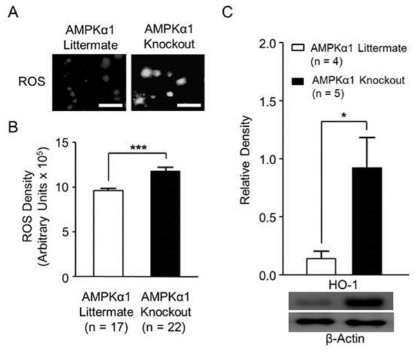
Figure 3. AMPKα1 deletion resulted in increased levels of ROS and HO-1 protein expression in the spinal dorsal horn.
(A) shows representative images of ROS intensity in the spinal dorsal horn of AMPKα1 knockout and AMPKα1 littermate mice. Scale bar = 40 µm. Bar graphs (B) show summaries (mean + S.E.) of ROS density in the spinal dorsal horn obtained from AMPKα1 knockout and AMPKα1 littermate mice. The mean (+ S.E.) relative density of HO-1 protein expressions to β-actin in the spinal dorsal horn, obtained from AMPKα1 knockout and AMPKα1 littermate mice, are shown in (C). Samples of Western blot gels of HO-1 are shown below. *, p < 0.05; ***, p < 0.001.

