Abstract
The plant hormone auxin is perceived by a family of F-box proteins called the TIR1/AFBs. Phylogenetic studies reveal that these proteins fall into four clades in flowering plants called TIR1, AFB2, AFB4, and AFB6. Genetic studies indicate that members of the TIR1 and AFB2 groups act as positive regulators of auxin signaling by promoting the degradation of the Aux/IAA transcriptional repressors. In this report, we demonstrate that both AFB4 and AFB5 also function as auxin receptors based on in vitro assays. We also provide genetic evidence that AFB4 and AFB5 are targets of the picloram family of auxinic herbicides in addition to indole-3-acetic acid. In contrast to previous studies we find that null afb4 alleles do not exhibit obvious defects in seedling morphology or auxin hypersensitivity. We conclude that AFB4 and AFB5 act in a similar fashion to other members of the family but exhibit a distinct auxin specificity.
Keywords: Arabidopsis, F-box protein, auxin
The plant hormone auxin is a small indolic molecule with an important role in virtually every aspect of plant growth and development from embryogenesis to senescence (Woodward and Bartel 2005). Auxin regulates transcription by promoting the degradation of a family of transcriptional repressors called the Aux/IAA proteins (Hagen 2015; Salehin et al. 2015). These proteins repress transcription by binding to transcription factors called AUXIN RESPONSE FACTORs (ARFs), and recruiting the corepressor protein TOPLESS to the chromatin. In the presence of auxin, the AUXIN/INDOLE-3-ACETIC ACID (Aux/IAA) proteins are degraded through the action of a ubiquitin protein ligase (E3) called SCFTIR1. This results in activation of complex transcriptional networks that lead to context-dependent changes in cell growth and behavior.
The SCFs are a subgroup of a large family of E3 ligases called Cullin Ring Ligases (CRL) conserved in all eukaryotes (Pickart 2001; Petroski and Deshaies 2005). SCFs consist of CULLIN1, S-phase kinase associated protein 1 (SKP1, ARABIDOPSIS SKP1 HOMOLOUGE, or ASK in plants), the RING-BOX1 (RBX1) protein, and one of a family of substrate adaptor proteins called F-box proteins (Pickart 2001; Petroski and Deshaies 2005). The F-box protein recruits substrates to the SCF and promotes ubiquitination, typically resulting in degradation by the proteasome. Several years ago, we discovered that SCFTIR1 and the related SCFAFBs function as auxin sensors (Dharmasiri et al. 2005; Kepinski and Leyser 2005; Tan et al. 2007). The TRANSPORT INHIBITOR RESPONSE1/AUXIN F-BOX (TIR1/AFB) proteins consist of the F-box domain and a Leucine Rich Repeats (LRRs) domain. Auxin binds directly to the LRR domain, but rather than causing a conformational change, typical for most hormone receptors, auxin promotes the interaction between SCFTIR1 and the Aux/IAA substrate.
There are six members of the TIR1/AFB group of F-box proteins in Arabidopsis. TIR1 and AFB1 through AFB3 as well as AFB5 have been shown to function as auxin receptors (Dharmasiri et al. 2005; Calderon Villalobos et al. 2012). The loss of a single member of TIR1 through AFB3 has a slight effect on auxin response and plant growth, but higher order combinations of these genes have a much more severe phenotype (Dharmasiri et al. 2005). Of these four proteins TIR1 and AFB2 appear to have major roles in seedling development, while AFB3 has a less significant role. The loss of AFB1 has a very minor effect in the seedling (Dharmasiri et al. 2005). This appears to be due to the fact that AFB1 does not assemble into an SCF complex efficiently (Yu et al. 2015). In this study we focus on the AFB4 and AFB5 genes. We describe the characterization of two new AFB4 mutants called afb4-8 and afb4-9. Both of these mutations appear to be null alleles, but neither has an obvious effect on growth of the seedling. We confirm that both AFB4 and AFB5 function as auxin receptors. In addition, we show that the afb4 and afb5 mutants are resistant to the synthetic auxin picloram indicating that these two proteins are selective for picloram.
Materials and Methods
Plant material and growth conditions and treatments
Arabidopsis thaliana mutants and transgenic lines used in this study were all in the Columbia (Col-0) ecotype. The Salk T-DNA insertion lines afb4-8 (Salk_201329) and afb4-9 (Salk_083223) were identified in the Salk-seq data (http://signal.salk.edu/cgi-bin/tdnaexpress). The afb4-9 line originally contained four additional T-DNA insertions. A previously described At5g27570/cdc20.5 insertion (Kevei et al. 2011) and an insertion in the At1g11340 gene were removed by backcrossing, but two intergenic insertions near genes At3g09720 (535 bp upstream of AT3g09720 and 219 bp upstream of AT3g09730) and At4g22160 (immediately after the stop codon) remained present in the afb4-9 and afb4-9 afb5-5 lines used in this study. The afb5-5 (Salk_110643) was obtained from the Arabidopsis Biological Resource Center at Ohio State University. The plant T-DNA junction sequences were determined for each insertion. The afb4-8 insertion is associated with a 20-bp deletion, while those of afb4-9 and afb5-5 are associated with 10-bp and 32-bp deletions, respectively. Seeds were surface sterilized either by vapor-phase sterilization (Clough and Bent 1998) or by treating for 2 min in 70% (v/v) ethanol followed by 10 min in 30% commercial bleach. Seeds were plated on medium containing 1/2 × Murashige and Skoog (MS) media, 1% sucrose, 0.8% agar, and stratified for 2−4 d at 4°.
Growth assays
All root assays were completed under long-day photoperiods (16:8) and hypocotyl assays were performed under short-day photoperiods (8:16). For auxin-inhibited root growth assays, 5-day-old seedlings were transferred onto fresh MS media ± auxin for 3 additional days after which root length was measured. Hypocotyl assays were performed similarly except the seedlings were transferred at day 4 for a 2-day treatment.
Pathogen infection assays
To assay the afb4-8 and afb5-5 mutants for altered disease responses, the mutants were grown on soil and inoculated at 4 wk of age with the bacterial pathogen Pseudomonas syringae pv. tomato strain DC3000. Bacteria were grown on NYG agar media with 100 μg/ml rifampicin at 30°. Inoculation was performed by syringe infiltration, as described previously (Mutka et al. 2013).
Generation of transgenic lines
The TIR1-Myc line was described previously (Gray et al. 1999). The pAFB5:AFB4- and pAFB5:AFB5-4×Myc lines (AFB4-Myc and AFB5-Myc) were generated using a 2-kb 5′ upstream region of the AFB5 gene with the AFB4 and AFB5 cDNA in binary vector pGW16. The AFB5 promoter was used for expressing AFB4 due to the low activity of the AFB4 promoter. The pAFB5:AFB5-mCitrine (AFB5-mCitrine) construct contained the entire genomic region between adjacent genes, from 1267 bp upstream of the start codon to 1139 bp downstream from the stop codon in the pMP535 binary vector (Prigge et al. 2005). The stop codon was mutated to a NheI site in order to insert a 27-bp linker and the mCitrine coding region. Each construct was transformed into the afb5-5 mutant background. The pAFB4:AFB4-2×Venus-3×HA (AFB4-Venus) construct contained a genomic fragment from -1235 (relative to ATG) to just before the stop codon fused in frame with a tag encoding two copies of Venus fluorescent protein and three copies of the hemagglutinin epitope in the pMLBART vector (Gleave 1992). After transformation into the afb4-2 background, the transgene was moved to an afb4-8 background by crossing. Roots of the mCitrine and Venus lines were observed using a Zeiss LSM 710 confocal microscope after staining with propidium iodide.
Protein expression and pulldown experiments
For pulldown assays, GST-IAA3 and GST-IAA7 were recombinantly expressed in Escherichia coli strains BL21 (DE3) (Figure 1B, Figure 4, or BL21-AI (Figure 6) and purified using Glutathione-Agarose (Sigma-Aldrich, G4510). For in vivo pulldown experiments, seedlings expressing Myc-tagged AFB4, AFB5, and TIR1 were grown for 8 d in liquid MS medium. TIR1-Myc expression was induced by treatment with 30 μM dexamethasone for 24 hr. The ASK1-antibody was generated as previously described (Gray et al. 1999). For the various auxin comparisons (Figure 4B) seedlings were incubated for 2 hr in 50 μM of the compounds or an equivalent volume of dimethyl sulfoxide (DMSO) prior to harvest. For all other in vivo pulldown experiments samples were incubated with auxin for 45 min following harvest. Tissue was harvested by grinding to a powder in liquid nitrogen and vortexed vigorously in extraction buffer [50 mM Tris pH 7.5, 150 mM NaCl, 10% glycerol, 0.1% NP-40, complete protease inhibitor (Roche), 50 μM MG-132]. Cellular debris was removed by centrifugation and total protein concentration was determined by Bradford assay. Each pulldown reaction included 1 mg total protein extract and equal volumes of GST-IAA protein for each sample in a 500-μl total volume. The pulldown reactions were incubated at 4° for 45 min with rocking and transferred to a Micro Bio-Spin Chromatography Column (Bio-Rad). Samples were washed three times in 1 ml extraction buffer without protease inhibitors or MG-132 in the presence or absence of auxin. Samples were eluted using reduced glutathione (Sigma) and separated on SDS-PAGE and stained with Ponceau [0.1% (w/v) Ponceau S in 5%(v/v) acetic acid] for loading control unless otherwise indicated. For Figure 4B, equivalent amounts were run on a separate SDS-PAGE gel and stained with Coomassie stain. AFB/TIR1-Myc proteins were detected by immunoblotting with anti-c-Myc-Peroxidase antibody (Roche). Proteins were visualized using the ECL Plus Western Blotting Detection System (Amersham).
Figure 1.
AFB4 and AFB5 are auxin receptors. (A−B) Pulldown experiments were carried out using crude plant extracts prepared from [tir1-1] GVG>>TIR1-Myc, [afb5-5] pAFB5:AFB5-Myc, and [afb5-5] pAFB5:AFB4-Myc seedlings and recombinant GST-IAA3. (A) TIR1-Myc, AFB4-Myc, and AFB5-Myc were immunoprecipitated with the anti-Myc antibody coupled to agarose beads, and ASK1 was detected with an anti-ASK1 antibody. (B) GST-IAA3 was immunoprecipitated with glutathione agarose beads, and AFB4-Myc and AFB5-Myc protein were detected with the anti-c-Myc-Peroxidase antibody. Pulldown reactions were incubated for 45 min in the presence or absence of 50 μM IAA.
Figure 4.
The AFB4 and AFB5 proteins respond to picloram. (A and B) Pulldown reactions were carried out as in Figure 1 with 50 μM of the indicated auxin. GST-IAA7 loaded was visualized by Coomassie staining. Pulldown experiments were repeated three times with similar results.
Figure 6.
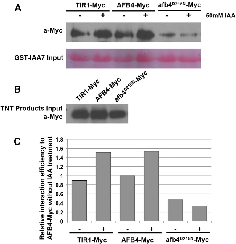
The afb4D215N protein has reduced affinity for IAA7. Equivalent amounts of in vitro translated Myc-tagged TIR1, AFB4, or afb4D215N proteins were incubated with GST-IAA7 protein attached to glutathione-agarose beads in the presence or absence of 50 µM IAA. After washing and elution from the agarose beads, the proteins were separated by polyacrylamide gel electrophoresis and blotted to nitrocellulose membranes. (A) The Myc-tagged receptor proteins were immunodetected using anti-c-Myc antibody and the GST-IAA7 input was visualized by Ponceau S staining. (B) The relative amounts of Myc-tagged proteins added. (C) The quantification of blot band density as presented in (A) by ImageJ. All the values were normalized to AFB4-Myc without IAA treatment. The experiment was repeated four times with similar results.
For the in vitro pulldown experiment, expression plasmids were made by adding the AFB4 and afb4D215N cDNA sequences to a pTNT vector (Promega) with a Gateway:4×Myc cassette via Gateway recombination (Invitrogen). AFB4-4×Myc, afb4D215N-4×Myc, and TIR1-Myc were produced from TNT T7 coupled wheat germ extract system (Promega, L4140). Comparable amounts of AFB4-Myc, afb4D215N-Myc, and TIR1-Myc were applied to each pulldown reaction as guided by western blot using anti-c-Myc-Peroxidase antibody (Roche, 11814150001). The pulldown assay was performed as described in Yu et al. (2013). TNT products and GST-IAA7 beads were incubated with or without the addition of 50 μM IAA. The eluted products were detected and visualized as with the in vivo pulldowns.
RNA extraction and quantitative PCR
Hypocotyl, cotyledon, and root tissue frozen in liquid N2 and ground using a mortar and pestle was used for RNA purification using the Invitrogen PureLink RNA minikit. RNA from whole 10-day-old seedlings (Figure 2C) was similarly ground and purified using RNeasy Plant Mini kit (Qiagen). RNA yield was quantified using the Thermo Scientific NanoDrop 2000. For quantitative RT-PCR, 1 μg RNA, pretreated with DNase using the DNA-free Kit (Ambion) according to manufacturer’s instructions, was used for generating cDNA with SuperScript IV (Figure 2) or SuperScript III (Figure 7A) Reverse Transcriptase (Invitrogen) and 20-mer oligo(dT) primers. Quantitative RT-PCR was performed using SyBR green and the primers listed in Table 1. Primer pairs were evaluated for specificity and efficiency using three serial dilutions of cDNA using the CFX96 Real-Time PCR Detection System (Biorad). Most data were normalized to the reference primer pair PP2AA3-S (Czechowski et al. 2005) according to the ΔΔCt method. Primer pairs AFB4-3 and AFB5-2 were normalized to the reference primer pair PP2AA3-L. All new primers were designed using QuantPrime (Arvidsson et al. 2008). Two biological replicates were performed, each replicate containing 95 to 100 mg whole seedlings (Figure 2C) or roughly 700 individual seedlings that were dissected into cotyledon, hypocotyl, and root samples (Figure 7A).
Figure 2.
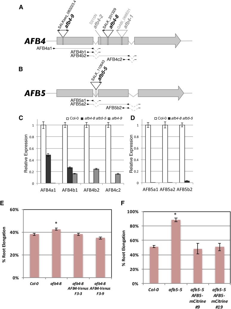
afb4-8, afb4-9, and afb5-5 mutants do not produce full-length transcripts. (A−B) Diagrams of the AFB4 and AFB5 genes. The positions of mutant lesions are shown above the genes with arrowheads indicating T-DNA left border sequences. Below the gene diagrams are the primers pairs used for qRT-PCR. Kinked dashed lines indicate spliced introns. (C−D) qRT-PCR of AFB4 and AFB5 transcripts in WT and mutants grown under LD conditions. Results from each AFB4 and AFB5 primer pairs were normalized relative to those to the PP2AA3 gene. AFB4a1, AFB4b2, and AFB5a2 were normalized to the longer PP2AA3-L amplicon (448 bp) while the rest used PP2AA3-S (59 bp). Error bars represent standard error. (E) Five-day-old seedlings were transferred to media with or without 8 µM picloram and grown for 4 more days before measuring. F3-3 and F3-9 are two independent F3 populations. n = 54, 56, 48, and 39, respectively. Error bars represent standard error. *P < 0.05 with Col and both transgenic lines. (F) Five-day-old seedlings for Col-0, afb5-5, and two afb5-5 pAFB5:AFB5-mCitrine lines (T2 generation) were transferred to media with or without 10 µM picloram and grown for 4 more days before measuring. The pAFB5:AFB5-mCitrine seedlings were then tested for sensitivity to basta herbicide; measurements from sensitive seedlings were excluded. Results are presented as the percent of the DMSO control treatment for each genotype. n = 12, 12, 5, and 5 for Col-0, afb5-5, line 9, and line 19, respectively. Error bars represent standard error. *P < 0.05 vs. Col-0 and both transgenic lines.
Figure 7.
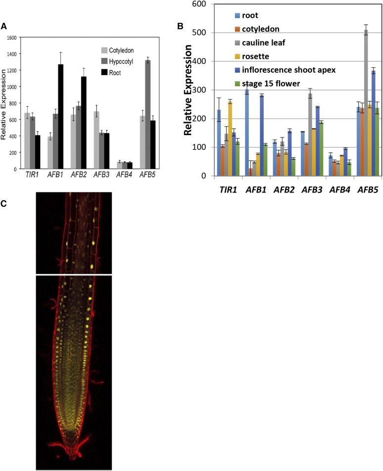
Expression of the AFB4 and AFB5 genes. (A) qRT-PCR of TIR1/AFB genes in 4-day-old WT seedling tissues grown under SD conditions. Primer pairs are listed in Table 1 with AFB4-4 and AFB5-4 being used for those respective genes. Expression is normalized to PP2AA3 using the PP2AA3-S primer pair. Error bars represent standard error. (B) TIR1/AFB expression levels in various tissues. Replotted from Winter et al. (2007). (C) mCitrine fluorescence (yellow) was visualized in roots of the AFB5-mCitrine line #9 using confocal microscopy. Cells were stained with propidium iodide (red).
Table 1. Primers used for quantitative RT-PCR.
| Target | Locus | Primer Sequence (5′ to 3′) |
|---|---|---|
| TIR1 | AT3G62980 | ATCGCTGCCACTTGCAGGAATC |
| TGGCCACTAACGTCGTCAACATC | ||
| AFB1 | AT4G03190 | GCTACTGTCCGAATGCCTGATCTTG |
| GCCTTGTTCCGTCAGAGGTATGTTG | ||
| AFB2 | AT3G26810 | GCCGCTAATTGCAGGCATCTTC |
| AGTCGTGCAAGTGTCTGGGAAAC | ||
| AFB3 | AT1G12820 | AGGTTGAAGCGGATGGTTGTAACAG |
| GCAAGTCCAGCTCACGAAGATGC | ||
| AFB4-a | AT4G24390 | CCAAGACCAGCTCCTTTTTCACCT |
| AFB4-1 | CAAGGACCTTTAGCTGcCTGCATT | |
| AFB4-b | TTGGTCTGCTGTGAAGGTTTTGG | |
| AFB4-2 | TCGAGTCAAGAgCCCAGAAGACTC | |
| AFB4-c | TGCTCAAGCCCATCATAAGCAAC | |
| AFB5-a | AT5G49980 | TCTTGGTTTGTTGTGAAGGTTTTGGT |
| AFB5-1 | AATCAAGCACTTTCAGCTTTcTGCAC | |
| AFB5-2 | GAATCAAGGGCcCAGAACACCT | |
| AFB5-b | AGCCCATCATACTCAATTGCCACA | |
| AFB5-c | TGCCAACAAGTGCAgAAAGCTG | |
| AFB5-3 | TCCACTTCATCATCCGTGACCTC | |
| PP2AA3-S | AT1G13320 | GTGGCCAAAATGATGCAATCTCTC |
| PP2AA3-L | AACTTGCTGAAGACAGGCACTGGA | |
| PP2AA3-R | ATGTTCTCCACAACCGCTTGGTC |
Data availability
Results and Discussion
A phylogenetic analysis revealed that the AFB4/AFB5 clade diverged from the TIR1/AFB1-3 clade ∼300−400 million yr ago whereas the AFB2/AFB3 clade diverged from TIR1/AFB1 ∼200 million yr ago (Parry et al. 2009). Genetic and biochemical studies have demonstrated that members of the TIR1 and AFB2 clades regulate auxin response but differ in their relative contributions to seedling development (Parry et al. 2009). However, the phylogenetically distinct AFB4 group comprised of AFB4 (At4g24390) and AFB5 (At5g49980) in Arabidopsis has not been characterized in as much detail. Since the corresponding genes have been retained in nearly every seed plant genome sequenced to date, it is likely that they have evolved distinct functions. To explore this possibility we performed a series of experiments focusing on the role of AFB4 and AFB5 during seedling development.
The AFB4 and AFB5 proteins are auxin receptors
Our first objective was to determine if AFB4 and AFB5 are subunits of SCF complexes. Transgenic lines expressing Myc-tagged versions of AFB4 and AFB5 under the control of the AFB5 promoter were generated for coimmunoprecipitation experiments. AFB4-Myc and AFB5-Myc were immunoprecipitated from plant extracts with the anti-c-Myc antibody coupled to agarose beads. After washing, the samples were resolved by SDS-PAGE, blotted, and probed with antibodies to the Arabidopsis SKP1-related protein ASK1 (Gray et al. 1999). A line expressing TIR1-Myc was included for comparison (Gray et al. 1999). Consistent with their similarity to the TIR1 and AFB1-3 proteins both AFB4 and AFB5 interact with ASK1 and presumably form an SCF complex (Figure 1A).
To determine whether AFB4 and AFB5 also exhibit the characteristics of auxin receptors, we performed pulldown experiments with the Aux/IAA protein IAA3. Equivalent amounts of total protein extract from AFB4-Myc and AFB5-Myc plants were incubated with GST-IAA3 bound beads in the presence or absence of 50 μM IAA. Both AFB4 and AFB5 interact with IAA3 in an auxin-dependent manner demonstrating that these proteins function as auxin receptors (Figure 1B).
AFB4 and AFB5 are the major targets of the picolinate class of auxinic herbicides
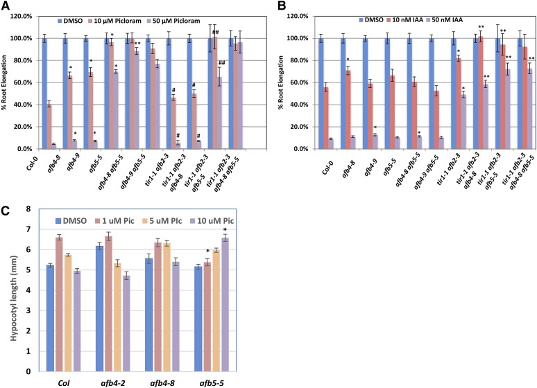
The synthetic auxin picloram (4-amino-3,5,6-trichloropicolinic acid) has been well studied for its auxinic herbicidal properties on a variety of plant species (Hamaker et al. 1963; Scott and Morris 1970; Chang and Foy 1983). To identify genes required for herbicide response, Walsh and colleagues screened EMS-mutagenized Arabidopsis seedlings to identify mutants that were specifically resistant to picolinate auxins (Walsh et al. 2006). One of the genes identified in this screen was AFB5. Further characterization revealed that the afb5 mutants were highly resistant to picloram but sensitive to 2,4-D (2,4-dichlorophenoxyacetic acid), a synthetic auxin from the aryloxyacetate class (Walsh et al. 2006). In addition, we recently showed that AFB5-Aux/IAA coreceptors selectively bind picloram (Calderon Villalobos et al. 2012). To further explore this specificity, we obtained a T-DNA insertion allele of AFB5 referred to as afb5-5. This allele has an insertion in intron 1 that results in the loss of full-length AFB5 mRNA (Figure 2, B and D). In addition, we identified two afb4 mutants with insertions in exon 2 (afb4-8) and exon 1 (afb4-9) (Figure 2A). Quantitative RT-PCR analysis shows that the afb4-8 does not produce transcript downstream of the insertion site while transcripts from afb4-9 plants do not include the first exon (Figure 2C). Thus both alleles are likely to be null mutants. The root growth response of these mutants to picloram was determined and compared to Col-0 and the tir1-1 afb2-3 double mutant. Consistent with Walsh et al. (2006), afb5-5 seedlings were strongly resistant to picloram-mediated root growth inhibition (Figure 3A). The afb4-8 and afb4-9 were slightly picloram-resistant while tir1-1 afb2-3 displayed very slight resistance compared to Col-0. We also tested both double mutant combinations and found that afb4-8 afb5-5 was slightly more resistant than afb5-5 alone. In addition, we tested the response of the afb4 and afb5 mutants to the naturally occurring auxin IAA (Figure 3B). In contrast to the tir1-1 afb2-3 mutant, afb4-8, afb5-5, and the double mutant did not display significant resistance to IAA.
Figure 3.
The afb4 and afb5 mutants are preferentially resistant to picloram. Five-day-old WT and mutant seedlings were transferred to media containing either picloram (A) or IAA (B) and grown another 4 d. Growth is presented as the percent of the DMSO control treatment for each genotype. Error bars represent standard error. (A) *P < 0.05 vs. Col-0, **P < 0.05 vs. afb5-5. # does not exhibit a significant difference with Col-0, ## does not exhibit a significant difference with afb5-5. (B) *P < 0.05 vs. Col-0, **P < 0.05 vs. tir1-1 afb2-3 Student’s t-test. (C) Four-day-old SD-grown seedlings were transferred to media containing 1 µM, 5 µM, or 10 µM picloram or the equivalent amount of DMSO and grown for 2 more days before measuring hypocotyl lengths. Error bars represent standard error. *P< 0.05 vs. Col-0 at the same concentration.
To confirm that these phenotypes are due to the T-DNA insertions we introduced pAFB4:AFB4-VENUS and pAFB5:AFB5-mCitrine constructs into afb4 and afb5 mutants respectively. The results shown in Figure 2, E and F show that the wild-type transgenes restore picloram sensitivity.
We also examined the effect of picloram on hypocotyl elongation. Seedlings were grown for 4 d under short day (SD) photoperiods before being transferred to fresh plates containing various concentrations of picloram. As expected based on previous studies, picloram stimulates elongation of Col-0 hypocotyls (Figure 3C) (Chapman et al. 2012). In contrast, both afb4-8 and afb5-5 are resistant to picloram with afb5-5 displaying a higher level of resistance. These results demonstrate that the picloram-dependent hypocotyl elongation is primarily AFB4/5-dependent.
In a previous study we showed that picloram binds to coreceptor complexes containing AFB5, but not TIR1 (Calderon Villalobos et al. 2012). To determine if AFB4 also displays this selectivity, pulldown assays were carried out as before but with the addition of 50 μM picloram. Both AFB4 and AFB5 interacted with IAA3 in a picloram-dependent manner whereas the interaction between TIR1 and IAA3 was only slightly affected by picloram (Figure 4A). We also examined the interaction of AFB4 and AFB5 with other auxins in a pulldown experiment (Figure 4B). The results indicate that both proteins also respond to IAA, 2,4-D, and 1-NAA. These results suggest a unique specificity of the AFB4 clade for picloram and presumably, related compounds and are consistent with our previous studies showing that the AFB5-IAA7 coreceptor displays selective binding for picloram.
Taken together, these data indicate that members of the AFB4 clade are the major targets of the picolinate herbicides in Arabidopsis. This finding is particularly important because of the broad use of picloram in agriculture. Identifying the genes that contribute to picloram sensitivity will provide the basis for the development of picloram resistant crops.
Loss of AFB4 does not result in an obvious seedling phenotype
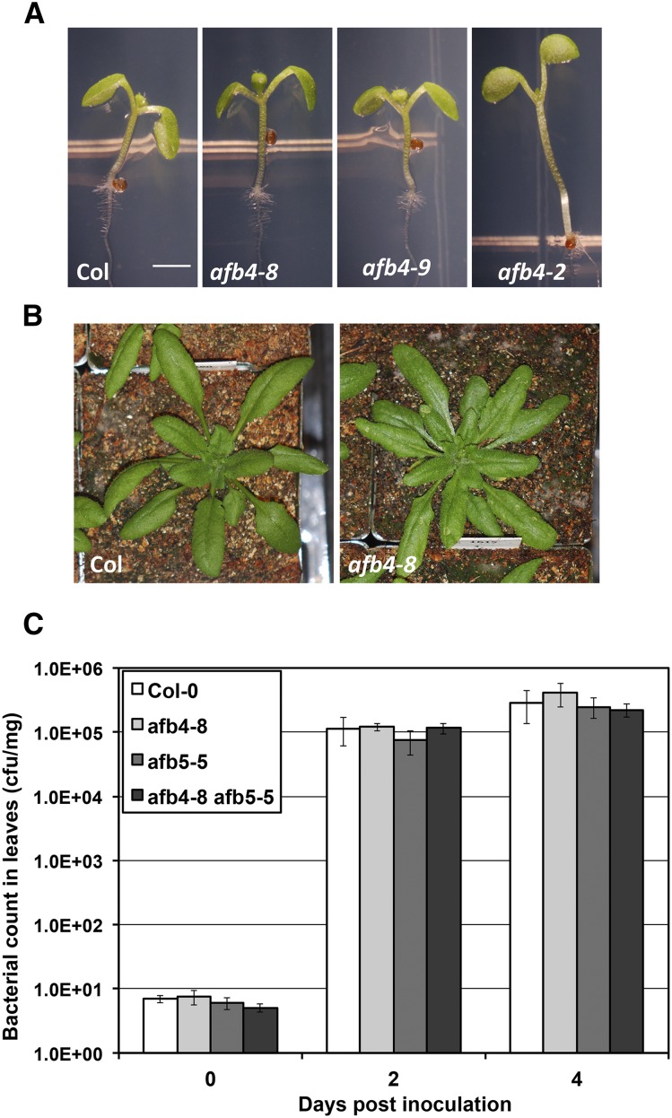
Previous studies have reported that mutations in AFB4 confer a pleiotropic phenotype. The afb4-1 allele was shown to exhibit a variety of growth defects as well as resistance to some pathogens (Hu et al. 2012). In another report, the afb4-2 mutant was reported to have a tall hypocotyl and be auxin hypersensitive (Greenham et al. 2011). In contrast afb4-8 and afb4-9 do not exhibit any of these qualities (Figure 5). The length of afb4-8 and afb4-9 hypocotyls is similar to wild type but clearly shorter than afb4-2 (Figure 3C and Figure 5A). Further afb4-8 and wild-type rosettes are similar in appearance (Figure 5B). Since these two alleles are nulls, it is clear that AFB4 is not a negative regulator of auxin response. In addition, we find that susceptibility to pathogen infection with a bacterial pathogen is similar to that of wild-type plants in the afb4-8 and afb5-5 single mutants, and in the afb4-8 afb5-5 double mutant (Figure 5C).
Figure 5.
The afb4-8 and afb4-9 do not have any obvious phenotype. Plants of the given genotypes were photographed after growing for 8 d under short-day conditions (A) or for 4 wk growing under long-day conditions (B). (C) Growth of Pseudomonas syringae strain DC3000 in the afb4-8, afb5-5, and afb4-8 afb5-5 mutants, following syringe infiltration (OD600 = 1 × 10⁻⁵). Values are the mean of 4 replicates on day 0, and 6 replicates on days 2 and 4 post inoculation. Error bars represent the standard error of the mean. Similar results were observed in four additional experiments.
Subsequent to obtaining these results we discovered that the tall hypocotyl phenotype in the afb4-2 mutant is genetically separable from AFB4. In addition, it is our experience that the severe phenotype of the afb4-1 mutant is unstable suggesting that other factors are contributing to the behavior of this line. Taken together our results indicate that AFB4 is an auxin receptor that behaves in a similar fashion to other members of the family.
The afb4-2 mutation does not confer auxin hypersensitivity. However, it is striking that the resulting amino acid substitution, D215N, affects the residue that corresponds to TIR1 D170. In a previous study we showed that the TIR1 D170E mutation does confer auxin hypersensitivity (Yu et al. 2013). Because D215N results in loss of a negatively charged residue, whereas D170E does not, we wondered if the afb4-2 mutation might disrupt AFB4 function. To test this, we performed an in vitro pulldown assay with AFB4 and afb4D215N proteins synthesized in a TNT extract. We used IAA7 protein synthesized in E. coli for the pulldown. The results shown in Figure 6, A and B show that the D215N substitution dramatically reduced recovery of the protein indicating that this mutation does affect function of AFB4.
Expression of the AFB4 and AFB5 genes
To investigate expression of the AFB4 and AFB5 genes we measured transcript levels for each of the TIR1/AFB genes in tissue collected from 4-day-old seedlings by quantitative RT-PCR. The results in Figure 7A indicate AFB4 and AFB5 are expressed in the root, hypocotyl, and cotyledon. AFB4 transcript levels are similar in the root, hypocotyl, and cotyledon, whereas the other members of the TIR1/AFB family exhibit different levels of expression in cotyledons, hypocotyls, and roots. In addition, published transcriptomic data show that AFB4 is expressed at a relatively low level in most tissues in the plant (Figure 7B) (Schmid et al. 2005; Winter et al. 2007).
We also used the AFB4-Venus and AFB5-mCitrine lines to determine expression of the respective genes in the root using confocal microscopy. We were not able to detect AFB4-Venus in any seedling tissue, consistent with the low expression level as observed in Figure 7B. In contract AFB5-mCitrine was detected in all cell types in the growing root (Figure 7C). As expected the protein was localized primarily to the nucleus of these cells.
Conclusions
In previous studies we demonstrated that TIR1, AFB1, AFB2, AFB3, and AFB5 all bind the Aux/IAA proteins in an auxin-dependent manner (Dharmasiri et al. 2005; Calderon Villalobos et al. 2012). Here we show that AFB4 also functions as an auxin receptor in a manner that is similar to the other members of the family. In addition, we present genetic evidence showing that both AFB4 and AFB5 respond to the synthetic auxin picloram, in addition to IAA, although the function of AFB5 in picloram response is much greater than that of AFB4. We expect that further genetic studies of the entire family of F-box protein auxin receptors may shed new light on the specialized functions of these proteins.
Acknowledgments
We thank Nikita Kadakia and Vanessa Peterson for technical assistance. Work in the authors’ lab was supported by a grant from the National Institutes of Health (GM43644 to M.E.) and from the National Science Foundation (MCB-1122250 to J.R.E.).
Footnotes
Communicating editor: B. Gregory
Literature Cited
- Arvidsson S., Kwasniewski M., Riano-Pachon D. M., Mueller-Roeber B., 2008. QuantPrime − a flexible tool for reliable high-throughput primer design for quantitative PCR. BMC Bioinformatics 9: 465. [DOI] [PMC free article] [PubMed] [Google Scholar]
- Calderon Villalobos L. I., Lee S., De Oliveira C., Ivetac A., Brandt W., et al. , 2012. A combinatorial TIR1/AFB-Aux/IAA co-receptor system for differential sensing of auxin. Nat. Chem. Biol. 8: 477–485. [DOI] [PMC free article] [PubMed] [Google Scholar]
- Chang I. K., Foy C. L., 1983. Growth responses of dwarf corn coleoptile sections to picloram. Pestic. Biochem. Physiol. 19: 203–209. [Google Scholar]
- Chapman E. J., Greenham K., Castillejo C., Sartor R., Bialy A., et al. , 2012. Hypocotyl transcriptome reveals auxin regulation of growth-promoting genes through GA-dependent and -independent pathways. PLoS One 7: e36210. [DOI] [PMC free article] [PubMed] [Google Scholar]
- Clough S. J., Bent A. F., 1998. Floral dip: a simplified method for Agrobacterium-mediated transformation of Arabidopsis thaliana. Plant J. 16: 735–743. [DOI] [PubMed] [Google Scholar]
- Czechowski T., Stitt M., Altmann T., Udvardi M. K., Scheible W. R., 2005. Genome-wide identification and testing of superior reference genes for transcript normalization in Arabidopsis. Plant Physiol. 139: 5–17. [DOI] [PMC free article] [PubMed] [Google Scholar]
- Dharmasiri N., Dharmasiri S., Weijers D., Lechner E., Yamada M., et al. , 2005. Plant development is regulated by a family of auxin receptor F box proteins. Dev. Cell 9: 109–119. [DOI] [PubMed] [Google Scholar]
- Gleave A. P., 1992. A versatile binary vector system with a T-DNA organisational structure conducive to efficient integration of cloned DNA into the plant genome. Plant Mol. Biol. 20: 1203–1207. [DOI] [PubMed] [Google Scholar]
- Gray W. M., del Pozo J. C., Walker L., Hobbie L., Risseeuw E., et al. , 1999. Identification of an SCF ubiquitin-ligase complex required for auxin response in Arabidopsis thaliana. Genes Dev. 13: 1678–1691. [DOI] [PMC free article] [PubMed] [Google Scholar]
- Greenham K., Santner A., Castillejo C., Mooney S., Sairanen I., et al. , 2011. The AFB4 auxin receptor is a negative regulator of auxin signaling in seedlings. Curr. Biol. 21: 520–525. [DOI] [PMC free article] [PubMed] [Google Scholar] [Retracted]
- Hagen G., 2015. Auxin signal transduction. Essays Biochem. 58: 1–12. [DOI] [PubMed] [Google Scholar]
- Hamaker J. W., Johnston H., Martin R. T., Redemann C. T., 1963. A picolinic acid derivative: a plant growth regulator. Science 141: 363. [DOI] [PubMed] [Google Scholar]
- Hu Z., Keceli M. A., Piisila M., Li J., Survila M., et al. , 2012. F-box protein AFB4 plays a crucial role in plant growth, development and innate immunity. Cell Res. 22: 777–781. [DOI] [PMC free article] [PubMed] [Google Scholar]
- Kepinski S., Leyser O., 2005. The Arabidopsis F-box protein TIR1 is an auxin receptor. Nature 435: 446–451. [DOI] [PubMed] [Google Scholar]
- Kevei Z., Baloban M., Da Ines O., Tiricz H., Kroll A., et al. , 2011. Conserved CDC20 cell cycle functions are carried out by two of the five isoforms in Arabidopsis thaliana. PLoS One 6: e20618. [DOI] [PMC free article] [PubMed] [Google Scholar]
- Mutka A. M., Fawley S., Tsao T., Kunkel B. N., 2013. Auxin promotes susceptibility to Pseudomonas syringae via a mechanism independent of suppression of salicylic acid-mediated defenses. Plant J. 74: 746–754. [DOI] [PubMed] [Google Scholar]
- Parry G., Calderon-Villalobos L. I., Prigge M., Peret B., Dharmasiri S., et al. , 2009. Complex regulation of the TIR1/AFB family of auxin receptors. Proc. Natl. Acad. Sci. USA 106: 22540–22545. [DOI] [PMC free article] [PubMed] [Google Scholar]
- Petroski M. D., Deshaies R. J., 2005. Function and regulation of cullin-RING ubiquitin ligases. Nat. Rev. Mol. Cell Biol. 6: 9–20. [DOI] [PubMed] [Google Scholar]
- Pickart C. M., 2001. Mechanisms underlying ubiquitination. Annu. Rev. Biochem. 70: 503–533. [DOI] [PubMed] [Google Scholar]
- Prigge M. J., Otsuga D., Alonso J. M., Ecker J. R., Drews G. N., et al. , 2005. Class III homeodomain-leucine zipper gene family members have overlapping, antagonistic, and distinct roles in Arabidopsis development. Plant Cell 17: 61–76. [DOI] [PMC free article] [PubMed] [Google Scholar]
- Salehin M., Bagchi R., Estelle M., 2015. SCFTIR1/AFB-based auxin perception: mechanism and role in plant growth and development. Plant Cell 27: 9–19. [DOI] [PMC free article] [PubMed] [Google Scholar]
- Schmid M., Davison T. S., Henz S. R., Pape U. J., Demar M., et al. , 2005. A gene expression map of Arabidopsis thaliana development. Nat. Genet. 37: 501–506. [DOI] [PubMed] [Google Scholar]
- Scott P. C., Morris R. O., 1970. Quantitative distribution and metabolism of auxin herbicides in roots. Plant Physiol. 46: 680–684. [DOI] [PMC free article] [PubMed] [Google Scholar]
- Tan X., Calderon-Villalobos L. I., Sharon M., Zheng C., Robinson C. V., et al. , 2007. Mechanism of auxin perception by the TIR1 ubiquitin ligase. Nature 446: 640–645. [DOI] [PubMed] [Google Scholar]
- Walsh T. A., Neal R., Merlo A. O., Honma M., Hicks G. R., et al. , 2006. Mutations in an auxin receptor homolog AFB5 and in SGT1b confer resistance to synthetic picolinate auxins and not to 2,4-dichlorophenoxyacetic acid or indole-3-acetic acid in Arabidopsis. Plant Physiol. 142: 542–552. [DOI] [PMC free article] [PubMed] [Google Scholar]
- Winter D., Vinegar B., Nahal H., Ammar R., Wilson G. V., et al. , 2007. An “Electronic Fluorescent Pictograph” browser for exploring and analyzing large-scale biological data sets. PLoS One 2: e718. [DOI] [PMC free article] [PubMed] [Google Scholar]
- Woodward A. W., Bartel B., 2005. Auxin: regulation, action, and interaction. Ann. Bot. (Lond.) 95: 707–735. [DOI] [PMC free article] [PubMed] [Google Scholar]
- Yu H., Moss B. L., Jang S. S., Prigge M., Klavins E., et al. , 2013. Mutations in the TIR1 auxin receptor that increase affinity for auxin/indole-3-acetic acid proteins result in auxin hypersensitivity. Plant Physiol. 162: 295–303. [DOI] [PMC free article] [PubMed] [Google Scholar]
- Yu H., Zhang Y., Moss B. L., Bargmann B. O., Wang R., et al. , 2015. Untethering the TIR1 auxin receptor from the SCF complex increases its stability and inhibits auxin response. Nat. Plants 1(3): 14030. [DOI] [PMC free article] [PubMed] [Google Scholar]
Associated Data
This section collects any data citations, data availability statements, or supplementary materials included in this article.


