Abstract
About two-thirds of the vital genes in the Drosophila genome are involved in eye development, making the fly eye an excellent genetic system to study cellular function and development, neurodevelopment/degeneration, and complex diseases such as cancer and diabetes. We developed a novel computational method, implemented as Flynotyper software (http://flynotyper.sourceforge.net), to quantitatively assess the morphological defects in the Drosophila eye resulting from genetic alterations affecting basic cellular and developmental processes. Flynotyper utilizes a series of image processing operations to automatically detect the fly eye and the individual ommatidium, and calculates a phenotypic score as a measure of the disorderliness of ommatidial arrangement in the fly eye. As a proof of principle, we tested our method by analyzing the defects due to eye-specific knockdown of Drosophila orthologs of 12 neurodevelopmental genes to accurately document differential sensitivities of these genes to dosage alteration. We also evaluated eye images from six independent studies assessing the effect of overexpression of repeats, candidates from peptide library screens, and modifiers of neurotoxicity and developmental processes on eye morphology, and show strong concordance with the original assessment. We further demonstrate the utility of this method by analyzing 16 modifiers of sine oculis obtained from two genome-wide deficiency screens of Drosophila and accurately quantifying the effect of its enhancers and suppressors during eye development. Our method will complement existing assays for eye phenotypes, and increase the accuracy of studies that use fly eyes for functional evaluation of genes and genetic interactions.
Keywords: ommatidia, Drosophila melanogaster, neurodevelopmental disorders, modifier screens, rough eye, human disease models
Current strategies for functional analysis of genes in various biological processes using animal models have been limited due to the lack of highly sensitive and quantitative assays. Drosophila melanogaster remains a powerful model for genetic studies, with about 75% of human disease genes having orthologs in flies (Reiter et al. 2001). Drosophila provides a wealth of genetic, cellular, and molecular biology tools, which have been instrumental in understanding basic biological processes (St Johnston 2002). With the availability of such tools, and high conservation of human disease-associated genes, the past decade has seen the growth of Drosophila models to study human diseases (Wangler et al. 2015). Specifically, the fly eye is an excellent experimental system for high throughput genetic screening, and for dissecting molecular interactions (Thomas and Wassarman 1999). Two-thirds of the vital genes in the Drosophila genome have been estimated to be required for eye development (Thaker and Kankel 1992). Although some genes are likely to be specific to eye development, other vital genes expressed in the eye are probably required for general cellular processes as well (Thomas and Wassarman 1999). Hence, phenotypic assessment of the eye can be extended to gene functions in other tissues. Since it is a dispensable organ for survival, the fly eye has been used for studies aimed at understanding basic biological processes, including cell proliferation and differentiation, neuronal connectivity, apoptosis, and tissue patterning (Karim et al. 1996).
The Drosophila compound eye is a simple nervous system consisting of a symmetrical organization of approximately 750 ommatidia (Ready et al. 1976). Each ommatidium contains eight photoreceptor neurons orchestrated in a trapezoid fashion, and surrounded by four lens-secreting cone cells and two primary pigment cells. The ommatidia are separated from one another by a lattice of 12 accessory cells that include six secondary pigment cells, three tertiary pigment cells, and three mechanosensory bristle complexes (Kumar 2012). Since the structure of the eye is ordered precisely, any subtle defect that alters the geometry of a single ommatidium, or disrupts the development of a single cell within the ommatidium, leads to observable morphological phenotypes such as the rough eye. Other commonly observed eye phenotypes can include small or large eye, change in size of individual ommatidia, changes in bristles, and loss of pigmentation. Genetic screens for modifiers of a phenotype caused by knockdown/mutation or misexpression of a gene in the developing eye have played a pivotal role in identifying novel genes interacting in the same or different biological pathways (Carrera et al. 1998; Cukier et al. 2008). The majority of genetic screens utilizing the eye phenotypes take advantage of the rough eye, or changes in the size of eye. The rough eye phenotype could arise due to lack of individual photoreceptor neurons or a change in the number, arrangement, or identity of photoreceptor neurons (Tomlinson et al. 1987, 1988; Van Vactor et al. 1991; Basler et al. 1991). Drosophila rough eye phenotypes have been utilized to identify genetic modifiers of several genes, including ras (Karim et al. 1996), ksr (Therrien et al. 2000), sina (Neufeld et al. 1998), sine oculis (Roederer et al. 2005), and humanized models of MECP2 (Cukier et al. 2008) and ATXN3 (Bilen and Bonini 2007). However, these studies assessing the rough eye morphology are qualitative in nature and hence open to varied interpretations. Usually, the different eye phenotypes are analyzed visually, and rank ordered manually based on their severity. While severe, overt eye phenotypes are readily recognizable to the naked eye, differentiating subtle alterations can be challenging. In the modifier screens, while strong enhancers and suppressors can be identified by qualitative analysis (i.e., visual inspection) of the eye phenotypes, weak modifiers may go undetected. Currently, no image analysis techniques are available that can automatically and accurately quantify the rough eye phenotypes observed in bright-field or scanning electron microscopy (SEM) images.
Here, we present a novel computational method that facilitates accurate analysis of Drosophila rough eye morphology from images obtained using bright-field microscopy or SEM. Using morphological transformation to detect the fly eye, and ommatidial measurements to quantify the disorderliness, this sensitive assay can detect morphological changes in Drosophila eyes. We tested our automated method by analyzing the morphological defects resulting from eye-specific knockdown of Drosophila orthologs of 12 genes known to be associated with neurodevelopmental disorders. We also validated our method by analyzing a representative group of eye images from six independent published and unpublished studies, including genetic screens for modifiers of tau-induced neurotoxicity (Ambegaokar and Jackson 2011), interactors of dFoxO (Kanao et al. 2010), interactors of DJ-1, a peptide library screen for Drosophila model of myotonic dystrophy type 1 (DM1) (Garcia-Lopez et al. 2011), and a genetic screen for interactors of Egfr (Mardon et al. 1994). We also validated the roughness of the fly eyes overexpressing C9orf72 pure repeats and RNA only (RO) repeats (Mizielinska et al. 2014). Our quantitative analysis and classification of these modifiers using Flynotyper was concordant with the authors’ original qualitative assessment. We further used this method for assessing the genetic modifiers of sine oculis (so), a key gene involved in eye formation, obtained from two genome-wide screens, and accurately classifying their effect on the so-associated eye phenotype. Our method provides a quantitative tool for dissecting phenotypic heterogeneity frequently observed in studies of genetic mutations, gene dosage alterations, and genetic modifiers in development and disease.
Materials and Methods
Drosophila stocks
The conditional knockdown of specific genes was achieved with the UAS-GAL4 system (Brand and Perrimon 1993), using w;GMR-GAL4; UAS-Dicer2 (Zhi-Chun Lai, Penn State University), and the UAS-RNAi transgenic lines. The following RNAi fly stocks from Vienna Drosophila Resource Center (Austria, Vienna) (Dietzl et al. 2007) were used in this study: UAS-ube3aRNAi (VDRC# 100130, 45876), UAS-prosapRNAi (VDRC# 103592, 21218), UAS-capsRNAi (VDRC# 25291, 25292), UAS-kismetRNAi (VDRC# 46685), UAS-paraRNAi (VDRC# 6132), UAS-ptenRNAi (VDRC# 101475, 35731), UAS-armRNAi (VDRC# 107344), UAS-rkRNAi (VDRC# 105360, 29931, 29932), UAS-tpc1RNAi (VDRC# 6005), UAS-ephRNAi (VDRC# 6545), UAS-nrx1RNAi (VDRC# 4306), and UAS-mcph1RNAi (VDRC# 106261, 28100). The following stocks were used in studying genetic interactions, ey-GAL4 and GMR-GAL4 with UAS-so and the Bloomington Drosophila deficiency kit (Bloomington Stock Center) (Cook et al. 2012). All stocks and crosses were cultured on conventional cornmeal/sucrose/dextrose/yeast medium at 25° unless otherwise indicated. A list of all genotypes obtained from the deficiency screen is given in Supplemental Material, Table S1.
Quantitative real-time PCR
We assessed mRNA expression using quantitative real-time PCR (qRT-PCR) by isolating RNA from fly heads expressing RNAi knockdown of specific genes. Groups of 40–50 female flies were frozen in liquid nitrogen and stored at −80°. For RNA extraction, fly heads were separated from bodies by repetitive cycles of freezing in liquid nitrogen and vortex mixing. Total RNA was isolated using TRIZOL (Invitrogen) and reverse transcribed using qScript cDNA synthesis kit (Quanta Biosciences). Quantitative RT-PCR was performed using an Applied Biosystems Fast 7500 system with SYBR Green PCR master mix (Quanta Biosciences). All SYBR green assays were performed in triplicate, and normalized to rp49 mRNA expression. Each qRT-PCR experiment was repeated twice with two independent RNA isolations and cDNA syntheses. A list of primers used for these experiments is provided in Table S2.
Eye imaging using bright-field microscope
For light microscope imaging of adult eyes, 2- to 3-d-old flies (GMR;Dicer2 >UAS-RNAi), reared at 28° and 30°, were immobilized by freezing at –80°, and then mounted on Blu Tack (Bostik Inc, Wauwatosa, WI). These flies were then imaged using a Semimotorized Olympus BX53 microscope with a LMPLFLN 20× objective, and a C-mount camera with 0.5× magnification (Olympus, Tokyo, Japan). Images were captured with CellSens Dimension software (Olympus Optical), and the slices stacked using Zerene Stacker (Zerene Systems, Richland, WA). All images shown in figures are maximum projections of 20 consecutive optical z-sections.
Eye imaging using SEM
Flies aged 2- to 3-d-old (GMR; Dicer2 >UAS-RNAi reared at 30°) were anesthetized with CO2, dissected midway through the abdomen, and fixed overnight at 4° in 0.2 M sodium cacodylate buffer containing 2.5% glutaraldehyde. The preparations were then washed three times with 0.1 M sodium cacodylate buffer for 5 min each at room temperature. The samples were then dehydrated through an ethanol series (50%, 70%, 85%, 95%, and 100%), and critically point dried using Leica CPD300 at the Penn State Microscopy and Cytometry Facility (The Pennsylvania State University, University Park, PA). The dried flies were mounted on carbon-taped SEM stubs, and sputter-coated with a 25-nm-thick gold coat. Samples were then imaged using Zeiss Field Emission-SEM (Carl Zeiss AG, Oberkochen, Germany).
The Flynotyper computational method
Detection of Drosophila eye from ommatidial clustering:
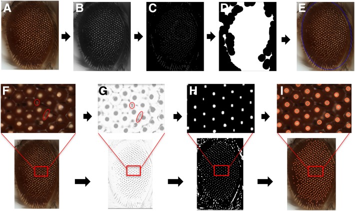
We obtained high quality images of eyes from individual fly genotypes using light microscopy or monotonic images from SEM. The fly eye is not flat but convex in shape, and each ommatidium is also convex (Figure 1, A–E). Therefore, in a bright-field image, when light falls on each ommatidium, there is a reflection spot in its center. The whole eye area is then presented as a region enriched for such bright spots, which also leads to a high contrast between the eye and the background in the image. Conversely, in a SEM image, there is a dark spot in the center of each ommatidium compared to the boundary, which might be due to variations in collection efficiency of the secondary electrons, owing to the convex shape of the ommatidium. First, the original images were converted to gray scale. A series of morphological transformations were applied to suppress the background and identify the fly eye based on the clustering of the ommatidia (Graham and Baldock 2000). The result of different morphological transformations depends on the different size of the matrix, and what operation was applied. The small pixel matrix is then referred to as the structuring element (SE). As the quality of the final output extracted from the image depends on the size of the SE, we tested different combination of SE sizes to optimize the pixel intensity of the circular shaped ommatidia (Figure S1). We performed a top-hat transformation to extract light objects from a dark background (Arulmozhi et al. 2012). As a result, the background is suppressed as pixel values are subtracted from the input frame, reducing its intensity and enhancing the eye region in the process. Edge detection was then applied over the resultant image to identify an approximate region with a clustering of ommatidia. Following edge detection, we used a morphological closing operation to expand the boundaries of the foreground area in the image by filling in the surrounding background to connect clusters of all edge-detected regions. By detecting the largest connected area, we were able to localize the position of the Drosophila eye as a region with ommatidial clustering.
Figure 1.
A computational strategy for automated assessment of Drosophila melanogaster eye morphology. Eye localization in a bright-field microscope image is carried out by first converting the original image (A) to grayscale (B), then morphological transformations are applied to suppress the background, followed by edge detection to identify an approximate region with ommatidial cluster (C), and finally morphological closing operation localizes the eye area (D), giving the final output image with the eye area localized (E). For detection of the ommatidial center, the original bright-field image (F) is converted to grayscale and inverted (G). Multiple filters and transformation operations further enhance the contrast of the inverted image and eliminate the noise from the ommatidial boundary (H). The centers of the bright spots due to light reflection are considered as ommatidial centers (I).
The images obtained from SEM were processed differently, as a low contrast between the foreground (eye) and the background limited the application of top hat transformation to suppress the background (Arulmozhi et al. 2012). In addition, the SEM images capture the details in the area surrounding the eye, thereby significantly increasing the background noise, and thus hindering the accurate detection of the ommatidial cluster. To circumvent this issue, we applied a thresholding operation for SEM images (Figure S2). The thresholding operation is a basic segmentation method performed on a gray-scale image to separate out objects of interest from the image background (Graham and Baldock 2000). This separation is based on the difference in pixel intensity between the object and the background. By using the mean intensity of the image as a determined cutoff, we could separate the region of interest from the background. The subsequent steps of the method are the same for images taken using both light microscope and SEM.
Detection of ommatidia by applying morphological transformation and searching local maxima:
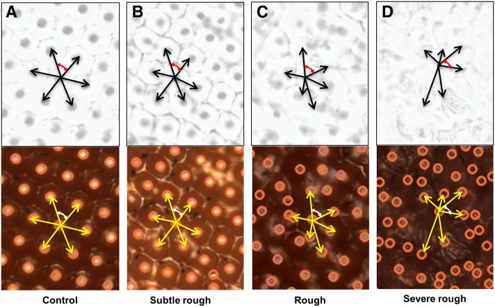
In a digital image, ommatidia appear as roughly circular shapes, also referred to as blobs. Due to the fusion of ommatidia observed in different genotypes, the boundary of each ommatidium can show nonuniform intensities. Our next step was to isolate each ommatidium as a single blob, and remove the noise caused by the boundary (Figure 1, F–I). The following steps were performed to extract ommatidia. We first enhanced the contrast between the ommatidia and the background. In a bright-field image, the contrast is between the reflection spot at the center of the ommatidium and the rest of the ommatidium, while, in a SEM image, this contrast is between a darker center and lighter boundary of the ommatidium. Then, by applying top hat transformation to the enhanced image, the ommatidia were highlighted from the background (Arulmozhi et al. 2012). However, noises such as light reflection on the boundary between neighboring ommatidia are also extracted. This noise was then removed by using the median filter. The median filter is used to blend the intensity of the pixels within a specified radius by calculating the median value of neighboring pixels in a specific SE, and assigning that value to each pixel in the element. Thus, the background noise was removed by smoothing the pixels in the neighboring region, resulting in bright blobs representing ommatidia. Because of the nonuniformity and reduced intensity of each blob, we further enhanced the contrast of the image using a combination of dilation and erosion operations. As a result, each ommatidium was isolated as a single blob, and its center was localized by searching for local maxima within the fly eye. Using the centers of ommatidia, phenotypic scores were then computed based on the distance and the angle between the ommatidia. Our algorithm robustly detected the ommatidial centers in a variety of eye phenotypes qualitatively classified as wild type-like, subtle rough, rough, and severe rough (Figure 2).
Figure 2.
A conceptual representation of Flynotyper algorithm for detection of ommatidial centers, and calculation of ommatidial disorderliness is shown for different classes of eye phenotypes. Grayscale inverted and bright field images of (A) wild type control eye, (B) subtle rough eye, (C) rough eye, and (D) severe rough eye phenotypes are shown. The ommatidial centers were accurately detected in all the four categories of eye phenotypes. In the algorithm, the six neighboring ommatidia are chosen based on distance; the closest six ommatidia are considered for calculation of phenotypic scores. The six local vectors are represented by black (for grayscale/inverted) and yellow arrows (for bright-field images). The red and white arrows represent the angle between two adjacent vectors in grayscale/inverted and bright-field images, respectively. Note the differences in the lengths of the local vectors and the angles between them in different classes of eye phenotypes.
Calculation of phenotypic score:
The Drosophila compound eye consists of a hexagonal array of packed ommatidia. In wild type compound eyes, ommatidia are ordered as mirror images that are radially symmetrical about an axis. The symmetric arrangement is disrupted in a defective eye, resulting in an irregular hexagonal arrangement. Based on this principle, we determine groups of six local vectors, v, with direction pointing from each ommatidium to six surrounding ommatidia. We quantified the disorganization of the ommatidia by applying the principle of entropy, a measure of disorderliness, in which a perfectly radial symmetry resulted in zero entropy. To quantify the disorderliness of the fly ommatidia, we calculated phenotypic scores based on three measures including distance ommatidial disorderliness index (ODID), angle ommatidial disorderliness index (ODIA), and ommatidial fusion index (Z). These indices essentially measure the level of disruption of symmetry of the ommatidia. The mathematical formulation to calculate ODID and ODIA is described as follows:
As illustrated in Figure 2, the distance ommatidial disorderliness index is defined as the difference between the lengths of each of the five local vectors, vi (I = 1…5), from the smallest vector, vmin. Distance ommatidial disorderliness index for each ommatidium is calculated as
| (1) |
Similarly, the angle ommatidial disorderliness index for each ommatidium is the difference among angles formed by pairs of adjacent local vectors to the smallest angle between the vectors. Thus, angle ommatidial disorderliness index for each ommatidium is calculated as:
| (2) |
Here, vi+1 indicates the vector adjacent to vi (note the vector adjacent to v6 is v1).
The total ommatidial disorderliness index, ODIT, is the sum of distance and angle ommatidial disorderliness indices using the number of most ordered ommatidia (N, a tunable parameter) and is determined by equations (1) and (2) as given below:
| (3) |
In this equation, N is the parameter defined by the user to be the number of most ordered ommatidia. If we let Fusion index Z represents the number of ommatidia identified by the detection algorithm, then phenotypic score P is computed as followed using (4)
| (4) |
The ommatidia were rank-ordered based on their total ommatidial disorderliness index values, and the distance to the center of the eye. As the phenotypic score is predominantly dependent on the ommatidial disorderliness index, higher phenotypic score represents increased disorderliness of the ommatidia, and thus increased severity of the eye phenotype. Please note that N is the most ordered ommatidia and is standard (and selected only once) for all the eyes used in the same experiment. The distance and angle disorderliness indices are calculated from N ommatidia. However, the number of detectable ommatidia or Fusion index, Z, can vary from eye to eye in the same experiment.
Data availability
Open source software available at http://flynotyper.sourceforge.net.
Results
Automated detection of fly eye and calculation of phenotypic score
We have developed an automated computational method to quantify observable morphological defects in the Drosophila eye from images taken using bright-field microscopy or SEM. The algorithm is implemented as Flynotyper software, and is available as an open-source package (http://flynotyper.sourceforge.net/) and ImageJ plugin (Figure S3). For each image, a series of morphological transformations was first applied to suppress the image background and identify the fly eye (Graham and Baldock 2000) (Figure 1, A–E). Then, each ommatidium was isolated and its center localized using image transformation and searching for local maxima (Figure 1, F–I). Based on the hexagonal arrangement of ommatidia, we determined groups of six local vectors directed from the center of each ommatidium to the centers of six neighboring ommatidia (Figure 2). We quantified the disorganization of the ommatidia by applying the principle of entropy—a measure of disorderliness—in which a perfectly radial symmetry will result in zero entropy. We calculated ommatidial disorderliness indices (ODI) of a fly eye using the cumulative differences in the lengths and angles formed by these adjacent local vectors (see Materials and Methods). Phenotypic scores were then determined using the ODI, and the estimated number of fused ommatidia (fusion index) (Figure S4). A higher phenotypic score represents increased disorderliness or altered symmetry of the ommatidial arrangement, and thus increased severity of the eye phenotype.
Assessing the performance of Flynotyper
We tested our algorithm on adult eye images from 21 lines with RNA-interference (RNAi) mediated eye-specific (GMR-GAL4/UAS-RNAi) knockdown of fly orthologs of human genes associated with neurodevelopmental disorders, including dpten, kismet, dube3a, prosap, arm, caps, para, rk, nrx-1, mcph1, tpc1, and eph (Table S3). We hypothesized that, when knocked down in the eye, these genes would show varying levels of severity in eye phenotypes based on their level of knock down. These neurodevelopmental genes were chosen after curating the mutations observed in exome sequencing studies of autism (Iossifov et al. 2014), intellectual disability (Gilissen et al. 2014), schizophrenia (Fromer et al. 2012, 2014), and epilepsy (Martin et al. 2014), their roles in CNS development (De Rubeis et al. 2014; Luo et al. 2012), and their conserved functions in model organisms such as mouse and fly. Further the genes were shortlisted based on the presence of fly orthologs, high sequence identity, and availability of RNAi stocks.
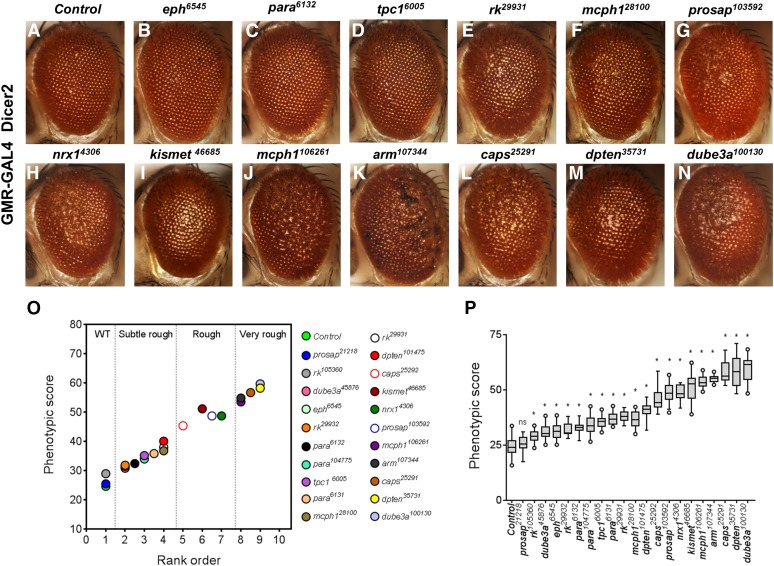
A wide range of defects in eye morphology was observed for these 21 fly lines compared to a control line derived from the same genetic background, but not expressing an RNAi construct (Figure 3, A–N). Based on visual assessments of two independent reviewers, we manually ranked the eye phenotypes from 1 to 10 based on severity, with rank 1 assigned to wild type-like, and rank 10 for the most severe phenotype (Table S4). These ranks were further classified into one of the following four broad qualitative categories: wild type-like, subtle rough, rough, and very rough (Figure 3O). A significant correlation (Pearson correlation coefficient, r = 0.99, P < 0.001) was observed when the manually determined ranks were compared to the corresponding phenotypic scores obtained from Flynotyper (Figure 3O). For example, knockdown of dpten, kismet, dube3a, prosap, arm, nrx-1, mcph1, and caps resulted in significantly high phenotypic scores compared to control flies (student t-test, corrected two-tailed P < 0.001), indicative of a very rough eye phenotype (Figure 3P and Table S5). Similarly, lower phenotypic scores were observed for knockdown of para, eph, tpc1, and rk, in agreement with the subtle rough eye phenotype observed in these flies (student t-test, corrected two-tailed P < 0.001). The phenotypic scores were also robust in distinguishing other qualitative categories of eye phenotypes, such as glossy, necrotic, and crinkled eyes (Figure S5). Notably, scores for the glossy eye phenotypes mapped well within these categories based on the extent of ommatidial fusion. For example, the glossy eyes with dpten knockdown was severe, and scored similar to that of very rough eye, while glossy eyes with kismet knockdown scored similar to the rough eye. For all the fly lines tested, ten or more eye images were used to run on Flynotyper.
Figure 3.
Analysis of Drosophila orthologs of human neurodevelopmental genes. (A–N) Representative bright-field microscope images of fly eyes displaying eye-specific knockdown of tpc1, eph, para, rk, mcph1, prosap, nrx-1, kismet, arm, caps, dpten, and dube3a genes from flies reared at 30°. Eyes of GMR-GAL4; Dicer2/+ control flies show normal ommatidial organization, while the eyes of flies with GMR-GAL4 driven RNAi knockdown of the 12 genes show disruption in the morphology of the eye. Note the variation in the severity of the eye phenotype for different genes. (O) Graph representing the mean phenotypic scores of control flies compared to the mean phenotypic scores for 21 RNAi lines with knockdown of neurodevelopmental genes (n = 9–30). The rank order of these fly lines was significantly correlated with the phenotypic scores (Spearman correlation coefficient = 0.99, P = 1.2 × 10⁻¹⁹). (P) Graph representing phenotypic scores of the 21 RNAi lines with GMR-GAL4 at 30°C is shown. The number of images analyzed for each genotype ranged from 9 to 30 (median of 20.5). Comparisons were made between each of the gene knockdowns to controls using a student t-test (*represents corrected two-tailed P < 0.001).
Flynotyper phenotypic scores also allowed us to distinguish between genotypes within a specific phenotypic category. While all of the different para RNAi lines (para6131, para6132, and para104775) showed subtle rough eye phenotype, our algorithm was able to accurately identify differing degrees of severity imposed by each RNAi construct (Figure S6). Further, phenotypic scores were consistent with rank order even when different numbers (n = 150, 200, 250, 300, and 350) of ommatidia were used for the analysis (Figure S7A). In the Flynotyper program, we chose N = 200 as the default number of ommatidia, since at this value the phenotypic distinction was robust, with less variation in phenotypic scores between genotypes within and across ranks (Figure S7B).
We also evaluated the robustness of Flynotyper at different resolutions of images (600 × 800, 1200 × 1600, and 1600 × 2400) taken using a bright-field microscope. Although greater number of ommatidia was detected for images acquired at higher resolutions (1200 × 1600 and 1600 × 2400) compared to those at a lower resolution (600 × 800), we found that the phenotypic scores were similar at all three resolutions tested (Figure S8). Thus, our method can reliably quantify eye morphology at different counts of ommatidia, and images taken at different resolutions. Traditionally, a SEM image is considered to be of higher quality than a bright-field image, due to its ability to resolve finer details of the specimen. To determine the performance of our framework for SEM and bright field images, we compared the phenotypic scores for the same genotypes using images obtained from these two techniques (Figure S9). A positive correlation (Pearson correlation coefficient, r = 0.95, P = 0.011) was observed between the phenotypic scores obtained from SEM images to that from bright field images (Figure S9C). Thus, Flynotyper processes images taken using SEM and bright-field microscopy with similar accuracies.
We further performed a sensitivity analysis to determine the minimum number of eye images required for Flynotyper to accurately quantify and distinguish between the different categories of phenotypes. We chose one control and four genotypes, one from each category (control like, subtle rough, rough, and very rough), imaged ten eyes for each category, and calculated their phenotypic scores. We then performed sampling with replacement with all possible combinations for n = 3 to n = 10 (where n is the number of eye images). Next, we calculated the mean phenotypic score for each combination of eye images, and generated plots for distribution of all possible mean scores. As expected, the spread of the distribution decreased as ‘n’ increased (Figure S10A). While no difference in the distribution of mean phenotypic scores was observed between controls and control-like eye images, significant differences (P < 0.001, Mann-Whitney test) were observed when each category was compared with each other, and with the control eye images for sample size as low as n = 3 (Figure S10B).
Testing dosage sensitivity of neurodevelopmental genes using Flynotyper
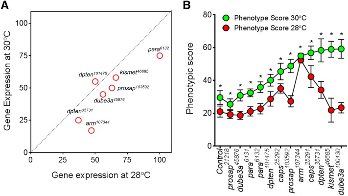
Next, we tested the ability of this method to detect subtle differences in phenotypic effects caused by changes in the gene dosage of neurodevelopmental genes. We generated flies with different levels of mRNA for the same gene by either using different RNAi lines for the same gene, or by rearing flies at different temperatures (28° and 30°). We performed qPCR experiments using fly heads for a subset of 10 RNAi lines to confirm a reduction in the mRNA levels of the targeted genes (Figure 4 and Table S2). The different RNAi lines of a gene were targeted to different sequences of the gene and may vary in their efficiency to knockdown, thereby resulting in different levels of mRNA. For example, knockdown of pten in two different RNAi lines, pten101475 and pten35731, resulted in mRNA expression of 55% and 25%, respectively. The phenotypic score of pten35731 was higher than that from pten101475, in agreement with the level of knockdown. For the seven genes tested, the expression level was lower in flies reared at 30° than in those reared at 28°, due to the temperature-dependent effect of the UAS-GAL4 expression system (Figure 4A and Table S6). The phenotypic scores of flies reared at 30° were correspondingly higher than those reared at 28°, indicating a gene dosage-dependent increase in the severity of eye phenotypes for these genes (Figure 4B). In fact, Flynotyper was also able to identify subtle changes in the phenotype due to dosage alteration where visual assessment failed. For example, a significant difference in phenotypic scores was observed for arm107344 flies reared at 28° and 30° and expressing 47% and 17% of mRNA, respectively (Student t-test, corrected two-tailed P = 4.09 × 10⁻¹⁸).
Figure 4.
Quantitative assessment of eye phenotypes due to gene-dosage alteration. (A) Plot of mean values of gene expression of RNAi lines with eye specific knockdown at 28° vs. 30°. Note that each experiment was conducted in triplicate, and then repeated using a fresh preparation of RNA and cDNA synthesis. Gene expression of flies reared at 30° is lower than that at 28° due to increased RNAi-mediated knockdown at 30° (note that most red circles are below the black diagonal line), indicating a temperature dependent effect of the UAS-Gal4 system. (B) A graph representing phenotypic scores for knockdown of prosap, dube3a, para, pten, arm, caps, and kismet at 30° and 28° is shown. A dosage dependent increase in severity was observed for all the genes tested. Asterisks (*) show significant P-values (student t-test, corrected two-tailed P < 0.001) when phenotypic scores at 30° was compared to that at 28°. The number of images processed for each genotype ranged from n = 14 to n = 30. A complete list of statistical analysis and n numbers for each of the genotype assessed is presented in Table S6.
Validation of Flynotyper using images from independent studies
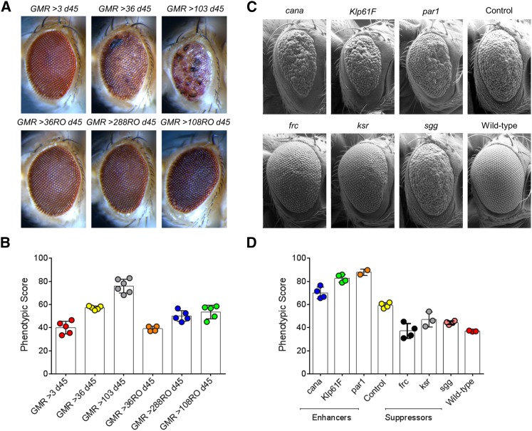
To validate the robustness of our software for images generated using different image acquisition set ups (both bright-field and SEM), we tested Flynotyper on adult eye images from six independent studies (both published and unpublished). We first tested the bright-field images of eyes overexpressing C9orf72 pure repeats, and RNA only (RO) repeats (Figure 5, A and B) (Mizielinska et al. 2014). Expanded repeats in C9orf72 is the most common genetic cause of frontotemporal dementia and amyotrophic lateral sclerosis (DeJesus-Hernandez et al. 2011; Renton et al. 2011; Majounie et al. 2012). While expression of 36 and 103 pure repeats caused neurodegeneration in the fly eye, 36 RO repeats did not show any degeneration similar to three pure repeats, and 288 RO and 108 RO repeats showed mild degeneration, in agreement with the original assessment (Mizielinska et al. 2014). We then tested a group of representative SEM images from a functional genetic screen for modifiers of tau-induced neurotoxicity (Ambegaokar and Jackson 2011). We were able to accurately identify and classify the different suppressors and enhancers of the human wild type full-length tau overexpression. While cana, Klp61F, and par1 enhanced the tau rough eye phenotype, frc, ksr, and sgg suppressed the rough eye phenotype (Figure 5, C and D). We also compared the phenotypic scores for each of these modifiers with the volumetric analysis reported in the published study (Ambegaokar and Jackson 2011), and observed a negative correlation (Pearson r = –0.71, two-tailed P = 0.049), with higher phenotypic scores corresponding to lower eye volumes (Figure S11). We also validated Flynotyper on images obtained from four additional studies, including a genetic screen for interactors of dFoxO (Kanao et al. 2010) (Figure S12, A and B), a screen for interactors of DJ-1, a gene mutated in familial Parkinson’s disease (Figure S12, C and D), a positional scanning combinatorial peptide library screen to identify molecules that reduced CUG-induced toxicity in a Drosophila model of myotonic dystrophy type 1 (DM1) (Garcia-Lopez et al. 2011) (Figure S13), and a classical study of a genetic screen for interactors of Egfr using P-element insertions (Mardon et al. 1994) (Figure S14). In the analysis of eye images from all these studies, Flynotyper was robust in accurately quantifying the severity of the eye phenotypes, and concordant with the results from the original assessments.
Figure 5.
Analysis of eye images obtained from independent studies. (A) Bright-field microscopy images of representative Drosophila eyes overexpressing C9orf72 pure or RO repeats using the GMR-GAL4 driver, imaged on d 45. While three pure repeats had no effect, 36 pure repeats were toxic and 103 pure repeats showed more overt toxicity; 36 RO repeats had no effect, and 108 RO and 288 RO repeats showed a mild effect. (B) Graph representing the phenotypic scores of C9orf72 pure or RO repeats using the GMR-GAL4 driver. The phenotypic scores are concordant with the visual assessment of the eye phenotypes (Mizielinska et al. 2014). The numbers of images used for these assays were n = 4 for GMR >36 RO d45, n = 5 each for GMR > 3 d45, GMR > 36 d45, GMR > 288 RO d45, and GMR > 108 RO d45, and n = 6 for GMR > 103 d45. (C) Scanning electron microscope images of genetic modifiers of tau-induced neurotoxicity. The control listed is w1118/+;gl-tau/+. All other panels, except wild type, contain one copy of gl-tau transgene in trans to one disrupted copy of the gene listed in the panel. (D) A graph representing the phenotypic scores of wild type, control, and the three enhancers and suppressors of w1118/+;gl-tau/+, is shown. The number of images used for these analyses were n = 2 for par1, n = 3 each for wild type, and ksr, n = 4 each for control, cana, Klp61F, sgg, and frc.
Discovery of novel interactors of sine oculis
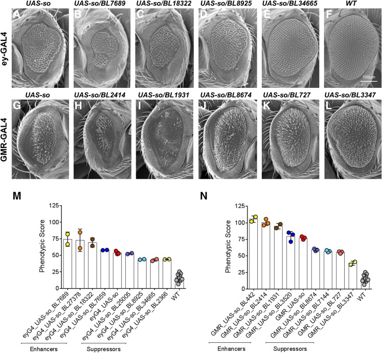
To illustrate the utility of our method in modifier screens, we used SEM images of candidate genetic interactors of sine oculis (so), a Drosophila gene involved in eye development (Cheyette et al. 1994; Serikaku and O’Tousa 1994; Pignoni et al. 1997). Overexpression of so using ey-GAL4 and GMR-GAL4 results in a rough eye that is also significantly smaller in size compared to the wild type eye. Two genetic screens using 425 deficiency lines were performed to identify regions of the Drosophila genome that potentially interact with sine oculis (Figure 6). A total of 16 novel modifiers were identified by qualitative assessment, when flies overexpressing so with ey-GAL4 and GMR-GAL4 were each crossed to 425 deficiency lines (Table S1). These modifiers varied in their level of suppression or enhancement of the so overexpression eye phenotype, and could be broadly classified as mild, moderate, and strong modifiers. We first assessed the phenotypic scores of flies overexpressing so, using ey-GAL4, with or without the candidate modifiers, and compared the effect of modifiers using student t-test (Table S7 and Figure S15). We identified four enhancers and four suppressors of so, and used Flynotyper to accurately classify these candidate modifiers based on their effects (Figure 6, B-E). For example, among the tested deficiency lines, BL25005 was identified as a mild suppressor, BL8925 and BL2366 as moderate suppressors, and BL34665 as a strong suppressor (Figure 6M). Although the phenotypic scores of BL34665 and BL2366 are similar, BL34665 can be classified as a strong suppressor since the eye area of BL34665 is similar to that of wild type. We note that, while our software does not directly calculate the size of the fly eye, a negative correlation exists between the size of the eye and the phenotypic score, as the calculation also accounts for the number of detectable ommatidia (Z). Further, methods to quantify the eye size are also available, which can be incorporated into the framework of ImageJ along with Flynotyper (Woodman et al. 2011; Posnien et al. 2012; Gonzalez-Bellido et al. 2011). Similar phenotypic assessments led to classification of BL7659 as a mild enhancer, BL18322 as a moderate enhancer, and BL27378 and BL7689 as strong enhancers (Figure 6M). Assessment of phenotypic scores of flies overexpressing so, using GMR-GAL4, with or without the candidate modifiers, led to the identification of four additional suppressors and four enhancers of so (Figure 6, H–L and N, and Table S7). Among the tested deficiency lines, BL8674, BL7144, BL727, and BL3347 were identified as strong suppressors, BL3520 as a mild enhancer and BL1931, BL2414, and BL442 as strong enhancers (Figure 6N). Thus, our method can be used to analyze the genetic interactions, and also accurately classify the genetic modifiers strong and weak interactors.
Figure 6.
Genetic modifiers of sine oculis. (A–L) Scanning electron microscope (SEM) images of adult compound eyes are shown. (A) ey-GAL4/UAS-so. Note that the eye is rough and smaller in size. (B–C) Enhancers of the rough eye phenotype due to overexpression of UAS-so with ey-GAL4. (D–E) Suppressors of the rough eye phenotype due to overexpression of UAS-so with ey-GAL4. Note that BL34665 rescues both the small eye, and the rough eye phenotype of ey-GAL4/UAS-so. (F) Wild type (WT). (G) GMR-GAL4/UAS-so. (H–I) Enhancers of the rough eye phenotype due to overexpression of UAS-so with GMR-GAL4. (J–L) Suppressors of the rough eye phenotype due to overexpression of UAS-so with GMR-GAL4. (M) A graph representing the phenotypic scores of WT, ey-GAL4/UAS-so, and the four enhancers and four suppressors of ey-GAL4/UAS-so. The number of images processed for each genotype ranged from n = 2 to n = 10. (N) Graph representing the phenotypic scores of WT, GMR-GAL4/UAS-so, and the four enhancers and four suppressors GMR-GAL4/UAS-so. The number of images processed for each genotype ranged from n = 2 to n = 10. Although the phenotypic scores distinguished the effect of most suppressors and enhancers on the eye phenotype, Flynotyper alone was not able to accurately classify BL34665 as a strong suppressor. Visual inspection of BL34665 images showed ommatidial organization and eye size comparable to that of wild type eyes.
Discussion
The Drosophila melanogaster eye has been used as a model to study various developmental processes (Thomas and Wassarman 1999; Kumar 2012). Classical studies on Drosophila have established a line of research using the fly eye as an experimental system for studying genetic effects (Meyerowitz and Kankel 1978; Moses and Rubin 1991; Ready et al. 1976; Thaker and Kankel 1992). For decades, the fly eye has been used as a system for functional screening of genes and genetic interactions involved in basic cellular processes, neuronal development and degeneration, and common complex diseases such as cancer and diabetes. For example, Alzheimer’s disease has been studied successfully using the Drosophila eye by transgenic expression of human β-amyloid and β-secretase genes. These transgenic flies displayed age-dependent neurodegeneration and β-amyloid plaque formation, both of which were rescued by addition of inhibitors of β-secretase and γ-secretase (Greeve et al. 2004). Similarly, Drosophila eye models have been used to characterize genetic interactions. For example, He and colleagues generated a fly model of protein-misfolding disease by misexpressing human proinsulin protein in the eye and identified novel genetic interactors (He et al. 2014). Another genetic screen for CLASP interactors using the Drosophila eye resulted in the identification of 36 genetic modifiers (Lowery et al. 2010). However, most studies using the fly eye are qualitative in nature, involving visual inspection of the phenotypes, and manual scoring of genetic interactors. In order to address this issue of a lack of highly sensitive and quantitative assay for scoring eye phenotypes, we developed a computational method for quantitative assessment of eye morphology in fruit flies. We have implemented our algorithm as a software package called Flynotyper, which is available as an open source software.
The following features of Flynotyper are noteworthy (Table S8). First, our algorithm is highly sensitive and robust across different types of eye morphological defects, including rough eye, glossy eye, crinkled eye, and necrotic eye. The results of the performance of Flynotyper indicate that this method accurately detects even subtle alterations in eye morphology, and provides a quantitative measure that can distinguish between varying severity of eye defects. This feature of the algorithm has allowed us to quantify the effect of dosage alteration of key neurodevelopmental genes. For example, Flynotyper accurately distinguished the severity of eye phenotypes for knockdown of prosap, dube3a, para, pten, caps, arm, and kismet, at 28° and 30°. In fact, the change in eye phenotype due to dosage alteration of arm at 28° and 30° was indistinguishable by visual assessment, but Flynotyper was able to identify the subtle changes. The subtle alterations in eye phenotypes between the different para and pten RNAi lines were also accurately detected by Flynotyper. Second, we validated the robustness of Flynotyper using different images generated using different image acquisition set ups. We tested Flynotyper on adult eye images from six independent studies (both published and unpublished), and found that our analysis was concordant with the original assessments. In fact, we found a negative correlation of phenotypic scores with the volumetric assessments reported by Ambegaokar and Jackson (2011), with higher phenotypic scores corresponding to lower eye volumes. Third, Flynotyper is capable of quantifying eye images taken from both bright-field and SEM, and is equally efficient in analyzing images of different resolutions. Performing two genome-wide screens using deficiency lines, we obtained a total of 16 novel candidate modifiers of an eye development gene, sine oculis. Flynotyper was able to quantify the effect of its enhancers and suppressors of so, and also classify them as mild, moderate, or strong modifiers, with the exception of BL34665 where the software performed suboptimally. Finally, this tool is fast (3 sec to process a 1800 × 2400 resolution image) and user friendly, rendering it ideal for use by nonexperts to quickly quantify images from multiple genotypes associated with various developmental and degenerative processes.
While we provide the first quantitative method for assessment of eye morphology and demonstrate its robustness using images from independent studies, in its current version, Flynotyper cannot automatically quantify certain eye phenotypes such as bristle integrity, loss of pigmentation, overall eye size, ommatidial size, and altered photoreceptor integrity that is not visible on the surface (Table S8). We note that Flynotyper is not an alternative to visual inspection or other phenotypic methods of the eye. Rather, it is a tool that will complement other existing assays, such as the thin sectioning of the eye (Jenny 2011), pseudopupil analysis (Warrick et al. 1998), and semiautomated methods to measure eye size (Woodman et al. 2011; Posnien et al. 2012; Gonzalez-Bellido et al. 2011), neurodegeneration (Diez-Hermano et al. 2015; Song et al. 2013), and pupal eye patterning (Johnson and Cagan 2009), to enable more accurate measurement of genetic effects (Figure S16). The strengths of Flynotyper will enhance the sensitivity of studies that use rough eye phenotypes for understanding the effects and interactions of genes involved in various biological processes.
Drosophila research has laid the ground for several highly impactful discoveries in neuroscience, and continues to do so (Wangler et al. 2015). With new technologies facilitating more accurate genetic manipulation (Bassett et al. 2013; Mohr et al. 2014; Venken and Bellen 2005; Cook et al. 2012) and sequencing studies providing an unprecedented number of candidate genes, there is now an increasing need for functional genomics. Our study will have a broader impact on quantitative genetic screens, and emphasizes the use of fly models for modeling human diseases. Our tool is an open source software available for free download at http://flynotyper.sourceforge.net.
Supplementary Material
Acknowledgments
The authors thank Drs. Michael Grotewiel, Shashikant Cooduvalli, Richard Ordway, Fumiko Kawasaki, Francesca Chiaromonte, Claire Reynolds, Scott Selleck, and Zhi-Chun Lai for useful discussions and comments on the manuscript. We thank the Microscopy and Cytometry Facility at The Huck Institutes of Life Sciences, Penn State University for assistance with SEM images. This work was supported in part by a Basil O’Connor Award from the March of Dimes Foundation (#5-FY14-66), and a NARSAD Young Investigator Grant from the Brain and Behavior Research Foundation to Santhosh Girirajan. The authors declare that no conflict of interest exists in relation to this work.
Footnotes
Supplemental material is available online at www.g3journal.org/lookup/suppl/doi:10.1534/g3.116.027060/-/DC1
Communicating editor: R. Kulathinal
Literature Cited
- Ambegaokar S. S., Jackson G. R., 2011. Functional genomic screen and network analysis reveal novel modifiers of tauopathy dissociated from tau phosphorylation. Hum. Mol. Genet. 20(24): 4947–4977. [DOI] [PMC free article] [PubMed] [Google Scholar]
- Arulmozhi K., Perumal S. A., Sanooj P., Nallaperumal K., 2012. Application of Top Hat transform technique on Indian license plate image localization, pp. 708–711 in 2012 Ieee International Conference on Computational Intelligence and Computing Research (Iccic). [Google Scholar]
- Basler K., Christen B., Hafen E., 1991. Ligand-independent activation of the sevenless receptor tyrosine kinase changes the fate of cells in the developing Drosophila eye. Cell 64(6): 1069–1081. [DOI] [PubMed] [Google Scholar]
- Bassett A. R., Tibbit C., Ponting C. P., Liu J. L., 2013. Highly efficient targeted mutagenesis of Drosophila with the CRISPR/Cas9 system. Cell Reports 4(1): 220–228. [DOI] [PMC free article] [PubMed] [Google Scholar]
- Bilen J., Bonini N. M., 2007. Genome-wide screen for modifiers of ataxin-3 neurodegeneration in Drosophila. PLoS Genet. 3(10): 1950–1964. [DOI] [PMC free article] [PubMed] [Google Scholar]
- Brand A. H., Perrimon N., 1993. Targeted gene expression as a means of altering cell fates and generating dominant phenotypes. Development 118(2): 401–415. [DOI] [PubMed] [Google Scholar]
- Carrera P., Abrell S., Kerber B., Walldorf U., Preiss A., et al. , 1998. A modifier screen in the eye reveals control genes for Kruppel activity in the Drosophila embryo. Proc. Natl. Acad. Sci. USA 95(18): 10779–10784. [DOI] [PMC free article] [PubMed] [Google Scholar]
- Cheyette B. N., Green P. J., Martin K., Garren H., Hartenstein V., et al. , 1994. The Drosophila sine oculis locus encodes a homeodomain-containing protein required for the development of the entire visual system. Neuron 12(5): 977–996. [DOI] [PubMed] [Google Scholar]
- Cook R. K., Christensen S. J., Deal J. A., Coburn R. A., Deal M. E., et al. , 2012. The generation of chromosomal deletions to provide extensive coverage and subdivision of the Drosophila melanogaster genome. Genome Biol. 13(3): R21. [DOI] [PMC free article] [PubMed] [Google Scholar]
- Cukier H. N., Perez A. M., Collins A. L., Zhou Z., Zoghbi H. Y., et al. , 2008. Genetic modifiers of MeCP2 function in Drosophila. PLoS Genet. 4(9): e1000179. [DOI] [PMC free article] [PubMed] [Google Scholar]
- DeJesus-Hernandez M., Mackenzie I. R., Boeve B. F., Boxer A. L., Baker M., et al. , 2011. Expanded GGGGCC hexanucleotide repeat in noncoding region of C9ORF72 causes chromosome 9p-linked FTD and ALS. Neuron 72(2): 245–256. [DOI] [PMC free article] [PubMed] [Google Scholar]
- De Rubeis S., He X., Goldberg A. P., Poultney C. S., Samocha K., et al. , 2014. Synaptic, transcriptional and chromatin genes disrupted in autism. Nature 515(7526): 209–215. [DOI] [PMC free article] [PubMed] [Google Scholar]
- Dietzl G., Chen D., Schnorrer F., Su K. C., Barinova Y., et al. , 2007. A genome-wide transgenic RNAi library for conditional gene inactivation in Drosophila. Nature 448(7150): 151–156. [DOI] [PubMed] [Google Scholar]
- Diez-Hermano S., Valero J., Rueda C., Ganfornina M. D., Sanchez D., 2015. An automated image analysis method to measure regularity in biological patterns: a case study in a Drosophila neurodegenerative model. Mol. Neurodegener. 10: 9. [DOI] [PMC free article] [PubMed] [Google Scholar]
- Fromer M., Moran J. L., Chambert K., Banks E., Bergen S. E., et al. , 2012. Discovery and statistical genotyping of copy-number variation from whole-exome sequencing depth. Am. J. Hum. Genet. 91(4): 597–607. [DOI] [PMC free article] [PubMed] [Google Scholar]
- Fromer M., Pocklington A. J., Kavanagh D. H., Williams H. J., Dwyer S., et al. , 2014. De novo mutations in schizophrenia implicate synaptic networks. Nature 506(7487): 179–184. [DOI] [PMC free article] [PubMed] [Google Scholar]
- Garcia-Lopez A., Llamusi B., Orzaez M., Perez-Paya E., Artero R. D., 2011. In vivo discovery of a peptide that prevents CUG-RNA hairpin formation and reverses RNA toxicity in myotonic dystrophy models. Proc. Natl. Acad. Sci. USA 108(29): 11866–11871. [DOI] [PMC free article] [PubMed] [Google Scholar]
- Gilissen C., Hehir-Kwa J. Y., Thung D. T., van de Vorst M., van Bon B. W., et al. , 2014. Genome sequencing identifies major causes of severe intellectual disability. Nature 511(7509): 344–347. [DOI] [PubMed] [Google Scholar]
- Gonzalez-Bellido P. T., Wardill T. J., Juusola M., 2011. Compound eyes and retinal information processing in miniature dipteran species match their specific ecological demands. Proc. Natl. Acad. Sci. USA 108(10): 4224–4229. [DOI] [PMC free article] [PubMed] [Google Scholar]
- Graham J., Baldock R., 2000. Image processing and analysis: a practical approach, Oxford University Press, Oxford. [Google Scholar]
- Greeve I., Kretzschmar D., Tschape J. A., Beyn A., Brellinger C., et al. , 2004. Age-dependent neurodegeneration and Alzheimer-amyloid plaque formation in transgenic Drosophila. J. Neurosci. 24(16): 3899–3906. [DOI] [PMC free article] [PubMed] [Google Scholar]
- He B. Z., Ludwig M. Z., Dickerson D. A., Barse L., Arun B., et al. , 2014. Effect of genetic variation in a Drosophila model of diabetes-associated misfolded human proinsulin. Genetics 196(2): 557–567. [DOI] [PMC free article] [PubMed] [Google Scholar]
- Iossifov I., O’Roak B. J., Sanders S. J., Ronemus M., Krumm N., et al. , 2014. The contribution of de novo coding mutations to autism spectrum disorder. Nature 515(7526): 216–221. [DOI] [PMC free article] [PubMed] [Google Scholar]
- Jenny A., 2011. Preparation of adult Drosophila eyes for thin sectioning and microscopic analysis. J. Vis. Exp. 54: pii: 2959. [DOI] [PMC free article] [PubMed] [Google Scholar]
- Johnson R. I., Cagan R. L., 2009. A quantitative method to analyze Drosophila pupal eye patterning. PLoS One 4(9): e7008. [DOI] [PMC free article] [PubMed] [Google Scholar]
- Kanao T., Venderova K., Park D. S., Unterman T., Lu B., et al. , 2010. Activation of FoxO by LRRK2 induces expression of proapoptotic proteins and alters survival of postmitotic dopaminergic neuron in Drosophila. Hum. Mol. Genet. 19(19): 3747–3758. [DOI] [PubMed] [Google Scholar]
- Karim F. D., Chang H. C., Therrien M., Wassarman D. A., Laverty T., et al. , 1996. A screen for genes that function downstream of Ras1 during Drosophila eye development. Genetics 143(1): 315–329. [DOI] [PMC free article] [PubMed] [Google Scholar]
- Kumar J. P., 2012. Building an ommatidium one cell at a time. Dev. Dyn. 241(1): 136–149. [DOI] [PMC free article] [PubMed] [Google Scholar]
- Lowery L. A., Lee H., Lu C., Murphy R., Obar R. A., et al. , 2010. Parallel genetic and proteomic screens identify Msps as a CLASP-Abl pathway interactor in Drosophila. Genetics 185(4): 1311–1325. [DOI] [PMC free article] [PubMed] [Google Scholar]
- Luo R., Sanders S. J., Tian Y., Voineagu I., Huang N., et al. , 2012. Genome-wide transcriptome profiling reveals the functional impact of rare de novo and recurrent CNVs in autism spectrum disorders. Am. J. Hum. Genet. 91(1): 38–55. [DOI] [PMC free article] [PubMed] [Google Scholar]
- Majounie E., Renton A. E., Mok K., Dopper E. G., Waite A., et al. , 2012. Frequency of the C9orf72 hexanucleotide repeat expansion in patients with amyotrophic lateral sclerosis and frontotemporal dementia: a cross-sectional study. Lancet Neurol. 11(4): 323–330. [DOI] [PMC free article] [PubMed] [Google Scholar]
- Mardon G., Solomon N. M., Rubin G. M., 1994. dachshund encodes a nuclear protein required for normal eye and leg development in Drosophila. Development 120(12): 3473–3486. [DOI] [PubMed] [Google Scholar]
- Martin H. C., Kim G. E., Pagnamenta A. T., Murakami Y., Carvill G. L., et al. , 2014. Clinical whole-genome sequencing in severe early-onset epilepsy reveals new genes and improves molecular diagnosis. Hum. Mol. Genet. 23(12): 3200–3211. [DOI] [PMC free article] [PubMed] [Google Scholar]
- Meyerowitz E. M., Kankel D. R., 1978. A genetic analysis of visual system development in Drosophila melanogaster. Dev. Biol. 62(1): 112–142. [DOI] [PubMed] [Google Scholar]
- Mizielinska S., Gronke S., Niccoli T., Ridler C. E., Clayton E. L., et al. , 2014. C9orf72 repeat expansions cause neurodegeneration in Drosophila through arginine-rich proteins. Science 345(6201): 1192–1194. [DOI] [PMC free article] [PubMed] [Google Scholar]
- Mohr S. E., Smith J. A., Shamu C. E., Neumuller R. A., Perrimon N., 2014. RNAi screening comes of age: improved techniques and complementary approaches. Nat. Rev. Mol. Cell Biol. 15(9): 591–600. [DOI] [PMC free article] [PubMed] [Google Scholar]
- Moses K., Rubin G. M., 1991. Glass encodes a site-specific DNA-binding protein that is regulated in response to positional signals in the developing Drosophila eye. Genes Dev. 5(4): 583–593. [DOI] [PubMed] [Google Scholar]
- Neufeld T. P., Tang A. H., Rubin G. M., 1998. A genetic screen to identify components of the sina signaling pathway in Drosophila eye development. Genetics 148(1): 277–286. [DOI] [PMC free article] [PubMed] [Google Scholar]
- Pignoni F., Hu B., Zavitz K. H., Xiao J., Garrity P. A., et al. , 1997. The eye-specification proteins So and Eya form a complex and regulate multiple steps in Drosophila eye development. Cell 91(7): 881–891. [DOI] [PubMed] [Google Scholar]
- Posnien N., Hopfen C., Hilbrant M., Ramos-Womack M., Murat S., et al. , 2012. Evolution of eye morphology and rhodopsin expression in the Drosophila melanogaster species subgroup. PLoS One 7(5): e37346. [DOI] [PMC free article] [PubMed] [Google Scholar]
- Ready D. F., Hanson T. E., Benzer S., 1976. Development of the Drosophila retina, a neurocrystalline lattice. Dev. Biol. 53(2): 217–240. [DOI] [PubMed] [Google Scholar]
- Reiter L. T., Potocki L., Chien S., Gribskov M., Bier E., 2001. A systematic analysis of human disease-associated gene sequences in Drosophila melanogaster. Genome Res. 11(6): 1114–1125. [DOI] [PMC free article] [PubMed] [Google Scholar]
- Renton A. E., Majounie E., Waite A., Simon-Sanchez J., Rollinson S., et al. , 2011. A hexanucleotide repeat expansion in C9ORF72 is the cause of chromosome 9p21-linked ALS-FTD. Neuron 72(2): 257–268. [DOI] [PMC free article] [PubMed] [Google Scholar]
- Roederer K., Cozy L., Anderson J., Kumar J. P., 2005. Novel dominant-negative mutation within the six domain of the conserved eye specification gene sine oculis inhibits eye development in Drosophila. Dev. Dyn. 232(3): 753–766. [DOI] [PMC free article] [PubMed] [Google Scholar]
- Serikaku M. A., O’Tousa J. E., 1994. sine oculis is a homeobox gene required for Drosophila visual system development. Genetics 138(4): 1137–1150. [DOI] [PMC free article] [PubMed] [Google Scholar]
- Song W., Smith M. R., Syed A., Lukacsovich T., Barbaro B. A., et al. , 2013. Morphometric analysis of Huntington’s disease neurodegeneration in Drosophila. Methods Mol. Biol. 1017: 41–57. [DOI] [PubMed] [Google Scholar]
- St Johnston D., 2002. The art and design of genetic screens: Drosophila melanogaster. Nat. Rev. Genet. 3(3): 176–188. [DOI] [PubMed] [Google Scholar]
- Thaker H. M., Kankel D. R., 1992. Mosaic analysis gives an estimate of the extent of genomic involvement in the development of the visual system in Drosophila melanogaster. Genetics 131(4): 883–894. [DOI] [PMC free article] [PubMed] [Google Scholar]
- Therrien M., Morrison D. K., Wong A. M., Rubin G. M., 2000. A genetic screen for modifiers of a kinase suppressor of Ras-dependent rough eye phenotype in Drosophila. Genetics 156(3): 1231–1242. [DOI] [PMC free article] [PubMed] [Google Scholar]
- Thomas B. J., Wassarman D. A., 1999. A fly’s eye view of biology. Trends Genet. 15(5): 184–190. [DOI] [PubMed] [Google Scholar]
- Tomlinson A., Bowtell D. D., Hafen E., Rubin G. M., 1987. Localization of the sevenless protein, a putative receptor for positional information, in the eye imaginal disc of Drosophila. Cell 51(1): 143–150. [DOI] [PubMed] [Google Scholar]
- Tomlinson A., Kimmel B. E., Rubin G. M., 1988. rough, a Drosophila homeobox gene required in photoreceptors R2 and R5 for inductive interactions in the developing eye. Cell 55(5): 771–784. [DOI] [PubMed] [Google Scholar]
- Van Vactor D. L., Jr, Cagan R. L., Kramer H., Zipursky S. L., 1991. Induction in the developing compound eye of Drosophila: multiple mechanisms restrict R7 induction to a single retinal precursor cell. Cell 67(6): 1145–1155. [DOI] [PubMed] [Google Scholar]
- Venken K. J., Bellen H. J., 2005. Emerging technologies for gene manipulation in Drosophila melanogaster. Nat. Rev. Genet. 6(3): 167–178. [DOI] [PubMed] [Google Scholar]
- Wangler M. F., Yamamoto S., Bellen H. J., 2015. Fruit Flies in biomedical research. Genetics 199(3): 639–653. [DOI] [PMC free article] [PubMed] [Google Scholar]
- Warrick J. M., Paulson H. L., Gray-Board G. L., Bui Q. T., Fischbeck K. H., et al. , 1998. Expanded polyglutamine protein forms nuclear inclusions and causes neural degeneration in Drosophila. Cell 93(6): 939–949. [DOI] [PubMed] [Google Scholar]
- Woodman P. N., Todd A. M., Staveley B. E., 2011. Eyer: automated counting of ommatidia using image processing techniques. Drosoph. Inf. Serv. 2011(94): 142–146. [Google Scholar]
Associated Data
This section collects any data citations, data availability statements, or supplementary materials included in this article.
Supplementary Materials
Data Availability Statement
Open source software available at http://flynotyper.sourceforge.net.


