Introduction
Schwannoma (neurilemmoma) is one of the few truly encapsulated benign neoplasm seen in humans [1]. This tumor is derived from Schwann cells that produce collagen and myelin of the nerves. The tumor is almost always solitary and the most common locations are the flexor surfaces of the extremities, neck, mediastinum, retroperitoneum, posterior spinal roots and the cerebellopontine angle [2]. It can arise along the course of any myelinated nerve in the mentioned common sites, and the nerve of origin can be demonstrated in the periphery of the capsule. Any site in the body can be affected by schwannoma as seen rarely in the vagina. We report a case of schwannoma arising from the anterior vaginal wall.
Case Report
A 40 years female, gravida 3 para 3, presented to us with history of menorrhagia since last 2 months. Patient had no significant family history of cancer or hereditary disease.
Ultrasonography abdomen report showed a left adnexal mass /submucosal fibroid.
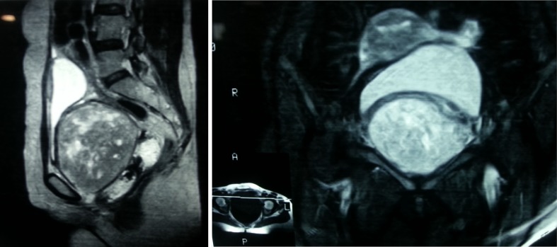
MRI SCAN of the pelvis (Figs. 1 and 2) showed a 10CM * 9 CM mass occupying the pelvis? Arising from vagina, abutting the rectum and urinary bladder but no obvious infiltration of rectal or bladder wall seen.
Figs. 1-2.
MRI scan of the pelvis showing a large homogenous mass ? arising from the vagina
On Examination
General physical and systemic examination was normal, on per vaginum examination a huge submucosal mass was felt, finger could not be passed beyond the mass, cervix could not be examined. Vaginal mucosa was mobile. On per rectal examination an extra luminal huge mass was palpable anteriorly and rectal mucosa was free.
Routine blood investigations were normal. CT thorax was normal.
On cystoscopy both ureteric orifices and bladder mucosa were normal. Patient was planned for exploratory laparotomy.
On opening the abdomen a huge mass was palpable coming from the pelvis. A simple hysterectomy was done to get access; the submucosal mass was arising from the anterior vaginal wall, firm in consistency. Excision of the mass with wide excision of anterior vaginal wall was done. Vaginal vault was repaired and hemostasis secured. A drain was kept in the pelvis and abdominal wall closed in layers. Patient had an uneventful recovery and was discharged on the 8th post operative day. Patient is on regular follow up since the last 9 months and has recovered well.
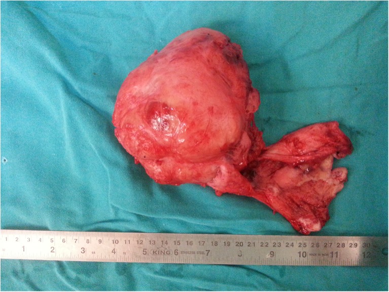
FINAL HISTOPATHOLOGY REPORT- GROSS (Fig. 3)—17*14*9 cm tumor with attached soft tissue with intact capsule. Cut surface is fleshy whorled appearance with small hemorrhagic areas.
Fig. 3.
Gross pathological specimen of hysterectomy and vaginal mass excision
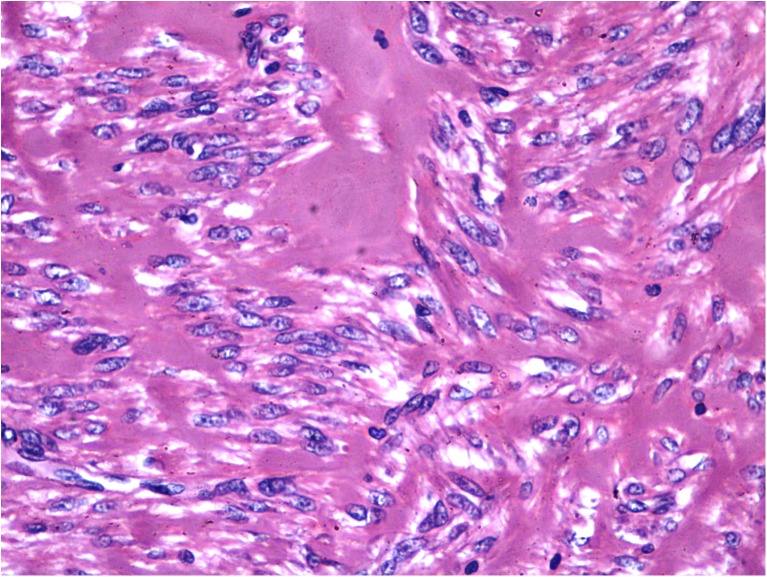
MICROSCOPIC (Fig. 4)—shows presence of hypocellular and hypercellular areas (ANTONY A and ANTONY B) forming verocay bodies. Individual cells have elongated curved nuclei.
Fig. 4.
Microscopic picture showing classical antony a and antony b areas (verocay bodies) confirming the diagnosis of neurilemmoma
IMMUNOHISTOCHEMISTRY- tumor cells express Desmin, SMA and h-Caldesmon and are immunonegative for S-100 and HMB-45.
Discussion
Schwannoma arise from the small to medium size nerves. The tumor is almost always solitary, circumscribed, encapsulated and eccentrically located. It is disease of adulthood and occurs in (20–60) years of age [3]. The tumor does not destroy or affect the nerve because of its peripheral location. It is a slow growing tumor and its early detection is difficult [4]. The symptoms occur late when the tumor becomes large and depends on its location. [1, 4] Retroperitoneal schwannoma may arise from the cervix, vagina,vulva and ureter as reported in few case reports [4–7]. The symptoms of it may be pain especially if it is large, urinary retention, constipation or as in this case, bleeding from vagina [1, 4–8]. Sometimes it is asymptomatic and discovered on routine examination [9]. Imaging (CT scan &MRI) are useful in the diagnosis [10]. Surgery by laparotomy or laparoscopy is the treatment of choice [4, 11]. But incomplete excision may lead to recurrences which occurred in 10% of reported cases. Hence long term follow up is necessary.
Acknowledgments
Conflict of Interest
None declared
Funding
None
Contributor Information
Shashikant Saini, Email: sksaini04@yahoo.co.in.
Mishal Shah, Email: shah_mishal@yahoo.co.in.
Sanjeev Patni, Email: sanjeevnidhi@yahoo.com.
References
- 1.Brooks JS. Disorders of soft tissue (chapter 5) In: Sterberg SS Antonioli DA, Carter D, editors. Diagnostic surgical pathology. 3. USA: Lippincott William andWilkins; 1999. p. 185. [Google Scholar]
- 2.Vogel F, Bouldin TW. The nervous system (chapter28) In: Rubin E, Farber JL, editors. Pathology. 1. USA: Lippincott William and Wilkins; 1988. p. 1474. [Google Scholar]
- 3.Morris JH. The nervous system (chapter29) In: Cortan RS, Kumar V, Robbins SL, editors. Robbins pathologic basis of diseases. 4. USA: W,B.Company; 1989. p. 1445. [Google Scholar]
- 4.Terada S, Suzuki N, Tomimatsu N, Akasofu K. Vaginal schwannoma. Arch Gynecol Obstet. 1992;251(4):203–206. doi: 10.1007/BF02718388. [DOI] [PubMed] [Google Scholar]
- 5.Obeidat BR, Amarin ZO, Jallad MF. Vaginal schwannoma. a casereport. J Reprod Med. 2007;52(4):341–342. [PubMed] [Google Scholar]
- 6.Inoue T, Kato H, Yoshikawa K, Adachi T, Etoh K, Wake N. Retroperitoneal schwannoma bearing at the right vaginal wall. J Obstet Gynaecol Res. 2004;30(6):454–457. doi: 10.1111/j.1447-0756.2004.00230.x. [DOI] [PubMed] [Google Scholar]
- 7.LeMaire WJ, Kreiss C, Commodore A, Barker EA. Neurilemmoma: an unusual benign tumor of the cervix. Alaska Med. 2002;44(3):63–65. [PubMed] [Google Scholar]
- 8.Droegeueller W,Herbst AL,Mishell DR and Stenchever MA.In:Benign gynecological lesions (chapter17). Comprehensive Gynecol. Mosby: USA;1987,447
- 9.Ooi LL, Mack PO. Benign retroperitoneal neurilemmoma–a case report. Ann Acad Med Singapore. 1990;19(3):410–412. [PubMed] [Google Scholar]
- 10.Nasu K, Arima K, Yoshimatsu J, Miyakawa I. CT and MRI findings in a case of pelvic schwannoma. Gynecol Obstet Invest. 1998;46(2):142–144. doi: 10.1159/000010003. [DOI] [PubMed] [Google Scholar]
- 11.Melvin WS. Laparoscopic resection of a pelvic schwannoma. Surg Laparosc Endosc. 1996;6(6):489–491. doi: 10.1097/00019509-199612000-00015. [DOI] [PubMed] [Google Scholar]


