Question
A 74-year-old woman with no significant past medical history presented with nail discoloration for 5 years. The discoloration began on her right thumb and progressed to involve all 10 fingernails, sparing the toenails. The patient stated she has been using a variety of ‘natural supplements’ for decades, but denied taking any medications.
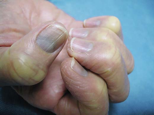
Examination of the nails revealed longitudinal ridging and nonblanching, bluish-gray pigmentation of the proximal nail bed following the shape of the lunula. This was more evident on the thumbs (fig. 1). Examination of the skin, oral and ocular mucosa was negative for dyspigmentation. No organomegaly, neuropsychiatric dysfunction or ocular rings were identified.
Fig. 1.

Longitudinal ridging and nonblanching, bluish-gray pigmentation of the proximal nail bed following the shape of the lunula.
What is your diagnosis?
Answer
Azure lunulae of argyria.
Upon further questioning, the patient revealed that in addition to more than ten over-the-counter supplements, she had been taking roughly 0.5 mg per serving of 400 ppm silver dilutions (Water Oz, Grangeville, Idaho, USA) to treat occasional sore throats for 20 years and discontinued use of this product approximately 2 years prior to presentation.
Blue lunulae of the fingernails are a common, cardinal feature of argyria; toenails are usually spared. The appearance of a bluish-gray discoloration following the distal margin of the lunulae can be an early sign of argyria, along with similar discoloration of the hands and face, especially the conjunctivae [1]. The discoloration is most prominent in sun-exposed areas, as the silver compounds upregulate melanin production, and sunlight catalyzes the reduction of the colorless silver compounds in the dermis to elemental silver, which is then oxidized and bound to form the metallic silver sulfide [2].
The differential diagnosis includes gray nail pigmentation due to cyanosis, or methemoglobinemia, azure lunulae of Wilson's disease (hepatolenticular degeneration) and blue discoloration due to medications such as minocycline, quinacrine hydrochloride, phenolphthalein and local mercury application [2,3,4]. The key to the diagnosis is the patient's stating of her long-term silver supplement use, coupled with the classic physical findings of the nails. A nail biopsy would be diagnostic, but a nail clipping would not, as the nail matrix is not affected and no nail plate changes would be seen [1]. Although azure lunulae of Wilson's disease are similar in appearance to azure lunulae of argyria [5], the average age of diagnosis of Wilson's disease is 20.4 years [6], our patient had no other findings consistent with Wilson's disease such as Kayser-Fleischer rings, organomegaly or neuropsychiatric abnormalities, and the patient's nail findings chronologically coincided following chronic ingestion of a silver solution. Therefore, a slit-lamp examination is unnecessary. If the diagnosis was hepatolenticular degeneration, the diagnosis would need confirmation with serum ceruloplasmin, slit-lamp exam, urinary copper and/or mutation analysis [6].
There are no known chelating agents or other treatments for argyria [7]. 1,064-nm Q-switched Nd:YAG laser may be promising in cutaneous argyria, but it has not been utilized for treating azure lunulae of argyria [7,8]. We decided not to offer this treatment to our patient, as we were concerned of possible side effects. Recently, a case of partial necrosis of the hallux was reported in a patient with pigmented onychomycosis treated with 1,064-nm Q-switched Nd:YAG laser. The authors hypothesize that the nail pigment acted as a target for selective photothermolysis [9].
Patients consume silver supplements for a wide variety of reasons ranging from ‘toxin elimination’ to ‘immune system strengthening’ [10]. Argyria is permanent, and silver toxicity can rarely cause retinal toxicity, hepatitis, nephritis and neurotoxicity [10]. Therefore, clinicians should be aware of signs of argyria, as alternative medicine is increasingly popular amongst patients who may consume silver supplements knowingly, or unknowingly secondary to unregulated, silver-tainted supplements [10].
Patient Outcome
For our patient, we ordered a serum silver level to assess the patient's current level of silver exposure and instructed her to discontinue the use of any silver-containing products as well as supplements that are not overseen by a quality control-ensuring, external regulating body. We have decided not to pursue laser therapy in light of its possible risks.
Statement of Ethics
The patient kindly provided us with the consent to describe her case and publish the depicted image.
Disclosure Statement
The authors have no conflicts of interest. This project received no funding for its production.
References
- 1.Koplon BS. Azure lunulae due to argyria. Arch Dermatol. 1966;94:333–334. [PubMed] [Google Scholar]
- 2.Kim Y, Suh HS, Cha HJ, Kim SH, Jeong KS, Kim DH. A case of generalized argyria after ingestion of colloidal silver solution. Am J Ind Med. 2009;52:246–250. doi: 10.1002/ajim.20670. [DOI] [PubMed] [Google Scholar]
- 3.Whelton MJ, Pope FM. Azure lunules in argyria. Corneal changes resembling Kayser-Fleischer rings. Arch Intern Med. 1968;121:267–269. [PubMed] [Google Scholar]
- 4.Nisar MS, Iyer K, Brodell RT, Lloyd JR, Shin TM, Ahmad A. Minocycline-induced hyperpigmentation: comparison of 3 Q-switched lasers to reverse its effects. Clin Cosmet Investig Dermatol. 2013;6:159–162. doi: 10.2147/CCID.S42166. [DOI] [PMC free article] [PubMed] [Google Scholar]
- 5.Bearn AG, McKusick VA. Azure lunulae; an unusual change in the fingernails in two patients with hepatolenticular degeneration (Wilson's disease) J Am Med Assoc. 1958;166:904–906. doi: 10.1001/jama.1958.62990080001010. [DOI] [PubMed] [Google Scholar]
- 6.Merle U, Schaefer M, Ferenci P, Stremmel W. Clinical presentation, diagnosis and long-term outcome of Wilson's disease: a cohort study. Gut. 2007;56:115–120. doi: 10.1136/gut.2005.087262. [DOI] [PMC free article] [PubMed] [Google Scholar]
- 7.Griffith RD, Simmons BJ, Bray FN, Falto-Aizpurua LA, Yazdani Abyaneh MA, Nouri K. 1064 nm Q-switched Nd:YAG laser for the treatment of Argyria: a systematic review. J Eur Acad Dermatol Venereol. 2015;29:2100–2103. doi: 10.1111/jdv.13117. [DOI] [PubMed] [Google Scholar]
- 8.Bristow IR. The effectiveness of lasers in the treatment of onychomycosis: a systematic review. J Foot Ankle Res. 2014;7:34. doi: 10.1186/1757-1146-7-34. [DOI] [PMC free article] [PubMed] [Google Scholar]
- 9.Leverone AP, Guimaraes DA, Bernardes-Engemann AR, Orofino-Costa R. Partial necrosis of the hallux in a patient treated with laser for onychomycosis: is this procedure really worthwhile? Dermatol Surg. 2015;41:869–872. doi: 10.1097/DSS.0000000000000383. [DOI] [PubMed] [Google Scholar]
- 10.Griffith RD, Simmons BJ, Yazdani Abyaneh MA, Bray FN, Falto-Aizpurua LA, Nouri K. Colloidal silver: dangerous and readily available. JAMA Dermatol. 2015;151:667–668. doi: 10.1001/jamadermatol.2015.120. [DOI] [PubMed] [Google Scholar]


