Abstract
Background
Serum and urine Blastomyces antigen concentrations can be used to diagnose blastomycosis in dogs.
Objectives
Blastomyces antigen concentrations correlate with clinical remission in dogs during antifungal treatment, and detect disease relapse after treatment discontinuation.
Animals
21 dogs with newly diagnosed blastomycosis monitored until clinical remission (Treatment Phase), and 27 dogs monitored over 1 year from the time of antifungal discontinuation or until clinical relapse (After Treatment Phase).
Methods
Prospective study. Dogs were monitored monthly during treatment and every 3 months after treatment discontinuation, with a complete history, physical exam, chest radiographs, and ocular exam. Urine and serum Blastomyces antigen concentrations were measured at each visit using a quantitative enzyme immunoassay.
Results
At enrollment in the Treatment Phase, Blastomyces antigen was positive in all 21 urine samples (100% sensitivity; 95% CI 85–100%), and in 18 of 20 serum samples (90% sensitivity; 95% CI 70–97%). At 2–4 months of treatment, urine antigen was more sensitive for clinically detectable disease (82%; CI 60–94%) than serum antigen (18%; CI 6–41%). The sensitivity of the urine test for clinical relapse was 71% (CI 36–92%), with close to 100% specificity (CI 84–100%) during after treatment surveillance in this population.
Conclusions
Urine Blastomyces antigen testing has high sensitivity for active disease at the time of diagnosis and during treatment, and moderate sensitivity but high specificity for clinical relapse. Urine testing should be useful at the time of diagnosis, when treatment discontinuation is being considered, and anytime there is poor clinical response or suspicion of relapse.
Keywords: Canine, Monitoring, Systemic fungal infection
Abbreviation
- UW‐VMTH
University of Wisconsin Veterinary Medical Teaching Hospital
Blastomycosis is a frequently diagnosed systemic fungal infection of dogs. The causative agent is Blastomyces dermatitidis, a dimorphic fungus that exists in an environmental mycelial form and a mammalian host‐associated yeast form. The disease is endemic in several regions within the United States, including the Ohio and Mississippi River Valleys, Mid‐Atlantic states, and regions of upstate New York.1, 2, 3
The gold standard for diagnosis of blastomycosis in dogs is either cytologic or histopathologic identification of the organism. In the absence of organism identification, diagnosis has been accomplished historically through a combination of clinical signs, radiographic findings, and serum antibodies.2, 4, 5, 6 Recently an antigen test has been developed, which detects a cell wall galactomannan, and has been found to have a high degree of both sensitivity and specificity for the diagnosis of blastomycosis in dogs and humans.7, 8, 9, 10 Although both urine and serum can be assayed to evaluate the presence of Blastomyces antigen, urine antigen measurement appears to have an increased sensitivity relative to serum antigen for the diagnosis of blastomycosis in dogs.8
In human case reports,11, 12 and in a pilot study in dogs,8 Blastomyces antigen concentrations decreased during antifungal treatment, suggesting that antigen concentrations could be used to monitor the progression or remission of clinical disease. We hypothesized that Blastomyces antigen concentrations would correlate with clinical remission in infected dogs treated with oral antifungal drugs, and that Blastomyces antigen testing would be useful in detecting clinical disease relapse after treatment discontinuation. Therefore, the purpose of this study was to prospectively monitor serum and urine Blastomyces antigen concentrations and clinical status during treatment in dogs with newly diagnosed blastomycosis, and to monitor dogs in clinical remission for 1 year after treatment discontinuation, to determine the utility of these tests for detecting clinically active disease.
Methods
Inclusion Criteria
Dogs evaluated at the University of Wisconsin Veterinary Medical Teaching Hospital (UW‐VMTH) with a diagnosis of blastomycosis, as confirmed by cytology or histopathology, were prospectively enrolled in this study. All dogs had a complete history, physical exam, ophthalmic and dermatologic evaluation, and thoracic radiographs performed; additional testing, to include neurologic exam, long bone radiographs, or prostatic ultrasound, was performed as indicated by clinical presentation. Board‐certified radiologists interpreted all imaging modalities, and board‐certified ophthalmologists performed all complete ophthalmic exams. Dogs were treated with either fluconazole (5 mg/kg PO q12h) or itraconazole (5 mg/kg PO q24h) at the discretion of the attending clinician.
Clinical Monitoring
Dogs were monitored during 2 phases: from the time of diagnosis until treatment discontinuation (Treatment Phase), for 12 months after treatment discontinuation (After Treatment Phase), or during both the phases. Dogs in the Treatment Phase were evaluated at the UW‐VMTH either before treatment or within 2 weeks of initiation of systemic antifungal treatment by the primary care veterinarian, and every month throughout the treatment period. During the After Treatment Phase, dogs were evaluated every 3 months for 1 year after the time of treatment discontinuation. At each recheck, a complete history, physical exam with indirect fundic exam, and thoracic radiographs were performed; additional specialty exams and imaging (eg, long bone radiographs) were performed as indicated by clinical presentation. At the time of each evaluation, dogs were categorized either as having clinically active disease or no clinically detectable disease (ie, in clinical remission), defined as the absence of clinical signs referable to Blastomyces infection, to include fever, cough, tachypnea, skin lesions, lameness, or neurologic signs, and lung or bone lesions that were resolved or static on imaging over 2 rechecks. Treatment with antifungal medication was continued for 1 month beyond the date of clinical remission, or for a minimum of 3 months after diagnosis, whichever was longer.
In addition to clinical staging tests, serum and urine samples were obtained at the initial evaluation and at every monthly recheck during the Treatment Phase, or at every 3‐month recheck during the After Treatment Phase. Samples were frozen at −20°C for Blastomyces antigen testing.
Blastomyces Urine and Serum Antigen Assays
Frozen serum and urine samples were shipped in batches on dry ice to MiraVista Diagnostics (Indianapolis, IN). Blastomyces antigen concentrations were determined using an enzyme immunoassay,8 which had been modified to yield quantitative results.10 Briefly, samples were pretreated with EDTA at 104°C in order to dissociate antigen‐antibody complexes before immunoassay, which increased the sensitivity of the assay.10 The linear range of quantitation for the assay was 0.20–14.70 ng/mL in both serum and urine. Concentrations between the assay cut off for the day and 0.2 ng/mL were reported as positive but below the limit of quantitation (BLQ); these were encoded as 0.19 ng/mL for the purposes of statistical analyses. Concentrations above 14.70 ng/mL were reported as above the limit of quantitation, and were encoded as 14.71 ng/mL for analyses.
Statistical Analyses
Population characteristics, duration and dosages of antifungal treatment, time until clinical remission, and Blastomyces serum and urine antigen measurements at specific time points are reported as medians with observed ranges. Antigen measurements at the time of diagnosis, 2–4 months after starting treatment, and at the time of clinical remission were compared using the Friedman test for repeated measures, with a Dunn's multiple comparison posthoc test. Antigen status at drug discontinuation was correlated with clinical relapse during the After Treatment Phase by a Fisher's exact test with P < .05 considered significant. The correlation between baseline urinary antigen concentrations and time for clinical remission was determined using a Spearman rank correlation coefficient.
Sensitivity and specificity of both urine and serum antigen tests for active clinical disease were determined both at 2–4 months of treatment and at last follow‐up during the After Treatment Phase; specificity was further evaluated for all dogs at the time of clinical remission. Dogs were excluded from analyses if they died or were lost to follow‐up before remission, or if they missed an evaluation such that the month of clinical remission could not be determined.
Results
Enrolled Patients
Thirty dogs were prospectively enrolled at the time of blastomycosis diagnosis, and 21 of these dogs completed the Treatment Phase of the study (Fig 1). Nine dogs did not complete the Treatment Phase because of protocol violations, to include incomplete follow‐up to remission (n = 4); drug discontinuation before confirmation of clinical remission (n = 1), inability to determine the month of remission (CNS blastomycosis followed with MRI, but not monthly; n = 1), and death or euthanasia before remission (n = 3 dogs, 1 each with histiocytic sarcoma, osteosarcoma, and sudden death at home without postmortem). In the 21 dogs, followed in the Treatment Phase, definitive diagnosis was made by cytology of a skin lesion or peripheral lymph node (n = 15 dogs), cytology of an endotracheal wash or lung aspirate (n = 2), or histopathology of the eye or bone (n = 4). Eight of these 21 dogs were included in a previous retrospective study comparing the efficacy of fluconazole and itraconazole for the treatment of blastomycosis.13
Figure 1.

Enrollment overview of dogs with blastomycosis infection recruited for serum and urine Blastomyces antigen monitoring.
Fourteen additional dogs were enrolled prospectively at the time of treatment discontinuation after clinical remission; of these dogs, 12 completed the After Treatment Phase (1 dog was lost to follow‐up and 1 was euthanized for congestive heart failure). In addition, 15 dogs that completed the Treatment Phase of the study were also monitored after treatment (Fig 1). Therefore, a total of 27 dogs were evaluated during the After Treatment Phase. The characteristics of dogs enrolled in each Phase of the study are shown in Table 1.
Table 1.
Clinical characteristics of 21 dogs monitored during treatment for blastomycosis (Treatment Phase) and 27 dogs monitored after completing antifungal treatment for blastomycosis (After Treatment Phase).
| Treatment Phase (n = 21) | After Treatment Phase (n = 27) | |
|---|---|---|
| Age | 4.8 years (1.0–11.0) | 3.5 years (1.0–11.0) |
| Sex | MN (8) | MN (14) |
| MI (3) | MI (3) | |
| FS (8) | FS (8) | |
| FI (2) | FI (2) | |
| Breed | Labrador Retriever (8) | Labrador Retriever (5) |
| Great Dane (2) | Mixed breed (4) | |
| Border Collie, Boxer, Cocker Spaniel, English Setter, Golden Retriever, Greyhound, Mastiff, Newfoundland, Peekapoo, Weimaraner, Wheaten Terrier (1 each) | Wirehaired Pointing Griffon (2) | |
| Great Dane (2) | ||
| Border Collie, Border Terrier, Boxer, Cocker Spaniel, English Bulldog, English Setter, Golden Retriever, Greyhound, Leonberger, Newfoundland, Peekapoo, Vizsla, Weimaraner, Wheaten Terrier (1 each) | ||
| BW | 32.8 kg (6.0–53.4) | 32.8 kg (6.0–56.0) |
| Treatment | Fluconazole (14) | Fluconazole (16) |
| Itraconazole (3) | Itraconazole (7) | |
| Itraconazole, then fluconazole (4) | Itraconazole, then fluconazole (4) |
Median (range).
Baseline Blastomyces Antigen Results
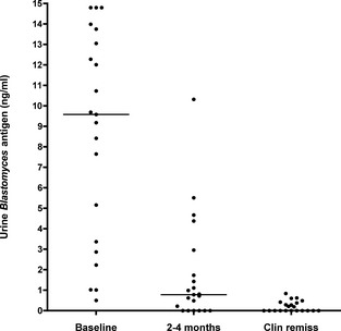
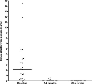
At the time of enrollment in the Treatment Phase, urine samples from all 21 dogs were positive for Blastomyces antigen (100% sensitivity; 95% CI: 85–100%), with a median concentration of 9.58 ng/mL (range, 0.50 to >14.70 ng/mL). Eighteen of 20 available serum samples were also positive (90.0% sensitivity; 95% CI: 70–97%), although two of these were BLQ, with a median serum Blastomyces antigen of 2.15 ng/mL (range, 0.00–14.57).
Treatment
Most dogs in the Treatment Phase were given fluconazole as the sole or primary antifungal agent (n = 18 dogs, median dosage 10.0 mg/kg/day, range 6.6–13.3 mg/kg/day). Four of these dogs were initially treated with itraconazole by the primary care veterinarian but were switched to fluconazole shortly after diagnosis because of lower cost.13 Only 3 dogs in the Treatment Phase were given itraconazole alone (4.1–10.5 mg/kg/day).
Urine Blastomyces Antigen Concentrations during Treatment
During the Treatment Phase, the median time to clinical remission was 154 days (range, 35–249 days), and the median duration of antifungal treatment was 199 days (range, 90–297 days). There was a modest but significant positive correlation between baseline urinary antigen concentrations and time to clinical remission (r = 0.49, P = .023; n = 21).
Urine Blastomyces antigen levels decreased dramatically over time during treatment, with median concentrations significantly lower at 2–4 months of treatment (0.78 ng/mL) and at clinical remission (0.00 ng/mL), compared with baseline (P < .01 and P < .001, respectively; Fig 2). At or around the 3‐month recheck (the minimum treatment time recommended by many clinicians),2, 5, 13 only 4 of 21 dogs were in clinical remission when both physical exam and chest radiographs were considered. The sensitivity of the urine antigen test for clinically active disease at 2–4 months of treatment was 82% (95% CI: 60–94%), with a specificity of 75% (albeit with low numbers of dogs in remission; CI: 30–95%). At the time of clinical remission, 10 of 21 dogs (48%) had detectable residual Blastomyces urinary antigen concentrations, ranging from 0.19 to 0.84 ng/mL, for a urine antigen specificity of 52% (CI: 32–72%) when clinically detectable disease is taken as the gold standard.
Figure 2.

Urine Blastomyces antigen concentrations over time in 21 infected dogs treated with fluconazole or itraconazole. Urine concentrations were significantly lower at 2–4 months of treatment (median 0.78 ng/mL, range 0.0–10.32; P < .01) and at clinical remission (median 0.0 ng/mL, range 0.0–0.84; P < .001), compared with baseline.
Serum Blastomyces Antigen Concentrations during Treatment
Serum Blastomyces antigen levels also decreased during the Treatment Phase, with significantly lower median concentrations at 2–4 months of treatment (0.0 ng/mL, range 0.0–0.56) and at clinical remission (all values negative, median concentration 0.0 ng/mL), compared with baseline (P < .001; Fig 3). At the 2–4 month recheck, the sensitivity of the serum antigen test for clinically active disease was only 18% (CI: 6–41%). At the time of clinical remission, none of the 21 dogs had detectable residual Blastomyces serum antigen concentrations (100% specificity; CI: 51–100%).
Figure 3.

Serum Blastomyces antigen concentrations over time in 21 infected dogs treated with fluconazole or itraconazole. Serum concentrations were significantly lower at both 2–4 months of treatment and at clinical remission, compared with baseline (P < .001).
Blastomyces Antigen Concentrations after Treatment Discontinuation
Twenty‐seven dogs were followed in the After Treatment Phase for a median of 12 months from the time of treatment discontinuation (range, 2–12 months, including dogs with early clinical relapse). Twenty of the 27 dogs had been treated solely or primarily with fluconazole (median dosage 10.0 mg/kg/day, range 6.6–13.3) until clinical remission (Table 1). The urine and serum antigen tests were negative at all time points in all dogs that stayed in clinical remission throughout follow‐up, except for a single dog that had 1 BLQ urine antigen sample found at the 9 month recheck, which was again undetectable at the 12 month recheck (ie, close to 100% specificity for both urine and serum during after treatment surveillance).
Seven of 27 dogs (26%) showed clinical evidence of relapse within a median of 4 months after stopping treatment (range, 2–12 months). These dogs had been treated with fluconazole (n = 4), itraconazole (n = 2), or itraconazole followed by fluconazole (n = 1). Clinical signs at the time of relapse included subcutaneous nodules (n = 5), tachypnea and recurrent pulmonary lesions (n = 1), and chorioretinitis (n = 1). The urinary antigen test was positive at the time of clinical relapse in 5 of 7 cases (71% sensitivity; CI: 36–92%), whereas the serum antigen test was positive in only 3 of 7 relapsed cases (43% sensitivity; CI: 16–75%). Of the 7 dogs that relapsed, only 1 had a rising urinary antigen concentration at the preceding recheck (at 3 months) before clinical relapse (noted at 4 months). Furthermore, for dogs that completed both phases of the study, the baseline urine antigen concentration at the time of diagnosis was not apparently higher between dogs that later relapsed (median 9.18 ng/mL, range 1.01–12.28, n = 5) and dogs that did not relapse over the 12 month observation period after treatment (median 12.53 ng/mL, range 0.50 to >14.70, n = 10).
There was no statistical correlation between residual positive urinary antigen concentrations at the time of drug discontinuation and later clinical relapse (P = 1.00). Of the 7 dogs that relapsed, only 2 had positive urine antigen concentrations at the time of drug discontinuation; conversely, of 8 dogs with clinically detectable urinary Blastomyces antigen at the time of drug discontinuation (range 0.2–1.44 ng/mL), only 3 dogs relapsed over the subsequent year. The only dog with a urine antigen concentration >1 ng/mL at treatment discontinuation (1.44 ng/mL) relapsed within 2 months.
Discussion
The purpose of this study was to monitor serum and urine Blastomyces antigen concentrations and clinical status during antifungal treatment in dogs with newly diagnosed blastomycosis, and to follow dogs in clinical remission for 1 year after treatment discontinuation, to determine the utility of Blastomyces antigen tests for detecting clinically active disease. Similar to prior reports in people and dogs,7, 8, 9, 10 we found urine Blastomyces antigen to be highly sensitive for the diagnosis of blastomycosis (100% sensitivity in this population). The sensitivity for serum antigen test was also quite high (about 90%), which is similar to previous reports (87%).8 The magnitude of urinary antigen concentration at diagnosis was modestly correlated with time to clinical remission, which suggests that dogs with higher urinary antigen concentrations may require longer duration of treatment.
We found a significant decrease in both the urine and serum Blastomyces antigen concentrations during treatment. This is consistent with case reports in humans11, 12 and in a retrospective study in dogs, using an older version of the same assay.8 In our study, serum antigen concentrations had a sensitivity of only 18% for active disease during treatment, as assessed 2–4 months into treatment. Therefore, serum Blastomyces antigen monitoring is not as helpful as urine antigen testing as a means to guide treatment duration.
Urine antigen concentrations showed better sensitivity for clinically detectable disease (82%) than did serum antigen concentrations. There were 3 dogs that had clinically detectable disease but negative urine Blastomyces antigen results during treatment; however, the clinically detectable disease in all cases was based on abnormal chest radiographs that were found to be static at the next monthly recheck. Taking this into consideration, the sensitivity of the urine antigen for clinically active disease during treatment approaches 100%. Even with this consideration, a urine antigen test alone, without clinical exam or chest radiographs, cannot be recommended for sole monitoring during treatment. The urine antigen test had a specificity of 75% during treatment, and a specificity of 52% at the time of clinical remission, ie, it was positive in some dogs with no clinically detectable disease. This may reflect the detection of continually excreted antigen from the cell wall of deceased organisms, or could be a true positive given the limitations of clinical exam to detect residual infection.
The percentage of dogs with a relapse of clinical signs after stopping antifungal treatment (26%) is comparable to that reported in retrospective studies of dogs treated with itraconazole and other antifungal drug combinations (20–24%).2, 5 Blastomyces urine antigen (71% sensitivity) appeared to be more useful than serum antigen (43% sensitivity) to confirm clinical relapse, although these numbers are imprecise, given the relatively small number of dogs that relapsed. Importantly, a consistently negative urine antigen was close to 100% specific for inactive disease during follow‐up monitoring. Only 1 dog showed a rise in urine antigen before clinically detectable relapse, but we only monitored dogs every 3 months during the period after treatment. It is possible that more frequent monitoring of urinary antigen might have been more sensitive to predict relapse before overt clinical disease.
The results of this study should be interpreted in light of several limitations. First of all, not all dogs were treated with the same antifungal drug protocol, and the sample size was too small to observe a large number of dogs with clinical relapse. Second, because not all baseline samples could be obtained before any antifungal treatment, a 2‐week grace period was allowed so that referred cases that had recently begun treatment could be included. This likely had little effect on urine antigen testing, since all dogs had positive results whether or not they had received initial doses of antifungal treatment before referral. It is possible that recent treatment led to false negative results on the serum antigen test; however, of 2 dogs with negative serum results at baseline, only 1 had recently started antifungal drugs. Third, the time of clinical remission for lung disease was based on the second of 2 static chest radiographs taken 1 month apart (as is standard clinical practice). If “actual” clinical remission were taken retrospectively as the time of the initial static chest radiograph, assay performance improves. In addition, dogs followed in the After Treatment Phase were only evaluated every 3 months. More frequent testing may have demonstrated a rising antigen concentration before the detection of clinical relapse in more dogs. Finally, these remission outcomes may not be representative of the experience in primary care practice, because clients willing to enroll their dogs in a research study may be more motivated to comply with medications and adhere to clinical recommendations.
Overall, these data indicate that urine Blastomyces antigen testing has a high degree of sensitivity for active disease both at the time of diagnosis (100%) and during treatment (at least 82%), and moderate sensitivity for clinical relapse (71%). Urine antigen results can be weakly positive (<1 ng/mL) in dogs that are in clinical remission at the time of drug discontinuation, and this finding is not necessarily predictive of relapse. An overtly positive urinary Blastomyces antigen test is quite specific for relapse, at least as observed in this small population of dogs. On the basis of these results, the authors recommend that Blastomyces urinary antigen concentrations be monitored, at minimum, at the time of diagnosis and when treatment discontinuation is being considered, as well as at any time during treatment when clinical efficacy is in doubt. We recommend the following goals before treatment discontinuation: a normal physical exam including fundic evaluation, normal or static chest radiographs, and, to be conservative, a negative urinary antigen concentration. The urine test should be repeated in any dogs with clinical suspicion of relapse, especially if Blastomyces organisms cannot yet be detected.
Acknowledgment
Funding for this study was provided by an investigator‐driven grant from MiraVista Diagnostics, Indianapolis, IN.
Conflict of Interest: Dr Wheat and Ms Kirsch are employees of MiraVista Diagnostics, which provided funding for this study, and which developed and offers Blastomyces antigen testing for dogs. This study was originated and designed at the University of Wisconsin‐Madison, where all data analyses and interpretations were performed.
A portion of this work was presented in abstract form at the 2010 ACVIM Forum in Anaheim CA
References
- 1. Legendre AM. Blastomycosis In: Greene CE, ed. Infectious Diseases of the Dog and Cat, 3rd ed St. Louis, MO: Saunders Elsevier; 2006:569–576. [Google Scholar]
- 2. Legendre AM, Rohrbach BW, Toal RL, et al. Treatment of blastomycosis with itraconazole in 112 dogs. J Vet Intern Med 1996;10:365–371. [DOI] [PubMed] [Google Scholar]
- 3. Werner A, Norton F. Blastomycosis. Compend Contin Educ Vet 2011;33:E1–E5. [PubMed] [Google Scholar]
- 4. Crews LJ, Feeney DA, Jessen CR, et al. Radiographic findings in dogs with pulmonary blastomycosis: 125 cases (1989‐2006). J Am Vet Med Assoc 2008;232:215–221. [DOI] [PubMed] [Google Scholar]
- 5. Arceneaux KA, Taboada J, Hosgood G. Blastomycosis in dogs: 115 cases (1980‐1995). J Am Vet Med Assoc 1998;213:658–664. [PubMed] [Google Scholar]
- 6. Crews LJ, Feeney DA, Jessen CR, et al. Utility of diagnostic tests for and medical treatment of pulmonary blastomycosis in dogs: 125 cases (1989‐2006). J Am Vet Med Assoc 2008;232:222–227. [DOI] [PubMed] [Google Scholar]
- 7. Durkin M, Witt J, Lemonte A, et al. Antigen assay with the potential to aid in diagnosis of blastomycosis. J Clin Microbiol 2004;42:4873–4875. [DOI] [PMC free article] [PubMed] [Google Scholar]
- 8. Spector D, Legendre AM, Wheat J, et al. Antigen and antibody testing for the diagnosis of blastomycosis in dogs. J Vet Intern Med 2008;22:839–843. [DOI] [PubMed] [Google Scholar]
- 9. Bariola JR, Hage CA, Durkin M, et al. Detection of Blastomyces dermatitidis antigen in patients with newly diagnosed blastomycosis. Diagn Microbiol Infect Dis 2011;69:187–191. [DOI] [PubMed] [Google Scholar]
- 10. Connolly P, Hage CA, Bariola JR, et al. Blastomyces dermatitidis antigen detection by quantitative enzyme immunoassay. Clin Vaccine Immunol 2012;19:53–56. [DOI] [PMC free article] [PubMed] [Google Scholar]
- 11. Mongkolrattanothai K, Peev M, Wheat LJ, et al. Urine antigen detection of blastomycosis in pediatric patients. Pediatr Infect Dis J 2006;25:1076–1078. [DOI] [PubMed] [Google Scholar]
- 12. Tarr M, Marcinak J, Mongkolrattanothai K, et al. Blastomyces antigen detection for monitoring progression of blastomycosis in a pregnant adolescent. Infect Dis Obstet Gynecol 2007;2007:89059. [DOI] [PMC free article] [PubMed] [Google Scholar]
- 13. Mazepa AS, Trepanier LA, Foy DS. Retrospective comparison of the efficacy of fluconazole or itraconazole for the treatment of systemic blastomycosis in dogs. J Vet Intern Med 2011;25:440–445. [DOI] [PubMed] [Google Scholar]


