Abstract
Desmoid tumors are rare potentially aggressive benign tumors. Various etiologies and recurrent factors have been presented and discussed. A case of an abdominal desmoid tumor with vascular mesenteric invasion in a 32-year-old female, over 2 years after pregnancy is presented. Pre-operative biopsy was not contributive, diagnosis was made after surgery. Resection required two vascular bypasses. Desmoid tumors appear frequently in women of child-bearing age (during or after pregnancy), hormonal signaling is probably involved, but pathways remain unknown. Multiple predictive factors of recurrence are discussed but not strongly identified due to underpowered studies: resection margins, age, sex, tumor’s size and location. Recent development is in favor of a non-aggressive treatment such as ‘wait and see’ procedures. Without radical treatment, these tumors could generate bowel compression or perforation. Due to their location and high risk of complication, surgery is the most fitted option.
Introduction
Desmoid tumors are rare benign and potentially locally aggressive and multifocal tumors. They may relapse but are never metastatic. Most of the time, treatment is conservative. In some rare cases, surgery must be performed. We report here the case of a 32 years old woman with no past medical history who was admitted for a sporadic intra-abdominal tumor.
Case report
The patient was a 32-year-old female with no medical past history outside a normal delivery history (58 kg, 172 cm, BMI 19.6). She fortuitously discovered an abdominal mass in her left hypochondria. Clinical examination was normal.
An abdominal echography showed a hypoechoic and heterogeneous lesion of 53 × 42 mm. Abdominal CT scan (Fig. 1) confirmed this tumor of the mesenteric root, well delineated and homogeneously enhanced with contrast administration. The lesion seemed to repress the superior mesenteric vein and invade the mesenteric artery. No metastases were found.
Figure 1:
Enhanced CT scan (arterial time) showing the tumor and its relations to the superior mesenteric artery (blue arrows).
Two weeks later, the MRI found tumoral progression (66 × 45 × 55 mm) hyper intense on T2-weighted images and enhanced by gadolinium. The Positon emission tomography - Fluorodeoxyglucose showed hypermetabolim of the lesion with 3.42 standard uptake value, and again an increase in tumor size (83 × 61 mm).
The first evoked diagnosis was a pseudopapillar pancreatic lesion. Echoendoscopy found a heterogeneous tumor, without calcification and no nodal involvement. Fine needle biopsy was performed, but not contributive.
As the tumor was highly proliferative, surgical resection was scheduled.
Due to high risk of vascular resection, laparoscopic approach was not considered as reasonable even if discussed in literature.
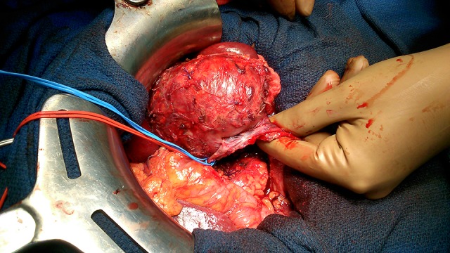
A voluminous lesion was found in the left hypochondra. Superior mesenteric vein and artery were invaded by the tumor and could not be dissociated from it (Fig. 2). The first jejunal segment and the angle of Treitz were incarcerated in the lesion. The only strategy was an en-bloc resection including mesenterical resection. We performed intra-operative control of the resection margins (vascular and intestinal) by frozen section exam that were free of tumor. Due to superior mesenteric artery’s diameter, splenic artery was used for arterial reconstruction. Besides, the superior mesenteric vein’s diameter imposed to take a superficial femoral vein graft to perform the veinous bypass.
Figure 2:

Intra-operative view: the tumor is invading mesenteric artery (red loop) and vein (blue loop) imposing their resection.
Because of the vascular grafts, an anticoagulant treatment was started. Vascular grafts were functional on day 1 control CT Scan.
Four days later, because of tachycardia, and abdominal pain, revisional surgery found an important hemoperitoneum with no active bleeding. Post-operative outcome was then simple and the patient was discharged on day 18 and is actually recurrence free.
The anatomopathological results determined that the tumor was a desmoid tumor measuring 7.5 cm. Immunohistochemistry revealed a nuclear staining of β-catenin. It was infiltrating the bowel external muscular layer. Estrogen receptors were not found on the surgical specimen. Resection was considered in tumoral zone because of the lack of encapsulation, whereas intra-operative controls were tumor free.
Discussion
Desmoid tumors are rare well-differentiated and aggressive musculoaponeurotic fibromatosis tumors, considered as grade 1 fibrosarcoma. Etiologic factors have been discussed such as hormonal influence (pregnancy), trauma and hereditary cancer syndrome. They are associated with familial adenomatous polyposis (Gardner’s syndrome, associated polyposis conditions mutation) [1] in 10–20% of patients, developing mainly after prophylactic colectomy. Most of them are sporadic: due to a β-catenine activating mutation.
Most of studies about desmoid tumors are underpowered and contradictive [2–4]. Nevertheless, the only constant finding is the increased risk of women of child-bearing age to develop these tumors, during or after pregnancy; suggesting that unknown hormonal pathways modulate those tumors [4–6].
Estrogen and antiestrogen binding sites were respectively found by Lim et al. [7] in 33% and 80% of these tumors. Sorensen and Keller [2] described the estrogenal regulation of desmoid tumor development over 72 patients. However, most of the patients were young female such as in the other studies [1, 3, 4, 8]. Efficiency of antiestrogenic treatment is not explained by estrogenic receptors’ presence, concluding that unknown factors are involved. Fiore et al. [6] concluded that desmoid tumors occurring during or after pregnancy could be safely treated, even with a wait and see procedure: 42% presented progression during pregnancy, 35% had a surgical resection, 20% medical therapy, 45% wait and see procedure. Four out of nine patients had a spontaneous regression.
Desmoid tumors do not develop metastasis but their recurrence rate is important: 19–57% [2, 5, 8]. Recurrence free survival is between 14 and 24 months [3, 4, 6]. Predictive factors of recurrence are: tumoral size (>4–5 cm) [2, 6], age (<37 years, for each 5 years increase of age [9] or <32 years [2]), tumor’s location (extra-abdominal desmoid tumors are associated with poorer prognosis [2, 8–9]) and past history of trauma in the area of primary tumor [9].
Some predictive factors are discussed such as the significance of microscopic positive margins after surgical resection. Salas et al. [8] found no different prognosis when margins were positive. According to Salas et al. [8] and Colombo and Gronchi [1], surgical treatment should not be aggressive, as locoregional infiltration would lead to a mutilating surgery with high risk of invaded margins. On the other hand, in a retrospective study on 177 patients in 2012 [10], margin status was the only significant independent prognostic factor. Five years recurrence-free survival with negative margins was 82% versus 52% with positive margins.
In our case, surgical margins were invaded: the tumor was not encapsulated. The surgery was already quite aggressive, involving vascular resections.
Until year 2000, the main treatment of desmoid tumors was surgical resection. The following medical treatments have been described but never compared: radiotherapy, chemotherapy, targeted treatment and antiestrogenics [3, 4]. Most recent development is in favor of a non-aggressive treatment, or ‘wait and see’ procedures. A retrospective study including 142 patients [4] aimed to evaluate the conservative approach. Selected patients for ‘wait and see’ procedure were younger women, with small asymptomatic desmoid tumors located predominantly on the thoracic wall. Fifty percentage of these patients had no tumoral progression after 5-year follow up and spontaneous regression concerned 5% of those lesions [3].
Sporadic intra-abdominal desmoid tumors are rare. Surgery remains the main option. Etiology and prognostic factors are not well known. Further research is needed to improve the knowledge of their physiopathology, and their medical management.
CONFLICT OF INTEREST STATEMENT
None declared.
Funding source
None.
References
- 1.Colombo C, Gronchi A.. Desmoid type fibromatosis: what works best. Eur J Cancer 2009;45:466–7 [DOI] [PubMed] [Google Scholar]
- 2.Sorensen A, Keller J.. Treatment of aggressive fibromatosis: a retrospective study of 72 patients followed for 1-27 years. Acta Orthop Scand 2002;73:213–9 [DOI] [PubMed] [Google Scholar]
- 3.Stoeckle E., Coindre JM, Longy M, Binh MB, Kantor G, Kind M, et al.. A critical analysis of treatment strategies in desmoids tumours: a review of a series of 106 cases. Eur J Surg Oncol 2009;35:129–34 [DOI] [PubMed] [Google Scholar]
- 4.Fiore M, Rimareix F, Mariani L, Domont J, Collini P, Le Péchoux C, et al.. Desmoid type fibromatosis: a front line conservative approach to select patients for surgical treatment. Ann Surg Oncol 2009;16:2587–93 [DOI] [PubMed] [Google Scholar]
- 5.Way JC, Culham B.. Desmoid tumour, the risk or recurrent or new disease with subsequent pregnancy: a case report. Can J Surg 1999;42:51–4 [PMC free article] [PubMed] [Google Scholar]
- 6.Fiore M, Coppola S, Cannell AJ, Colombo C, Bertagnolli MM, George S, et al.. Desmoid type fibromatosis and pregnancy: a multi institutional analysis of recurrence and obstetric risk. Ann Surg 2014;259:973–8 [DOI] [PubMed] [Google Scholar]
- 7.Lim CL, Walker MJ, Mehta RR, Das Gupta TK.. Estrogen and antiestrogen binding sites in desmoid tumors. Eur J Cancer Clin Oncol 1986;22:583–7 [DOI] [PubMed] [Google Scholar]
- 8.Salas S, Dufresne A, Bui B, Blay JY, Terrier P, Ranchere-Vince D, et al. Prognostic factors influencing progression free survival determined from a series of sporadic desmoid tumors: a wait and see policy according to tumor presentation. J Clin Oncol 2011;29:3553–8 [DOI] [PubMed] [Google Scholar]
- 9.Peng PD, Hyder O, Mavros MN, Turley R, Groeschl R, Firoozmand A, et al.. Management and recurrence patterns of desmoids tumors: a multi institutional analysis of 211 patients. Ann Surg Oncol 2012;19:4036–42\ [DOI] [PMC free article] [PubMed] [Google Scholar]
- 10.Mullen JT, Delaney TF.. Desmoid tumor: analysis of prognostic factors and outcomes in a surgical series. Ann Surg Oncol 2012;19:4028–35 [DOI] [PubMed] [Google Scholar]


