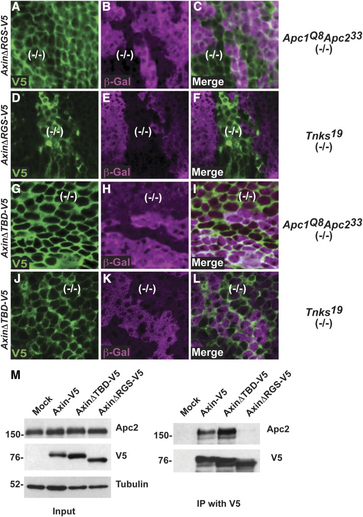
Figure 6.
APC- and Tnks-mediated regulation of Axin are achieved through partially separable mechanisms. Confocal images of third instar larval wing imaginal discs stained with antibodies indicated at bottom left; genotypes are indicated on the right. (A–L) Wing disc labeled with α-V5 (green), α-β-gal (magenta), and merge. Absence of β-gal staining marked Apc1Q8Apc233 mutant clones (A–C and G–I), and Tnks19 mutant clones (D–F and J–L). In contrast with Axin-V5 (Figure 3, A–C), the levels of AxinΔRGS-V5 did not increase inside Apc1Q8Apc233 mutant clones compared to the surrounding wild-type tissue (A–C). The levels of AxinΔRGS-V5 inside Tnks19 mutant clones were increased compared to that of the surrounding wild-type tissue (D–F). In contrast with Axin-V5 (Figure 3, A–C and Figure 2, L–N), the levels of AxinΔTBD-V5 did not increase in Apc1Q8Apc233 mutant clones (G–I) or in Tnks19 mutant clones (J–L) compared to the surrounding wild-type tissue. (M) Immunoprecipitation with V5 antibody from S2R+ cell lysates transfected with Axin-V5, AxinΔTBD-V5, and AxinΔRGS-V5. Apc2 was pulled down with Axin-V5. Deletion of the Tnks binding domain of Axin (AxinΔTBD-V5) had no effect in the interaction between Axin and Apc2. In contrast, deletion of the APC binding domain of Axin (AxinΔRGS-V5) inhibited the interaction with Apc2.

