Supplemental Digital Content is available in the text.
Abstract
Background:
The use of progressive tension sutures has been shown to be comparable to the use of abdominal drains in abdominoplasty. However, the use of barbed progressive tension sutures (B-PTSs) in deep inferior epigastric artery perforator (DIEP) flap donor-site closure has not been investigated.
Methods:
A retrospective chart review was performed on patients with DIEP flap reconstruction in a 3-year period at 2 institutions by 2 surgeons. Patients were compared by method of DIEP donor-site closure. Group 1 had barbed running progressive tension sutures without drain placement. Group 2 had interrupted progressive tension closure with abdominal drain placement (PTS-AD). Group 3 had closure with only abdominal drain placement (AD). Data collected included demographics, perioperative data, and postoperative outcomes.
Results:
Seventy-five patients underwent DIEP reconstruction (25 B-PTS, 25 PTS-AD, and 25 AD). Patient characteristics—age, body mass index, comorbidities, smoking status, and chemotherapy—were not significantly different between groups. Rate of seroma was 1.3% (B-PTS = 0%, PTS-AD = 4%, AD = 0%), wound dehiscence 16% (B-PTS = 8%, PTS-AD = 16%, AD = 24%), and umbilical necrosis 5.3% (B-PTS = 0%, PTS-AD = 0%, AD = 16%). No hematomas were observed in any patients. No statistically significant difference was found between complication rates across groups.
Conclusions:
Use of B-PTSs for abdominal closure after DIEP flap harvest can obviate the need for abdominal drains. Complication rates following this technique are not significantly different from closure using progressive tension suture and abdominal drain placement. This practice can prevent the use of abdominal drains, which can promote patient mobility, increase independence upon discharge, and contribute to patient satisfaction.
Breast reconstruction is one of the 5 most frequently performed reconstructive surgeries in the United States. The deep inferior epigastric artery perforator (DIEP) flap is a popular method of autologous breast reconstruction, with over 5000 procedures performed in 2010. This flap, first described by Koshima and Soeda1 in 1989, and by Allen and Treece2 for breast reconstruction in 1994, avoids some of the donor-site morbidities associated with the transverse rectus abdominis myocutaneous flap.3 However, it is not without complications. A recent meta-analysis comparing DIEP donor sites to elective abdominoplasty described DIEP seroma rates as high as 20% (average 3.7%) and wound dehiscence rates as high as 15% (average 7.2%).4 The DIEP flap and the abdominoplasty dermolipectomy excision territories are very similar, and similar techniques have been used to limit the incidence of postoperative complications in both. These techniques include tissue sealants, abdominal drains, progressive tension sutures (PTSs), and barbed sutures.5
Abdominal wall drains prevent fluid accumulation in the potential dead space created by tissue undermining and flap harvest. Although abdominal drains are effective, they are a potential portal for infection, limit patient mobility, require daily care upon discharge, and are a source of significant patient dissatisfaction. After bilateral DIEP breast reconstruction, a patient may have upwards of 6 drains, including 2 per breast. Avoiding abdominal drains could therefore improve patient satisfaction and decrease morbidity following DIEP flap harvest.
PTSs offer a reliable method for facilitating dead space closure. This technique was first described by Pollock and Pollock6,7 for closure of combined abdominal liposuction and abdominoplasty procedures. It has since been established that abdominal closure using PTS without drains has seroma rate comparable to that of traditional closure with drains.8,9 Additionally, a prospective, randomized double-blind clinical trial found no difference in the rate of seroma or other complications when comparing abdominoplasties performed using (1) drains alone, (2) PTS alone, and (3) PTS plus drains.10
Recently, a PTS technique using knotless running barbed suture has been described.11 Claimed benefits of this technique include prevention of knot complications, decreased operative time, and distribution of tension along the suture length. No-drain abdominoplasty closure using this barbed PTS (B-PTS) technique has been shown to have complication rate comparable to closure with PTS plus drains.12,13 Since B-PTS does not require knot-tying, operative time is either equal to or faster than the standard PTS technique.7,11,13 Furthermore, B-PTS has been shown to be a cost-effective option for skin closure in breast reconstruction.14 However, the technique has not yet been studied in DIEP flap donor-site closure.
The goal of this study was to compare clinical outcomes following DIEP donor-site closure using (1) abdominal drains, (2) PTS plus abdominal drains, and (3) B-PTS without drains. We hypothesize that the B-PTS technique is noninferior to the standard PTS technique and reduces the risk of dehiscence and other wound complications compared with traditional closure with drains alone.
MATERIALS AND METHODS
Clinical Study
Approval was obtained from the Institutional Review Boards at UT Southwestern Medical Center at Dallas and the Mayo Clinic, Rochester, Minn. Patients who underwent DIEP flap reconstruction by the 2 senior authors (M.S.-C. and S.T.) during a 3-year period at 2 institutions were identified. A retrospective chart review was performed on patients selected from this larger case series who had abdominal donor-site closures that could be clearly categorized into 1 of 3 groups based on method of DIEP donor-site closure. The first group had traditional closure with abdominal drain placement (AD) (Fig. 1). The second group had progressive tension suture closure with abdominal drain placement (PTS-AD) (Fig. 2). Finally, the third group (B-PTS) had barbed progressive tension suture closure without abdominal drain placement (Fig. 3). A chart review was conducted to collect data points including demographic information, comorbidities, systemic chemotherapy, and postoperative complications of seroma, hematoma, wound dehiscence, contour deformity, umbilical necrosis, and pain.
Fig. 1.
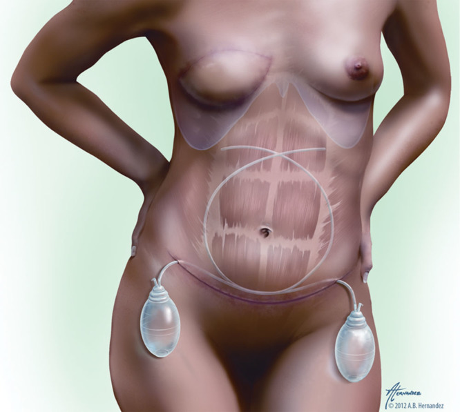
Illustration of traditional abdominal closure with 2 drains. Copyright © 2012 Alexandra Hernandez, M.A., of Gory Details Illustration. Printed with permission of Gory Details Illustration.
Fig. 2.
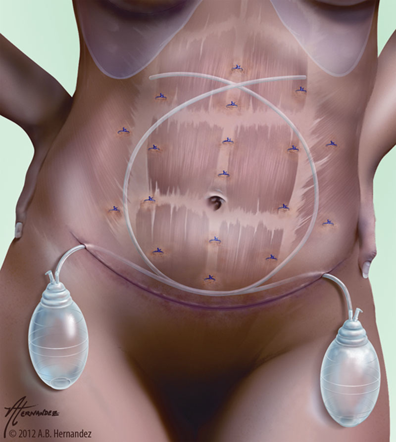
Illustration of interrupted progressive tension sutures with drains for closure of DIEP donor site. Copyright © 2012 Alexandra Hernandez, M.A., of Gory Details Illustration. Printed with permission of Gory Details Illustration.
Fig. 3.
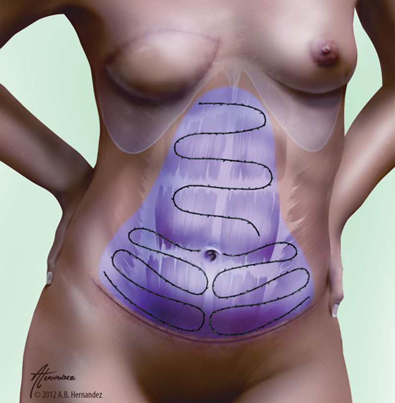
Illustration of no-drain DIEP donor-site closure with running, barbed progressive tension sutures and limited dissection shown in purple. Copyright © 2012 Alexandra Hernandez, M.A., of Gory Details Illustration. Printed with permission of Gory Details Illustration.
Surgical Technique
Patients underwent harvest of the deep inferior epigastric flap in the standard fashion. We prefer a minimal epigastric dissection to spare lateral perforators and maximize vascularity of the abdominal flap (Fig. 4). Donor-site closure in the B-PTS group was performed using a 3-0 V-LOC 180 (Covidien, Mansfield, Mass.), which is a knotless unidirectional barbed suture. The suture was placed in a horizontal running zigzag fashion, advancing the abdominal flap inferiorly with a progressive tension technique by securing Scarpa’s fascia to the anterior rectus sheath. Note that the V-LOC suture can easily break if excess tension is applied directly to it. Therefore, the tension must be applied to the abdominal flap by pulling inferiorly and then applying gentle steady traction to the suture to secure the flap to the abdominal wall. Second, the V-LOC needle is short—after placing it through Scarpa’s fascia, it must be regrasped with forceps before securing it to the abdominal wall. (See Video 1, Supplemental Digital Content 1, which demonstrates the use of running B-PTS for closure of the abdominal donor site without drains. This video is available in the “Related Videos” section of the full-text article on PRSGlobalOpen.com or available at http://links.lww.com/PRSGO/A195.)
Fig. 4.
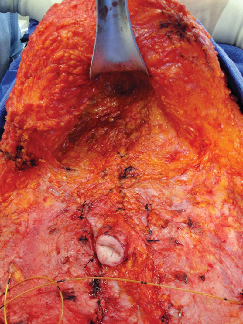
Intraoperative view demonstrating the use of limited epigastric dissection to preserve lateral perforators and maximize the vascularity of the abdominal flap.
Video 1.
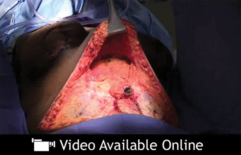
See video, Supplemental Digital Content 1, which demonstrates the use of running barbed progressive tension sutures for closure of the abdominal donor site without drains. This video is available in the “Related Videos” section of the full-text article on PRSGlobalOpen.com or available at http://links.lww.com/PRSGO/A195.
This is especially true when the skin flaps are thick. Once the skin flap was advanced appropriately with this technique, the wound edges were approximated without tension and only a few interrupted 2-0 Vicryl sutures were placed through Scarpa’s fascia and the anterior abdominal wall to minimize risks of fat necrosis or suture abscesses (Fig. 5). Finally, we used a single barbed deep dermal suture and applied Dermabond Prineo (Johnson & Johnson, New Brunswick, N.J.) for skin closure (Fig. 6A, B).
Fig. 5.
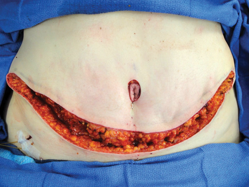
This patient is in a semiflexed position for closure, and the abdominal flap is secured to the abdominal wall with barbed, progressive tension sutures, which minimizes dead space and relieves tension on the wound edges. Note the close approximation of tissue along the left hemiabdominal closure which is achieved without any traditional sutures in Scarpa’s fascia. The right hemiabdominal closure has not been fully completed and a slight gap in the wound edges persists.
Fig. 6.
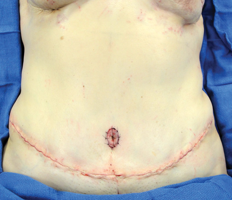
The same patient as in Figure 5 is shown following complete closure without drains using a running barbed suture in the deep dermis, which is subsequently covered with Dermabond Prineo dressing (not shown).
Closure in the PTS-AD group was performed using 2-0 Vicryl interrupted sutures, advancing the abdominal flap with a progressive tension technique. Two 15-French round Blake drains were placed in the PTS-AD and AD groups. In both the PTS-AD and AD groups, Scarpa’s fascia was reapproximated with 3-point sutures, followed by a few 3-0 Vicryl deep dermal interrupted sutures to approximate the skin and finally a single 2-0 V-LOC 90 running dermal suture. Figures 7 and 8 show the intraoperative view of an African American patient who underwent bilateral DIEP reconstruction and donor-site closure with B-PTS and no drains, as well as follow-up results 4 weeks later. Dermabond Prineo dressing was also used. Postoperatively, an abdominal binder was used by all patients for mild compression and support.
Fig. 7.
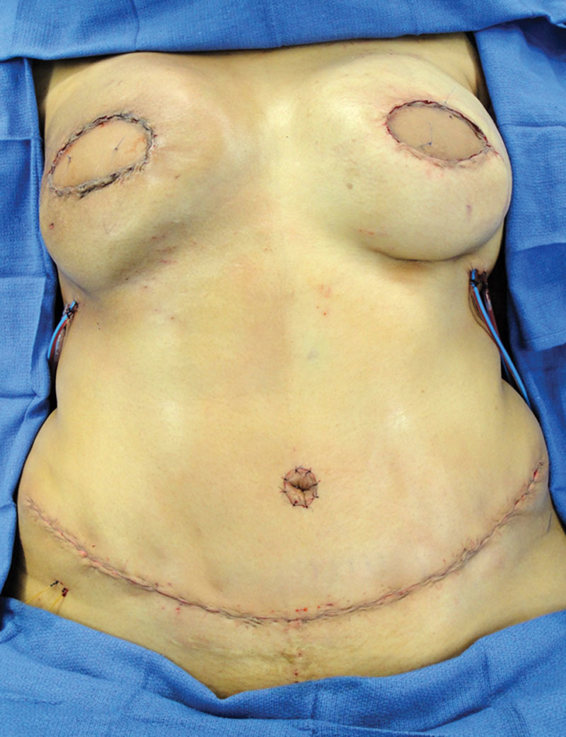
Intraoperative view of an African American patient who underwent bilateral DIEP reconstruction with prior radiation on the right chest and donor-site closure with B-PTS and no drains. The 2 catheters in the right lower quadrant are On-Q pain pump catheters. Note that some small surface contour irregularities and dimpling can occur where the B-PTS suture is more superficial but resolves without intervention once the suture is absorbed.
Fig. 8.
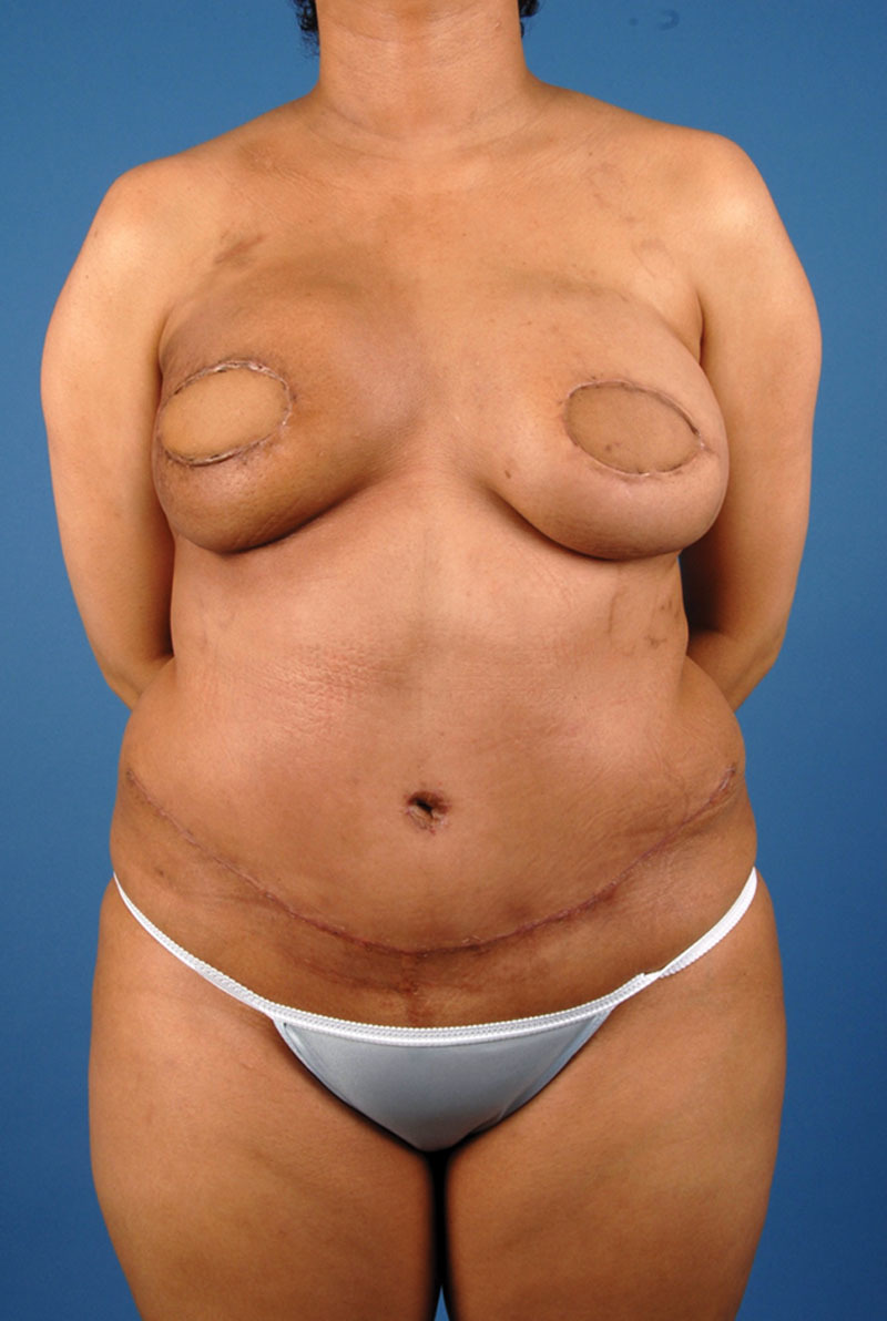
Early postoperative view (4 weeks) of the patient from Figure 7 demonstrating minimal abdominal scarring, resolution of the dimpling along the right lower quadrant, and no seroma formation, with an aesthetically pleasing abdominal contour. Future planned procedures include fat grafting to address contour irregularities in the breast, periareolar advancement flaps, and nipple-areolar complex reconstruction.
Power and Statistical Analysis
The nQuery Advisor program (nQuery Advisor version 7.0; Statistical Solutions Ltd., Boston, Mass.) was used to determine the required sample size for a power of 80%. A meta-analysis of DIEP flap donor-site complication rates showed a seroma rate of about 4% (range, 0–20%). Therefore, assumptions for power analysis were a seroma rate of 4% for the AD group, expected seroma rate of 4% for the PTS-AD and B-PTS groups, effect size of 16%, significance level of 0.05, and power of 80%. The required sample size was found to be 19 in each group. Statistical analysis of study results was performed with Microsoft Excel (Microsoft Corporation, Redmond, Wash.). Patient age, body mass index (BMI), follow-up, and intraoperative blood loss were compared using 2-sided independent t tests. Chi-square and Fisher’s exact t tests for contingency tables were used to examine nominal between-group patient characteristics and postoperative complications. Logistical regression analysis (Epi Info 7.1.2; Centers for Disease Control, Atlanta, Ga.) was used to analyze the correlation between wound dehiscence and patient factors (including wound closure type, age, BMI, diabetes, smoking, and adjuvant chemotherapy).
RESULTS
Clinical Study
Seventy-five patients were included in the study (25 AD, 25 PTS-AD, and 25 B-PTS). Patient characteristics including age, BMI, comorbidities, smoking status, and systemic therapy were not significantly different among the 3 groups (Table 1). However, the mean follow-up period was longer in the AD and PTS-AD groups (AD = 546 days; PTS-AD = 383 days; and B-PTS = 205 days). Follow-up period in the B-PTS group was significantly lower than that in the AD group (P = 0.0001) and the PTS-AD group (P = 0.001). The senior author (M.S.-C.) operated on 46 patients, whereas S.T. operated on 29 patients (Table 2). Both surgeons originally used only abdominal drains and then transitioned to incorporate the PTS technique. Finally, noting minimal drain output in the PTS-AD group, drains were completely omitted in the final group of patients. Closure was performed by residents, fellows, and senior surgeons using all 3 techniques.
Table 1.
Patient Characteristics

Table 2.
Distribution of Patients by Surgeon and Type of Abdominal Closure

No statistically significant difference was found between individual complication rates of the 3 groups (Table 3). Average rate of seroma was 1.3% (AD = 0%, PTS-AD = 4%, and B-PTS = 0%), rate of wound dehiscence was 16% (AD = 24%, PTS-AD = 16%, and B-PTS = 8%), and the rate of umbilical necrosis was 5.3% (AD = 16%, PTS-AD = 0%, and B-PTS = 0%). Adjuvant chemotherapy was significantly associated with the development of wound dehiscence across all groups (P = 0.016). Age, BMI, diabetes, and smoking status were not significantly associated with wound dehiscence. Severe postoperative pain (prompting a postoperative visit to the emergency department) was noted in 1 patient (1.3%) in the AD group and in none of the other groups. Hematoma and significant contour deformity were not observed in any patients.
Table 3.
Incidence of Postoperative Complications (No. Patients)

The overall donor-site complication rate was 24% (AD = 44%, PTS-AD = 20%, and B-PTS = 8%). Comparing to the AD group, this difference was significant for B-PTS (P = 0.008) but not for PTS-AD (P = 0.128). There was also no statistical difference in the complication rates between the B-PTS and the PTS-AD groups (P = 0.417).
Logistical regression analysis (Table 4) showed that overall donor-site complication rate was significantly correlated with wound closure type and adjuvant chemotherapy (P = 0.02) but not BMI, age, smoking status, or diabetes (P > 0.05). The wound dehiscence rate was significantly correlated only with adjuvant chemotherapy (P = 0.009) and was unaffected by wound closure type.
Table 4.
Logistic Regression Analysis of Overall Donor-site Complication and Wound Dehiscence Against Multiple Variables

DISCUSSION
Barbed sutures were first introduced in the 1990s, and today, there are 2 major available barbed sutures in the United States: Quill SRS (Angiotech Pharmaceuticals Inc., Vancouver, Canada) and V-LOC (Covidien, Mansfield, Mass.). The Quill SRS is a bidirectional double-arm suture, whereas the V-LOC is a unidirectional single-arm suture with a welded loop. Claimed benefits of barbed suture include prevention of knot complications, decreased operative time, and more even distribution of tension. It has been shown that PTSs obviate the need for drains in abdominoplasty closures,8 but this technique has not gained traction for DIEP donor-site closures. Therefore, we chose to review and compare a technique in common use (AD), the technique that the senior surgeon (M.S.-C.) had begun using (PTS-AD) and finally the natural evolution toward this novel technique (B-PTS). This design was chosen to mirror the study design chosen by Andrades et al10 in their prospective randomized study of abdominoplasty closure. They reported an additional 50 minutes of additional surgical time with PTS. To minimize the cost of additional operative time, we utilized the running barbed suture (V-LOC) and estimate that this technique required an additional 15–30 minutes in our early experience. However, with practice, the senior surgeon (M.S.-C.) now only requires 10–15 minutes. We did not find the B-PTS or PTS technique interfered with the overall case length, as flap insetting occurred simultaneously.
This study has demonstrated that DIEP donor-site closure with a no-drain B-PTS technique is comparable to the traditional closure drains. There were no instances of seroma in the B-PTS group. One instance of seroma was observed in the PTS-AD group (1.4%). This is consistent with what has been previously reported in the literature.4 These results reaffirm the work of others who have reported that the use of PTSs eliminates the needs for drains in abdominoplasty.8–10
The rate of wound dehiscence was lower in both PTS groups compared with the AD group. However, this difference was not statistically significant (PTS-AD vs AD, P = 0.73; B-PTS vs AD, P = 0.25; Fig. 9). Chemotherapy was significantly associated with wound dehiscence. However, it is difficult to extrapolate any generalizations on wound healing as related to chemotherapy from such a small subset of patients. As Pollock and Pollock6 noted, PTSs transfer wound tension in the flap to the fascial system. Furthermore, by the gradual advancement of the undermined abdominal flap, tension is distributed over the entire length of the flap, and the flap edge can therefore be closed without tension. The results of this study suggest that barbed progressive tension technique may limit the rate of wound dehiscence, possibly through this mechanism. As such, this technique should be considered in patients where the donor-site closing tension, or the risk of dehiscence, is high. Furthermore, our results support the work by Warner and Gutowski,13 who observed that using barbed sutures to perform progressive tension closure limited the development of seroma and skin necrosis. Further investigations with more patients are ongoing to further elucidate the potential role of progressive tension technique to prevent wound breakdown. Finally, our study did not examine this hypothesis, but it is possible that the decreased tension at the abdominal scar can potentially result in less scar widening and better long-term scar appearance. A long-term follow-up study with blinded rating of scar appearance could address this hypothesis.
Fig. 9.
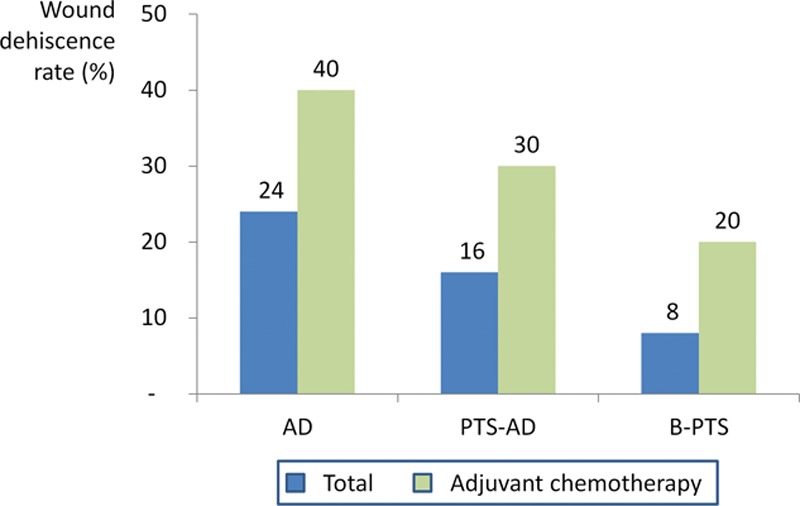
Rate of wound dehiscence stratified by study group and having received adjuvant chemotherapy. Differences between abdominal closure groups were not statistically significant although there was a potential clinical trend (B-PTS vs AD, P = 0.25). Having received adjuvant chemotherapy was significantly associated with developing wound dehiscence (P = 0.016).
There are several limitations to this study. The attending surgeon was likely to be more closely involved in the abdominal closure in patients in the B-PTS group, as this was a novel technique. This might cause some selection bias and drive some of the higher complication rates seen in the AD and PTS-AD groups. Most of the patients in the AD group were operated on by S.T., whereas most of the patients in the PTS-AD group were operated on by M.S.-C (Table 2). This is a potential source of bias, as we may be comparing the 2 surgeons’ abdominal closures rather than the specific techniques themselves. However, this technique was easy to teach and learn, and several cases were performed by residents and fellows after initial instruction and/or demonstration.
This study has shown that abdominal drains can be safely avoided in DIEP donor sites with no increase in postoperative complications. Drains limit patient mobility, require daily care upon discharge, and can be a portal for infection. Anecdotally, patient satisfaction is greatly improved by avoiding drain placement. As such, a technique that obviates the need for drain placement is a useful addition to the armamentarium of the reconstructive surgeon. Ongoing studies include the use of B-PTS for latissimus flap and transverse upper gracilis flaps to minimize drain usage and reduce postoperative complications.
CONCLUSIONS
No-drain DIEP flap donor-site closure using B-PTS is a safe and effective alternative to traditional abdominal closure using drains. Overall donor-site complication rates following this technique are significantly lower than with traditional donor-site closure. This technique may help to prevent abdominal wound dehiscence, especially in patients who will undergo adjuvant chemotherapy.
ACKNOWLEDGMENTS
Figures 1, 2, and 3: Copyright © Alexandra Hernandez, M.A., of Gory Details Illustration and printed with permission.
Supplementary Material
Footnotes
Disclosure: The authors have no financial interest to declare in relation to the content of this article. The Article Processing Charge was paid for by the authors.
Supplemental digital content is available for this article. Clickable URL citations appear in the text.
REFERENCES
- 1.Koshima I, Soeda S. Inferior epigastric artery skin flaps without rectus abdominis muscle. Br J Plast Surg. 1989;42:645–648. doi: 10.1016/0007-1226(89)90075-1. [DOI] [PubMed] [Google Scholar]
- 2.Allen RJ, Treece P. Deep inferior epigastric perforator flap for breast reconstruction. Ann Plast Surg. 1994;32:32–38. doi: 10.1097/00000637-199401000-00007. [DOI] [PubMed] [Google Scholar]
- 3.Garvey PB, Buchel EW, Pockaj BA, et al. DIEP and pedicled TRAM flaps: a comparison of outcomes. Plast Reconstr Surg. 2006;117:1711–1719. doi: 10.1097/01.prs.0000210679.77449.7d. [DOI] [PubMed] [Google Scholar]
- 4.Salgarello M, Tambasco D, Farallo E. DIEP flap donor site versus elective abdominoplasty short-term complication rates: a meta-analysis. Aesthetic Plast Surg. 2012;6:363–369. doi: 10.1007/s00266-011-9804-y. [DOI] [PubMed] [Google Scholar]
- 5.Bercial ME, Sabino Neto M, Calil JA, et al. Suction drains, quilting sutures, and fibrin sealant in the prevention of seroma formation in abdominoplasty: which is the best strategy? Aesthetic Plast Surg. 2012;36:370–373. doi: 10.1007/s00266-011-9807-8. [DOI] [PubMed] [Google Scholar]
- 6.Pollock H, Pollock T. Progressive tension sutures: a technique to reduce complications in abdominoplasty. Plast Reconstr Surg. 2000;105:2583–2586. doi: 10.1097/00006534-200006000-00047. [DOI] [PubMed] [Google Scholar]
- 7.Pollock T, Pollock H. Progressive tension sutures in abdominoplasty. Clin Plast Surg. 2004;31:583–589. doi: 10.1016/j.cps.2004.03.015. [DOI] [PubMed] [Google Scholar]
- 8.Pollock TA, Pollock H. No-drain abdominoplasty with progressive tension sutures. Clin Plast Surg. 2010;37:515–524. doi: 10.1016/j.cps.2010.03.004. [DOI] [PubMed] [Google Scholar]
- 9.Antonetti JW, Antonetti AR. Reducing seroma in outpatient abdominoplasty: analysis of 516 consecutive cases. Aesthet Surg J. 2010;30:418–425. doi: 10.1177/1090820X10372048. [DOI] [PubMed] [Google Scholar]
- 10.Andrades P, Prado A, Danilla S, et al. Progressive tension sutures in the prevention of postabdominoplasty seroma: a prospective, randomized, double-blind clinical trial. Plast Reconstr Surg. 2007;120:935–946. doi: 10.1097/01.prs.0000253445.76991.de. [DOI] [PubMed] [Google Scholar]
- 11.Wiener TC. Continuous running sutures: a modification for progressive tension abdominoplasty. Aesthet Surg J. 2012;32:248–249. doi: 10.1177/1090820X11433820. [DOI] [PubMed] [Google Scholar]
- 12.Rosen AD. Use of absorbable running barbed suture and progressive tension technique in abdominoplasty: a novel approach. Plast Reconstr Surg. 2010;125:1024–1027. doi: 10.1097/PRS.0b013e3181cb64f7. [DOI] [PubMed] [Google Scholar]
- 13.Warner JP, Gutowski KA. Abdominoplasty with progressive tension closure using a barbed suture technique. Aesthet Surg J. 2009;29:221–225. doi: 10.1016/j.asj.2009.01.009. [DOI] [PubMed] [Google Scholar]
- 14.Jandali S, Nelson JA, Bergey MR, et al. Evaluating the use of a barbed suture for skin closure during autologous breast reconstruction. J Reconstr Microsurg. 2011;27:277–286. doi: 10.1055/s-0031-1275491. [DOI] [PubMed] [Google Scholar]


