Abstract
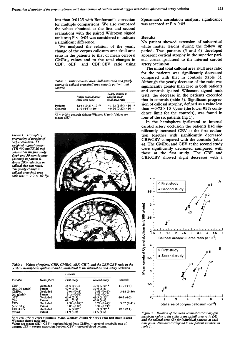
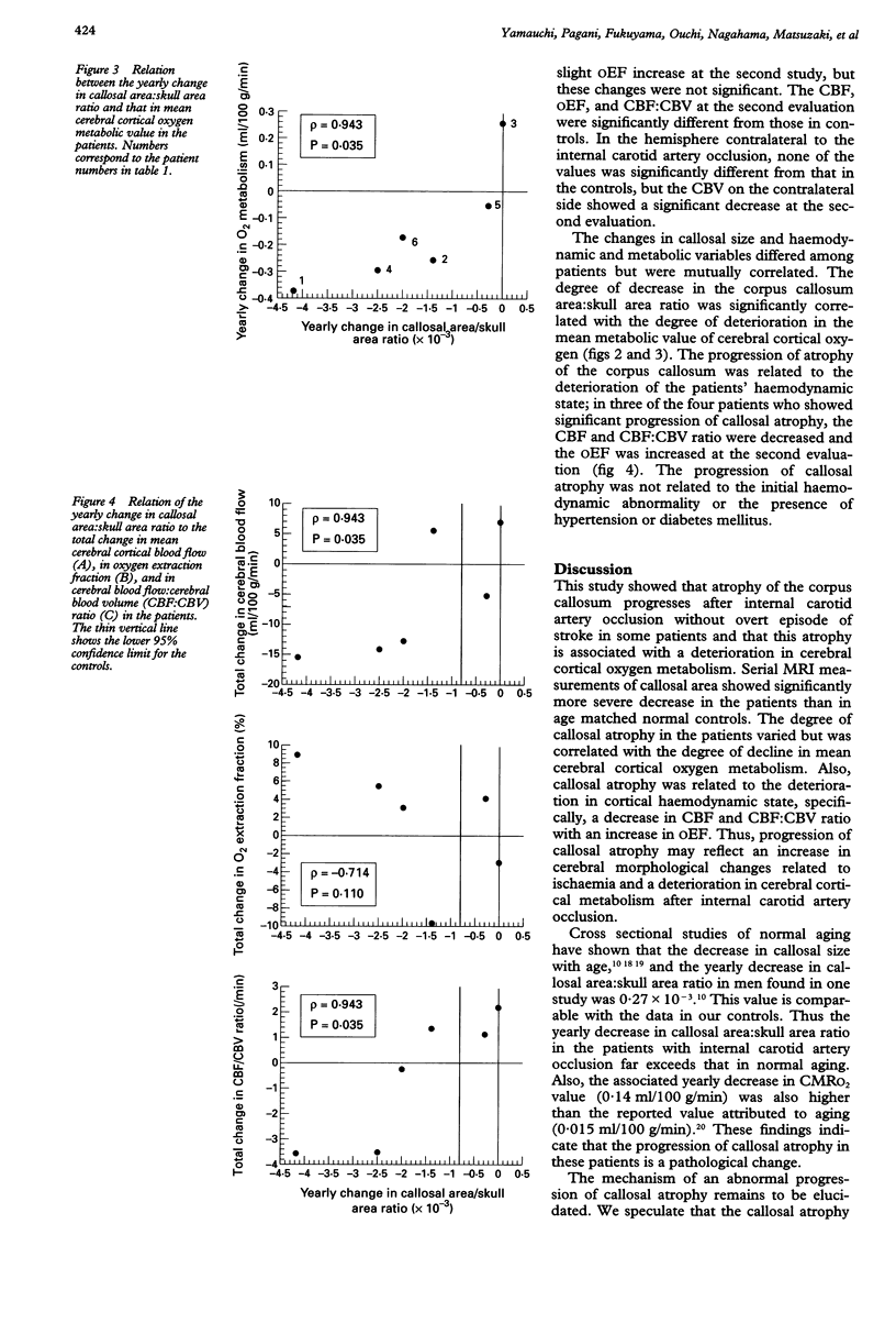
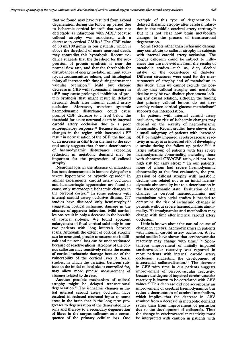
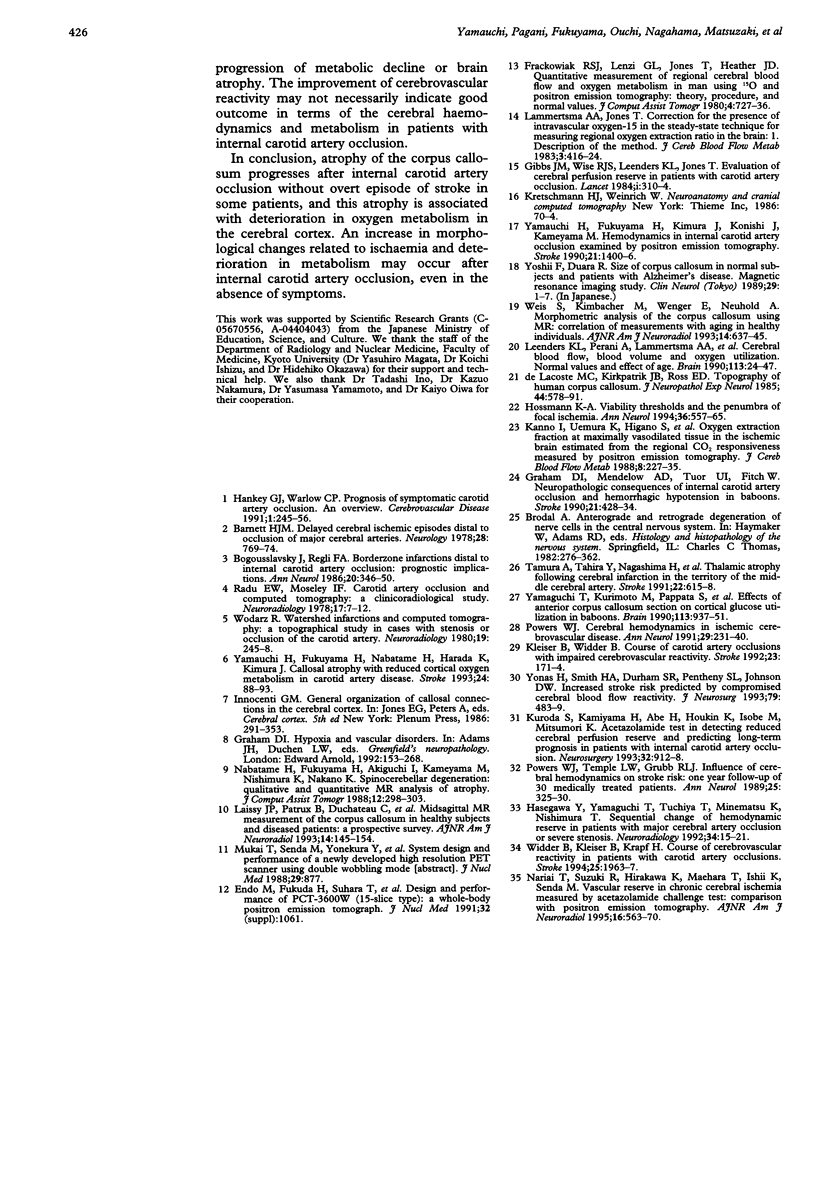
In cerebrovascular disease, progression of brain atrophy may reflect an increase in ischaemic changes. The purpose of this study was to determine whether atrophy of the corpus callosum progresses in association with a deterioration in cerebral cortical oxygen metabolism after occlusion of the carotid artery. Magnetic resonance imaging and PET were used to serially evaluate six patients with occlusion of the unilateral internal carotid artery at intervals ranging from 12 to 50 months. One patient had no symptoms, one had a transient ischaemic attack, and four had a minor stroke. All patients had presented at most only subcortical lesions at the first evaluation. During follow up, no patient showed extension of subcortical lesions or recurrent stroke. The initial total callosal area:skull area ratio for the patients was significantly less than that for 14 age matched normal control subjects. The yearly decrease of callosal size in the patients, which differed significantly from zero and exceeded that in the controls, was significantly correlated with the deterioration in mean cerebral cortical oxygen metabolism. Three of the four patients who showed significant progression of callosal atrophy presented deterioration in haemodynamic states as well. It is concluded that in some patients atrophy of the corpus callosum progresses after occlusion of the carotid artery even in the absence of any overt episode of stroke, and that this atrophy is associated with deterioration in cerebral cortical oxygen metabolism. An increase in cerebral morphological changes with deterioration in cerebral metabolism related to ischaemia may occur after occlusion of the carotid artery, even in the absence of symptoms.
Full text
PDF
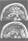
Images in this article
Selected References
These references are in PubMed. This may not be the complete list of references from this article.
- Barnett H. J. Delayed cerebral ischemic episodes distal to occlusion of major cerebral arteries. Neurology. 1978 Aug;28(8):769–774. doi: 10.1212/wnl.28.8.769. [DOI] [PubMed] [Google Scholar]
- Bogousslavsky J., Regli F. Borderzone infarctions distal to internal carotid artery occlusion: prognostic implications. Ann Neurol. 1986 Sep;20(3):346–350. doi: 10.1002/ana.410200312. [DOI] [PubMed] [Google Scholar]
- Gibbs J. M., Wise R. J., Leenders K. L., Jones T. Evaluation of cerebral perfusion reserve in patients with carotid-artery occlusion. Lancet. 1984 Feb 11;1(8372):310–314. doi: 10.1016/s0140-6736(84)90361-1. [DOI] [PubMed] [Google Scholar]
- Graham D. I., Mendelow A. D., Tuor U., Fitch W. Neuropathologic consequences of internal carotid artery occlusion and hemorrhagic hypotension in baboons. Stroke. 1990 Mar;21(3):428–434. doi: 10.1161/01.str.21.3.428. [DOI] [PubMed] [Google Scholar]
- Hasegawa Y., Yamaguchi T., Tsuchiya T., Minematsu K., Nishimura T. Sequential change of hemodynamic reserve in patients with major cerebral artery occlusion or severe stenosis. Neuroradiology. 1992;34(1):15–21. doi: 10.1007/BF00588426. [DOI] [PubMed] [Google Scholar]
- Hossmann K. A. Viability thresholds and the penumbra of focal ischemia. Ann Neurol. 1994 Oct;36(4):557–565. doi: 10.1002/ana.410360404. [DOI] [PubMed] [Google Scholar]
- Kanno I., Uemura K., Higano S., Murakami M., Iida H., Miura S., Shishido F., Inugami A., Sayama I. Oxygen extraction fraction at maximally vasodilated tissue in the ischemic brain estimated from the regional CO2 responsiveness measured by positron emission tomography. J Cereb Blood Flow Metab. 1988 Apr;8(2):227–235. doi: 10.1038/jcbfm.1988.53. [DOI] [PubMed] [Google Scholar]
- Kleiser B., Widder B. Course of carotid artery occlusions with impaired cerebrovascular reactivity. Stroke. 1992 Feb;23(2):171–174. doi: 10.1161/01.str.23.2.171. [DOI] [PubMed] [Google Scholar]
- Kuroda S., Kamiyama H., Abe H., Houkin K., Isobe M., Mitsumori K. Acetazolamide test in detecting reduced cerebral perfusion reserve and predicting long-term prognosis in patients with internal carotid artery occlusion. Neurosurgery. 1993 Jun;32(6):912–919. doi: 10.1227/00006123-199306000-00005. [DOI] [PubMed] [Google Scholar]
- Laissy J. P., Patrux B., Duchateau C., Hannequin D., Hugonet P., Ait-Yahia H., Thiebot J. Midsagittal MR measurements of the corpus callosum in healthy subjects and diseased patients: a prospective survey. AJNR Am J Neuroradiol. 1993 Jan-Feb;14(1):145–154. [PMC free article] [PubMed] [Google Scholar]
- Lammertsma A. A., Jones T. Correction for the presence of intravascular oxygen-15 in the steady-state technique for measuring regional oxygen extraction ratio in the brain: 1. Description of the method. J Cereb Blood Flow Metab. 1983 Dec;3(4):416–424. doi: 10.1038/jcbfm.1983.67. [DOI] [PubMed] [Google Scholar]
- Leenders K. L., Perani D., Lammertsma A. A., Heather J. D., Buckingham P., Healy M. J., Gibbs J. M., Wise R. J., Hatazawa J., Herold S. Cerebral blood flow, blood volume and oxygen utilization. Normal values and effect of age. Brain. 1990 Feb;113(Pt 1):27–47. doi: 10.1093/brain/113.1.27. [DOI] [PubMed] [Google Scholar]
- Nabatame H., Fukuyama H., Akiguchi I., Kameyama M., Nishimura K., Nakano Y. Spinocerebellar degeneration: qualitative and quantitative MR analysis of atrophy. J Comput Assist Tomogr. 1988 Mar-Apr;12(2):298–303. [PubMed] [Google Scholar]
- Nariai T., Suzuki R., Hirakawa K., Maehara T., Ishii K., Senda M. Vascular reserve in chronic cerebral ischemia measured by the acetazolamide challenge test: comparison with positron emission tomography. AJNR Am J Neuroradiol. 1995 Mar;16(3):563–570. [PMC free article] [PubMed] [Google Scholar]
- Powers W. J. Cerebral hemodynamics in ischemic cerebrovascular disease. Ann Neurol. 1991 Mar;29(3):231–240. doi: 10.1002/ana.410290302. [DOI] [PubMed] [Google Scholar]
- Powers W. J., Tempel L. W., Grubb R. L., Jr Influence of cerebral hemodynamics on stroke risk: one-year follow-up of 30 medically treated patients. Ann Neurol. 1989 Apr;25(4):325–330. doi: 10.1002/ana.410250403. [DOI] [PubMed] [Google Scholar]
- Radü E. W., Moseley I. F. Carotid artery occlusion and computed tomography. A clinicoradiological study. Neuroradiology. 1978 Nov 24;17(1):7–12. doi: 10.1007/BF00345262. [DOI] [PubMed] [Google Scholar]
- Tamura A., Tahira Y., Nagashima H., Kirino T., Gotoh O., Hojo S., Sano K. Thalamic atrophy following cerebral infarction in the territory of the middle cerebral artery. Stroke. 1991 May;22(5):615–618. doi: 10.1161/01.str.22.5.615. [DOI] [PubMed] [Google Scholar]
- Weis S., Kimbacher M., Wenger E., Neuhold A. Morphometric analysis of the corpus callosum using MR: correlation of measurements with aging in healthy individuals. AJNR Am J Neuroradiol. 1993 May-Jun;14(3):637–645. [PMC free article] [PubMed] [Google Scholar]
- Widder B., Kleiser B., Krapf H. Course of cerebrovascular reactivity in patients with carotid artery occlusions. Stroke. 1994 Oct;25(10):1963–1967. doi: 10.1161/01.str.25.10.1963. [DOI] [PubMed] [Google Scholar]
- Wodarz R. Watershed infarctions and computed tomography. A topographical study in cases with stenosis or occlusion of the carotid artery. Neuroradiology. 1980;19(5):245–248. doi: 10.1007/BF00347803. [DOI] [PubMed] [Google Scholar]
- Yamaguchi T., Kunimoto M., Pappata S., Chavoix C., Riche D., Chevalier L., Mazoyer B., Mazière M., Naquet R., Baron J. C. Effects of anterior corpus callosum section on cortical glucose utilization in baboons. A sequential positron emission tomography study. Brain. 1990 Aug;113(Pt 4):937–951. doi: 10.1093/brain/113.4.937. [DOI] [PubMed] [Google Scholar]
- Yamauchi H., Fukuyama H., Kimura J., Konishi J., Kameyama M. Hemodynamics in internal carotid artery occlusion examined by positron emission tomography. Stroke. 1990 Oct;21(10):1400–1406. doi: 10.1161/01.str.21.10.1400. [DOI] [PubMed] [Google Scholar]
- Yamauchi H., Fukuyama H., Nabatame H., Harada K., Kimura J. Callosal atrophy with reduced cortical oxygen metabolism in carotid artery disease. Stroke. 1993 Jan;24(1):88–93. doi: 10.1161/01.str.24.1.88. [DOI] [PubMed] [Google Scholar]
- Yonas H., Smith H. A., Durham S. R., Pentheny S. L., Johnson D. W. Increased stroke risk predicted by compromised cerebral blood flow reactivity. J Neurosurg. 1993 Oct;79(4):483–489. doi: 10.3171/jns.1993.79.4.0483. [DOI] [PubMed] [Google Scholar]
- Yoshii F., Duara R. [Size of the corpus callosum in normal subjects and patients with Alzheimer's disease--magnetic resonance imaging study]. Rinsho Shinkeigaku. 1989 Jan;29(1):1–7. [PubMed] [Google Scholar]
- de Lacoste M. C., Kirkpatrick J. B., Ross E. D. Topography of the human corpus callosum. J Neuropathol Exp Neurol. 1985 Nov;44(6):578–591. doi: 10.1097/00005072-198511000-00004. [DOI] [PubMed] [Google Scholar]


