Abstract
Introduction:
Hard tissues can be studied by either decalcification or by preparing ground sections. Various mounting media have been tried and used for ground sections of teeth. However, there are very few studies on the use of cyanoacrylate adhesive as a mounting medium.
Aims:
The aim of our study was to evaluate the efficacy of cyanoacrylate adhesive (Fevikwik™) as a mounting medium for ground sections of teeth and to compare these ground sections with those mounted with Canada balsam.
Materials and Methods:
Ground sections were prepared from twenty extracted teeth. Each section was divided into two halves and mounted on one slide, one with cyanoacrylate adhesive (Fevikwik™) and the other with Canada balsam. Scoring for various features in the ground sections was done by two independent observers.
Statistical Analysis Used:
Statistical analysis using Student's t-test (unpaired) of average scores was performed for each feature observed.
Results:
No statistically significant difference was found between the two for most of the features. However, cyanoacrylate was found to be better than Canada balsam for observing striae of Retzius (P < 0.0205), enamel lamellae (P < 0.036), dentinal tubules (P < 0.0057), interglobular dentin (P < 0.0001), sclerotic dentin – transmitted light (P < 0.00001), sclerotic dentin – polarized light (P < 0.0002) and Sharpey's fibers (P < 0.0004).
Conclusions:
This initial study shows that cyanoacrylate is better than Canada balsam for observing certain features of ground sections of teeth. However, it remains to be seen whether it will be useful for studying undecalcified sections of carious teeth and for soft tissue sections.
Keywords: Canada balsam, cyanoacrylate, dental enamel, dentin, tooth
INTRODUCTION
Hard tissues can be studied in two ways – one, by the process of decalcification and the other, by cutting and/or grinding them into thin sections. After decalcification, the hard tissues become soft and the subsequent tissue processing procedures are similar to that of any other soft tissue. However, in case of teeth, since enamel has the maximum amount of mineral content (93.6–98.5%), but only 0.05–0.8% of organic content and the rest water,[1] no enamel is left for observation on completion of decalcification. Though enamel has been studied by partial decalcification methods,[2] one of the ways to study tooth enamel and other hard tissues is to prepare thin sections by grinding or using a hard tissue microtome.[3,4] These sections are cleaned, dried and placed on a glass slide and a coverslip is placed over it by using a mounting medium. The commonly used mounting media for ground sections of teeth are Canada balsam and Di-n-butyl phthalate in xylene (DPX).[5]
Canada balsam has certain advantages: Permanence, perfect transparency and hardness, once it is set. However, Canada balsam also possesses certain inherent problems. It takes some time to set and usually yellows with age, which could be a problem with photography.[6] Cyanoacrylate has been used as a mounting medium for undecalcified bone sections and teeth.[7,8]
Cyanoacrylate adhesive [Fevikwik™] is commonly used to fix broken household appliances or to bond plastics, metal, rubbers, glass, china and other dissimilar substrates. It polymerizes very quickly, is ready to use and bonds almost anything to anything.[9] There are not many studies on the usage of cyanoacrylate adhesive for ground sections of teeth. Though the use of methyl 2-cyanoacrylate monomer as a quick setting mounting medium was first described by Molnar and Molnar in 1967,[10] it is only in the recent past that interest has renewed in its use for stained resin-embedded semithin sections.[11] To our knowledge, there is no study in the recent past on the use of cyanoacrylate glue for ground sections of teeth.
The aim of our study was to evaluate the efficacy of a cyanoacrylate adhesive [Fevikwik™] as a mounting medium for ground sections of teeth and to compare these ground sections with those mounted with Canada balsam.
MATERIALS AND METHODS
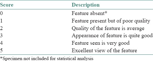
Twenty teeth extracted from patients visiting our dental college, with orthodontic or periodontic problems, were used for preparing ground sections. A 37C fine/coarse combination stone was used for grinding the teeth to uniform thickness. Each section was then split into two halves, as much as possible in equal proportions. The two halves of the section from the same tooth were then mounted on the same slide, one half with Canada balsam and the other with a cyanoacrylate adhesive [Fevikwik™]. All the mounted slides were then observed, by two independent observers, for various features in the ground sections under light microscope with an attached polarizer [Table 1]. Scoring was given, as shown in Table 2, by both observers and an average was taken for each feature observed. Statistical analysis was then performed using the average scores obtained for all the features observed.
Table 1.
List of features observed in the ground sections
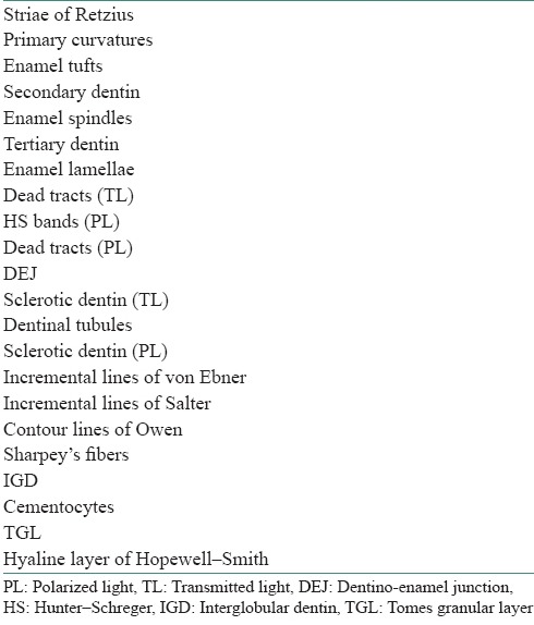
Table 2.
Observers’ scoring of microscopic features
RESULTS AND OBSERVATIONS
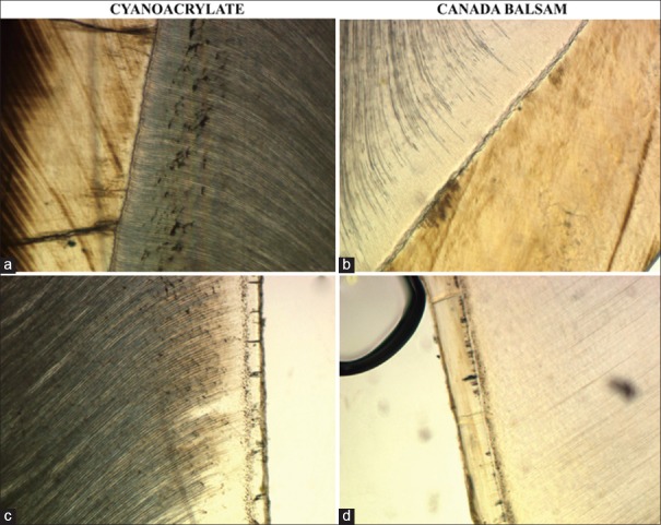
While observing the various features of enamel, dentin and cementum of ground sections of teeth, the mean values of scores were generally higher with cyanoacrylate adhesive than Canada balsam, except for incremental lines of Salter. However, there was no statistically significant difference between the two for most of the features. However, statistically, cyanoacrylate adhesive was found to be better than Canada balsam for observing striae of Retzius (P < 0.0205), enamel lamellae (P < 0.036), dentinal tubules (P < 0.0057), interglobular dentin (P < 0.0001), sclerotic dentin – transmitted light (TL) (P < 0.00001), sclerotic dentin – polarized light (PL) (P < 0.0002) and Sharpey's fibers (P < 0.0004). These are shown in Table 3. Figure 1 shows photomicrographs of ground sections of teeth mounted with Fevikwik™ and Canada balsam.
Table 3.
Comparison of mean scores of observation of ground sections mounted with cyanoacrylate and Canada balsam
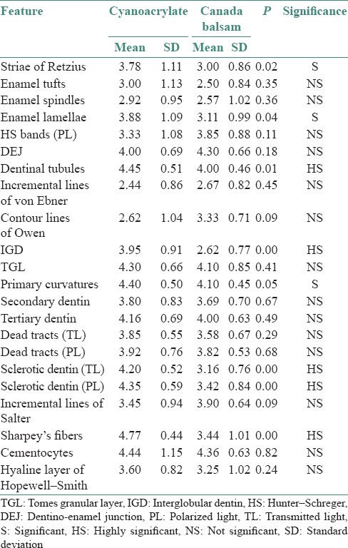
Figure 1.
(a) Striae of Retzius and areas of interglobular dentine are more prominently seen with ground sections mounted with cyanoacrylate glue (Ground sections, x100), (b) than those with Canada balsam (ground section, x100). (c) Sharpey's fibers are more noticeable in sections mounted with cyanoacrylate (ground section, x100) (d) than those with Canada balsam (ground section, x100) and note the presence of an air bubble, which was observed more often with Canada balsam
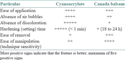
When technical aspects of the use of cyanoacrylate adhesive and Canada balsam were compared, it was found that the setting time, ease of application and absence of air bubbles were better with the former. However, Canada balsam was found to be less technique-sensitive and easier to remove (with xylene) when compared with cyanoacrylate adhesive [Table 4].
Table 4.
Comparison of technical aspects of application of cyanoacrylate and Canada balsam for ground sections of teeth
DISCUSSION
Traditionally, ground sections of teeth have been mounted with either Canada balsam or DPX. Canada balsam was preferred in this study because of its properties of permanence and similar refractive index (RI) as glass. Moreover, features of enamel such as striae of Retzius appear better with Canada balsam as compared to DPX due to its larger molecular size.[12] However, there are certain inherent problems associated with Canada balsam: it takes a long time to set and there is a tinge yellowish stain around the ground sections and on the border of the coverslip.[6]
Cyanoacrylate adhesive [Fevikwik™] is a commonly used household item which apparently bonds anything to anything. It is purported to set fast and the bond is supposed to be quite strong. Its main component is cyanoacrylate and is available as small 1 ml squeezable tubes.[9]
The aim of our study was to see whether cyanoacrylate adhesive can be used as mounting media for ground sections of teeth and whether it is as good as Canada balsam or better. Two aspects were compared: visualization of structures of teeth and technical aspects of application.
Although observation of most of the structures of teeth were not statistically different between the two, cyanoacrylate adhesive was found to be better than Canada balsam for observing striae of Retzius, enamel lamellae, dentinal tubules, interglobular dentin, Sharpey's fibers and sclerotic dentin in transmitted as well as PL.
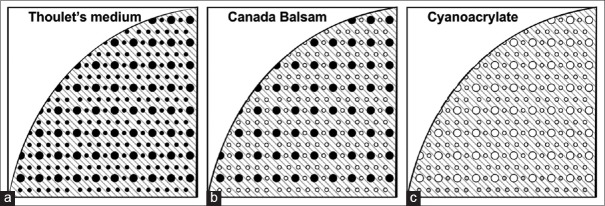
This better visualization could be related to the difference in molecular size of the components in the two mounting media. Sound tooth enamel has pores, the volume of which is <1%. These pores are of different sizes and depending on their diameter, they may or may not allow penetration of the molecules of the mounting media. Mounting media such as Thoulet's medium can penetrate these pores because of their small molecular size.[12]
This is schematically depicted in Figure 2a. If the molecular size of the mounting medium is smaller than the diameter of these pores, they penetrate and displace the gaseous content or debris within the pores and the structures appear translucent when observed under TL. However, if the pores are smaller than the molecular size of the mounting medium, the gaseous particles are not eliminated from these pores. This lack of penetration of mounting media is also responsible for the observation of striae of Retzius in enamel as prominent, brown-colored incremental lines because these pores are especially located near these structures.[12] Larger pores may be penetrated by Canada balsam, but smaller pores may not be [Figure 2b]. Since cyanoacrylate adhesive has larger molecular size when compared with Canada balsam, it is likely that there is a greater lack of penetration and therefore more prominence of these structures [Figure 2c].
Figure 2.
(a) Thoulet's medium – all pores are penetrated (b) Canada balsam – only larger pores are penetrated, but not the smaller pores (c) cyanoacrylate – all pores (large and small) are not penetrated
Moreover, the differences in refractive indices could also be responsible for the difference in observations. The RI of enamel is 1.63 and that of Canada balsam is 1.54 and close to that of glass. Hence, chances of refraction are minimized. However, the RI of cyanoacrylate adhesive is 1.45.[13]
The larger difference of RI between enamel and cyanoacrylate and between cyanoacrylate and glass and the lack of penetration due to larger molecular size provide greater contrast to the sections and hence hypomineralized structures are better visualized with cyanoacrylate glue. Since incremental lines of Salter are hypermineralized[14] and more glass-like, they appear better when Canada balsam (which has RI of 1.54) is used as mounting medium.
Regarding the technical aspects of the two mounting media, cyanoacrylate adhesive is known to set fast which is an advantage to observe ground sections earlier than Canada balsam. Because it sets very fast, it is highly possible that the chances of air bubble entrapment are very minimal, but one has to place the coverslip properly and at a rapid pace. Another added advantage is the fact that cyanoacrylate adhesive does not cause any discoloration, unlike Canada balsam. However, because cyanoacrylate adhesive results in a strong chemical bond of the coverslip to the glass slide, it is more difficult to remove. On the other hand, Canada balsam has the advantage of not being technique-sensitive, easier to remove and being more cost-effective.
SUMMARY AND CONCLUSIONS
There are not too many studies on the usage of mounting media for ground sections of teeth. Mounting media which are commonly used for soft tissue sections have been used for undecalcified sections of teeth and bone. Traditionally, ground sections of teeth have been mounted with DPX or Canada balsam. We have used a cyanoacrylate adhesive in our study for mounting ground sections of teeth. This preliminary study shows that it is better than Canada balsam in observation of certain features, rapidity of setting, minimal air bubble entrapment and lack of discoloration. However, it remains to be seen whether it is better than DPX and whether it is useful in studying undecalcified sections of carious teeth (to study the zones of enamel and dentin caries), for which further studies and more number of samples are required. Moreover, use of cyanoacrylate adhesive for soft tissue sections can also be explored.
Financial support and sponsorship
Nil.
Conflicts of interest
There are no conflicts of interest.
REFERENCES
- 1.Little MF, Cueto CS, Rowley J. Chemical and physical properties of altered and sound enamel. I. ASH, Ca, P, CO2, N, water, microradiolucency and density. Arch Oral Biol. 1962;7:173–84. [Google Scholar]
- 2.Liem RS, Jansen HW. Preparation of partially decalcified sections of human dental enamel for electron microscopy. Caries Res. 1982;16:217–26. doi: 10.1159/000260601. [DOI] [PubMed] [Google Scholar]
- 3.Donath K. Norderstedt: EXAKT-Kulzer Publication; 1995. Preparation of Histologic Sections. [Google Scholar]
- 4.Suvarna S, Layton C, Bancroft J. 7th ed. Churchill Livingstone - Elsevier; 2013. Bancroft's Theory and Practice of Histological Techniques; pp. 339–40. [Google Scholar]
- 5.Kumar G. 13th ed. New Delhi: Elsevier India Pvt. Ltd; 2013. Orban's Oral Histology and Embryology; p. 415. [Google Scholar]
- 6.Gibb T, Oseto C. San Diego, California: Elsevier Academic Press Publications; 2006. Arthropod Collection and Identification: Laboratory and Field Techniques; p. 75. [Google Scholar]
- 7.Smith P, Tchernov E. London, Tel Aviv: Freund Publishing House Ltd; 1992. Structure, Function and Evolution of Teeth; p. 316. [Google Scholar]
- 8.Usui M, Yoshizu T. Tokyo: Springer-Verlag; 2003. Experimental and Clinical Reconstructive Microsurgery. [Google Scholar]
- 9. [Last accessed on 2015 Mar 05]. Available from: http://www.ge.iic.com/files/fichas%20productos/cianoacrilato.pdf .
- 10.Molnar GW, Molnar MD. Methyl 2-cyanoacrylate monomer: A quick setting mounting medium. Stain Technol. 1967;42:106. [PubMed] [Google Scholar]
- 11.Liu PY, Phillips GE, Kempf M, Cuttle L, Kimble RM, McMillan JR. Cyanoacrylate glue as an alternative mounting medium for resin-embedded semithin sections. J Electron Microsc (Tokyo) 2010;59:87–90. doi: 10.1093/jmicro/dfp040. [DOI] [PubMed] [Google Scholar]
- 12.Silverstone LM, Hicks MJ, Featherstone MJ. Dynamic factors affecting lesion initiation and progression in human dental enamel. Part I. The dynamic nature of enamel caries. Quintessence Int. 1988;19:683–711. [PubMed] [Google Scholar]
- 13.Hillson S. 2nd ed. New York: Cambridge University Press; 2005. Teeth; p. 203. [Google Scholar]
- 14.Matsuo A, Yajima T. Comparative observations of the cement lamellae and incremental lines by light and scanning electron microscopy and microradiography. Jpn J Oral Biol. 1992;34:171–80. [Google Scholar]


