Dear editor
We read with great interest the article titled “Value of optical coherence tomography in the detection of macular pathology before the removal of silicone oil” by Rashad et al.1 The authors have evaluated the optical coherence tomography (OCT) findings before the removal of silicone oil (SiO). We congratulate the authors for this well-organized study and would like to contribute to their findings.
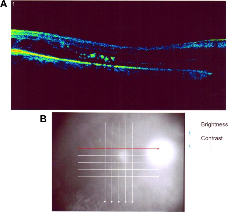
OCT is a revolutionary investigation in ophthalmology.2 It provides an opportunity to visualize the histological structure of the retina. SiO plays a very important role in ophthalmic surgery and is used as a tamponade agent in a lot of vitreoretinal surgeries.3 OCT findings of siliconized eyes may provide us with new insights about the proper timing of removal of SiO and deciding on additional treatments. Thus, findings discussed in this study are very important.
One of the significant findings of this study is the high percentage of macular edema (ME) in siliconized eyes. Being an important pathology that impairs visual acuity, we think the high percentage of ME in this study should be well analyzed. We wonder if ME was present before the injection of SiO or developed under SiO. The incidence of ME was found to be high, especially in diabetic patients (58.3%). Diabetic patients suffer from microcirculation problem.4 Existence of SiO may decrease oxygenation of the retina and increase the risk of developing ME. Thus, follow-up with OCT is especially important in diabetic patients.
Treatment of ME in siliconized eyes is also an important issue. We wonder which pharmacotherapy the authors used. Intravitreal anti-vascular endothelial growth factor, the most efficient drug for ME, is useless in this situation. While topical nonsteroidals are efficient in treating postoperative ME, we do not know if they would be efficient in siliconized eyes. Subtenon steroid injection may be an efficient treatment. Based on OCT findings, early removal of silicone and intravitreal anti-vascular endothelial growth factor may be applied.
Footnotes
Disclosure
The authors report no conflicts of interest in this communication.
References
- 1.Rashad MA, Mohamed AA, Ahmed AI. Value of optical coherence tomography in the detection of macular pathology before the removal of silicone oil. Clin Ophthalmol. 2016;10:121–135. doi: 10.2147/OPTH.S90449. [DOI] [PMC free article] [PubMed] [Google Scholar]
- 2.Huang D, Swanson EA, Lin CP, et al. Optical coherence tomography. Science. 1991;254:1178–1181. doi: 10.1126/science.1957169. [DOI] [PMC free article] [PubMed] [Google Scholar]
- 3.Cibis PA, Becker B, Okun E, Cowan S. The use of liquid silicone in retinal detachment surgery. Arch Ophthalmol. 1962;681:590–599. doi: 10.1001/archopht.1962.00960030594005. [DOI] [PubMed] [Google Scholar]
- 4.Ichiyama Y, Sawada O, Mori T, Fujikawa M, Kawamura H, Ohji M. The effectiveness of vitrectomy for diffuse diabetic macular edema may depend on its preoperative optical coherence tomography pattern. Graefes Arch Clin Exp Ophthalmol. 2016 Jan 18; doi: 10.1007/s00417-015-3251-4. Epub. [DOI] [PubMed] [Google Scholar]