Abstract
There has been a significant surge in aesthetic chest surgery for men in the last several years. Male chest enhancement is performed with surgical placement of a solid silicone pectoral implant. In the past, male chest correction and implantation were limited to the treatment of men who had congenital absence or atrophy of the pectoralis muscle and pectus excavatum deformity. But today, the popularization of increased chest and pectoral size fostered by body builders has more men desiring chest correction with implantation for non-medical reasons. We present a case of a 44-year-old, male with a displaced left pectoral implant with near extrusion and with an associated peri-implant soft tissue mass and fluid collection. While the imaging of these patients is uncommon, our case study presents the radiographic findings of male chest enhancement with associated complications.
Keywords: Pectoral implant, implant displacement, chest enhancement, male breast, mammography, ultrasound, computed tomography
CASE REPORT
A 44-year-old, human immunodeficiency virus positive (HIV +), male presented for management of a displaced and painful left pectoral implant. He was six years status post bilateral pectoral implant placement that was initially complicated by bilateral seromas that required percutaneous drainage at an outside institution. The patient reported that when he was five and a half year’s post-operative he experienced moderate pain along the left side of his chest of one week duration. He also stated that for the last two months he has had continued moderate swelling and displacement of his left implant towards the left side of his chest. Recently, he noticed skin changes with discoloration along the left lower chest wall without nipple involvement. The patient denied any history of trauma.
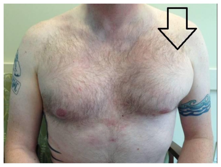
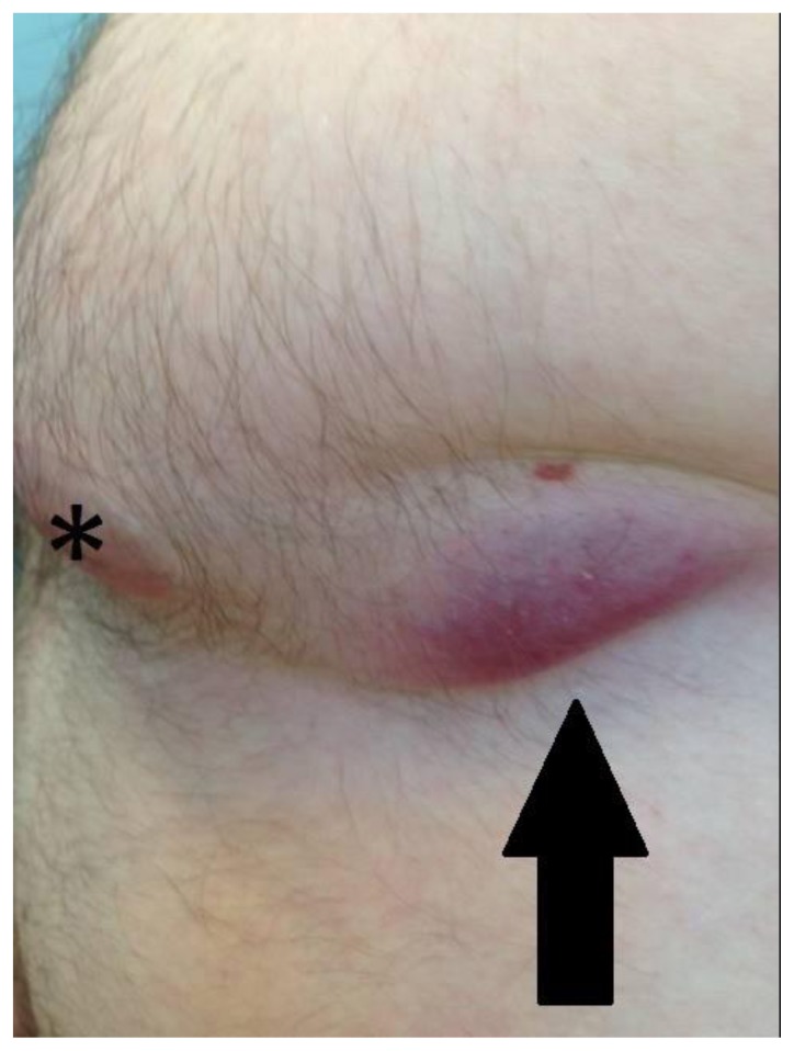
On physical exam, the patient had bilateral pectoral implants with gross asymmetry of the left side of his chest (Figure 1). The left side of the chest was about two times the size of the right. There was lateral displacement with overlying soft tissue swelling and near extrusion, a forcing out of a normal position, of left pectoral implant. There was a 4 cm × 3 cm area of inflamed and thinned skin laterally to the nipple level at the site of the impending extrusion (Figure 2). Since the soft tissue swelling of the chest was concerning for a mass in this HIV+ patient, the patient was sent for bilateral diagnostic mammography and ultrasound to exclude carcinoma or infection.
Figure 1.
44-year-old male with left pectoral implant displacement.
FINDINGS: Photograph demonstrates gross asymmetry and increase in size of the left side of the patient’s chest (open arrow) secondary to implant displacement and soft tissue swelling.
TECHNIQUE: Color photograph, Nikon Coolpix A, digital camera, 16.0 Megapixel.
Figure 2.
44-year-old male with left pectoral implant displacement with near extrusion of the implant through the skin.
FINDINGS: 4 cm × 3 cm area of inflamed and thinned skin (arrow) lateral to the nipple (*) at the site of the impending extrusion of the implant.
TECHNIQUE: Color photograph, Nikon Coolpix A, digital camera, 16.0 Megapixel.
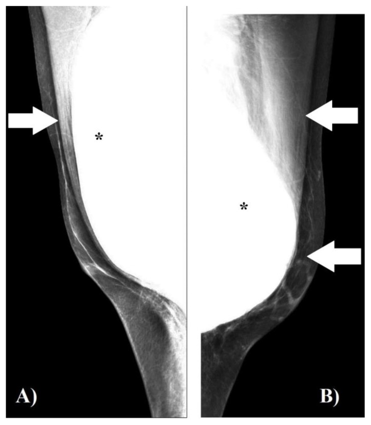
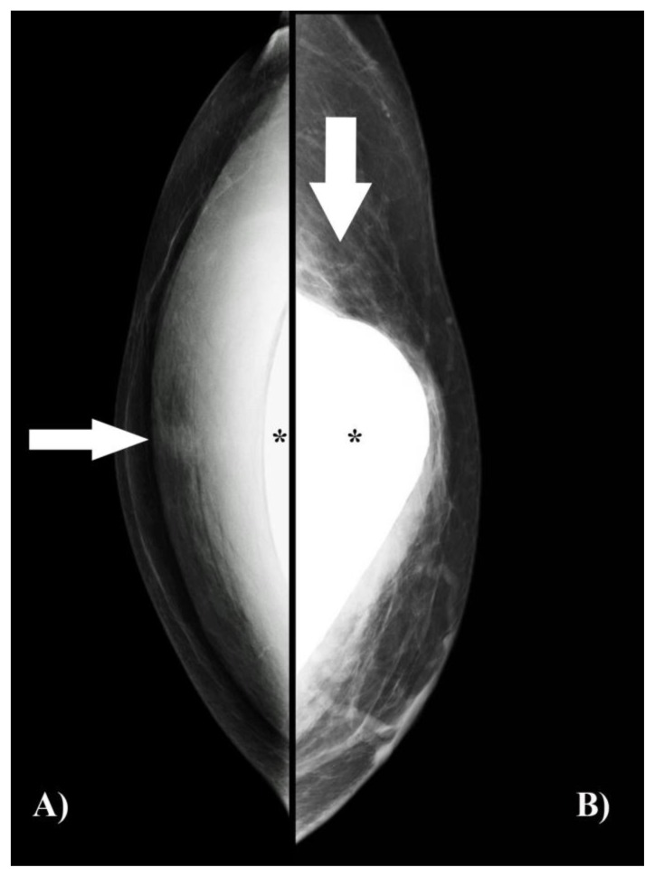
Mammography showed bilaterally fatty replaced breasts without mass, distortion or calcifications. Bilateral subpectoral silicone implants were present. There was asymmetric orientation of the left implant, displaced anterolateral and inferiorly, relative to the normally positioned right pectoral implant (Figure 3). Additionally, on the craniocaudal view of the left breast, the overlying left pectoralis muscle was thinned significantly and was compatible with loss of integrity (Figure 4).
Figure 3.
44-year-old male with left pectoral implant displacement.
FINDINGS: Mammographic mediolateral oblique “implant in-view” of both breasts. The implants (*) are composed of silicone and appear white on the mammogram due to attenuation of the x-ray beam.
A) Correct positioning of the right silicone implant posterior to the pectoralis muscle (arrow). B) Abnormal position of the left silicone implant. It is displaced anteriorly and inferiorly (arrows) compared to the right implant in A.
TECHNIQUE: Digital mammogram, mediolateral oblique “implant in-view” of both breasts.
A) Right mammogram - 110 milliamperage second (mAs), 29 kilovolts (kV), target/filter combination: molybdenum/rhodium, 10.5 cm compression thickness.
B) Left mammogram - 130 mAs, 28 kV, target/filter combination: molybdenum/rhodium, 10.4 cm compression thickness.
Figure 4.
44-year-old male with left pectoral implant displacement.
FINDINGS: Mammographic craniocaudal “implant in-view” of both breasts. The implants (*) are composed of silicone and appear white on the mammogram due to attenuation of the x-ray beam.
A) Correct positioning of the right silicone implant (*) posterior to the intact pectoralis muscle (arrow).
B) Abnormal position of the left silicone implant (*). The implant is displaced anteriorly and laterally. The overlying pectoralis muscle is thinned significantly (arrow) laterally. This is compatible with loss of pectoralis muscle integrity. The rest of the pectoral silicone implant is located posterior to the visualized pectoralis muscle.
TECHNIQUE: Digital mammogram craniocaudal “implant in-view” of both breasts.
A) Right mammogram -115 mAs, 28KV, target filter combination: molybdenum/rhodium, 5.5 cm compression thickness.
B) Left mammogram - 110 mAs, 28 kV, target filter combination: molybdenum/rhodium, 5 cm compression thickness.
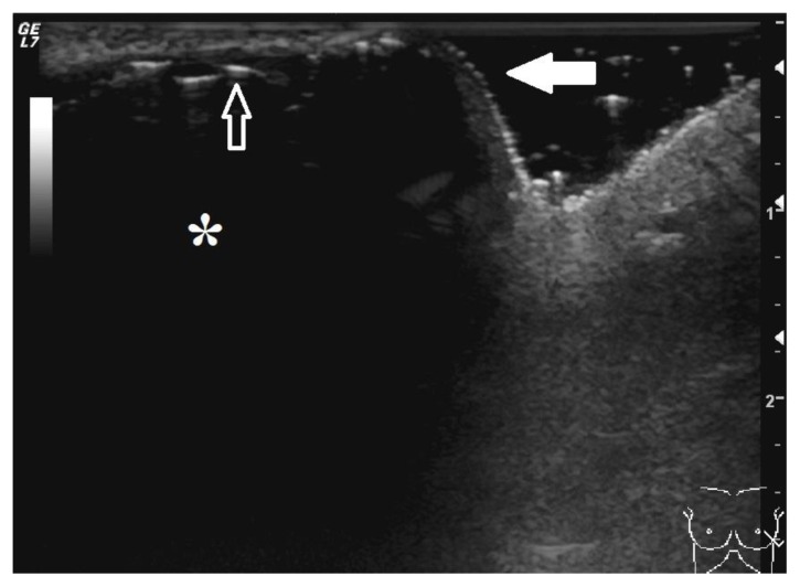
Sonographic imaging of the left breast and axilla were also performed to further evaluate for a soft tissue mass or fluid collection. At the 5 o’clock position of the left breast at the site of skin discoloration and pending extrusion, the overlying pectoralis muscle was not readily appreciated and the implant was directly subjacent to the skin (Figure 5). Sonographically, the implant was anechoic. The anterior margin of the implant was easily seen as a thin echogenic line. The posterior margin of the implant was not visualized due to attenuation of the ultrasound beam by the silicone. Sonographic evaluation of the upper outer quadrant of the patient’s chest at the site of the soft tissue asymmetry on the left was also performed. Only fatty tissue, subcutaneous fat and muscle were noted in this region. No definite mass or discrete fluid collections were identified. Multiple normal appearing lymph nodes were noted in the left axilla. Mammographic and sonographic imaging findings supported the diagnoses of a displaced left pectoral implant with impending extrusion.
Figure 5.
44-year-old male with left pectoral implant displacement.
FINDINGS: Sonographic image of the displaced implant at site of impending extrusion. The implant (*) is anechoic. The thin echogenic line (open arrow) that is present between the anechoic implant and skin represents the anterior margin of the implant. The posterior margin of the implant is not visualized due to attenuation of the ultrasound beam by the silicone. The overlying pectoralis muscle is not readily appreciated and the implant is directly subjacent to the skin (closed arrow).
TECHNIQUE: Ultrasound, radial plane, 14 megahertz transducer.
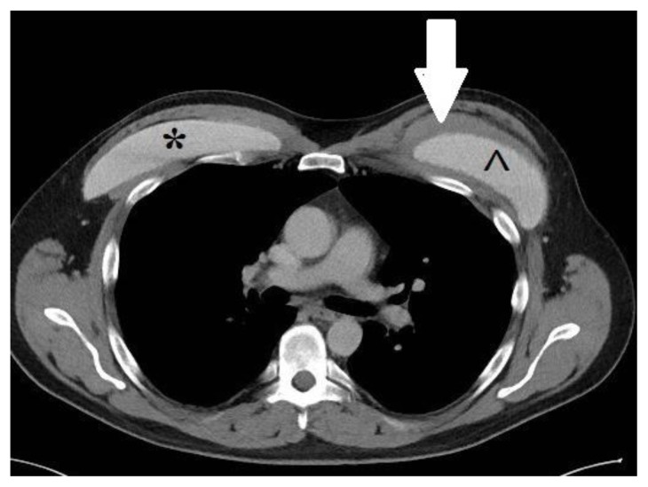
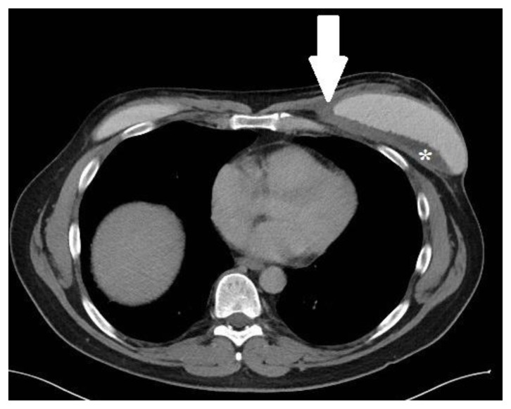
Due to clinical concerns, a computed tomography (CT) study of the chest with contrast was ordered and performed on another day to further evaluate this patient. On the day of the CT study, the patient received an appropriate bolus of intravenous contrast; however, there was too early of a scan time by the technologist. Consequently, it was a limited study and the images appear as though there is no contrast in the vascular and soft tissue structures. Regardless of the contrast scanning time, the CT study demonstrated bilateral subpectoral silicone implants. The normally positioned right pectoral implant was located between the right pectoralis major and minor muscles. No associated findings were seen involving the right implant. The left pectoral silicone implant was displaced inferiorly and laterally with pending extrusion, Figure 6. There was a soft tissue density that was equal to slightly lower in density to muscle located circumferentially around the left implant. The soft tissue density was thicker along its medial aspect (Figure 6). Also detected was a 6 cm × 2 cm × 6 cm, low density, lenticular-shaped fluid collection located between the soft tissue density surrounding the implant and the posterior aspect of the displaced left implant (Figure 7). The underlying ribs appeared normal and no adenopathy was seen.
Figure 6.
44-year-old male with left pectoral implant displacement.
FINDINGS: Axial contrast CT, with too early of a scan time by the technologist, of the chest at the level of the great vessels demonstrates the normally placed right silicone pectoral implant (*). The right implant is located between the right pectoralis major and minor muscles. The left silicone pectoral implant (^) is displaced inferiorly and laterally. The silicone implants are high in density compared to muscle on CT imaging. In addition, there is a soft tissue density that is equal in density to slightly less dense than muscle that surrounds the displaced left implant (arrow) on this image. The soft-tissue density is thicker medially. The soft tissue density histologically was dense fibrotic tissue with acute and chronic inflammation, histiocytic infiltrate, and granulation tissue consistent with inflamed implant capsule.
TECHNIQUE: Axial CT, 88 mAs, 140 kV, 5 mm slice thickness, contrast -with too early of a scan time by the technologist.
Figure 7.
44-year-old male with bilateral pectoral silicone implants with near extrusion of the left implant.
FINDINGS: Axial contrast CT, with too early of a scan time by the technologist, of the chest near the level of the diaphragm shows near extrusion of the left pectoral implant through the skin laterally at the level of the nipple with an associated soft tissue density (arrow). The soft tissue density is equal in density to slightly less dense than muscle and surrounds the displaced left implant. The soft-tissue density is thicker medially. Histologically it represented dense fibrotic tissue with acute and chronic inflammation, histiocytic infiltrate, and granulation tissue consistent with inflamed implant capsule. In addition, a 6 cm, low density, lenticular-shaped fluid collection (*) is located between the soft tissue density and the posterior aspect of the displaced left implant. On cytological examination, the peri-implant fluid represented fibrin, macrophages and acute inflammation.
TECHNIQUE: Axial CT, 88 mAs, 140 kV, 5 mm slice thickness, contrast -with too early of a scan time by the technologist.
Following imaging and further progression of the patient’s skin changes, he was scheduled for surgery. The working differential diagnosis of this HIV+ patient with the above imaging findings was left pectoral implant displacement with impending extrusion with possible malignancy, infection or inflammatory changes from migration of the implant.
At surgery, the left subpectoral silicone implant was removed without difficulty with wide excision of the thinned skin measuring 10 cm × 4 cm. There was evidence of peri-implant fluid and samples were sent for both cytology and microbiology. In addition, tissue samples were obtained for pathology from multiple sites including the left pectoralis muscle, left chest wall and lateral infra-areolar left breast tissue. Also, additional tissue samples were sent from a separate incision for the swelling at the site of palpable concern just superior to the implant near the left axillary line. Once hemostasis was ensured and the remaining tissue appeared healthy, the pocket that previous held the implant and skin incision were closed and a sterile compressive dressing was applied.
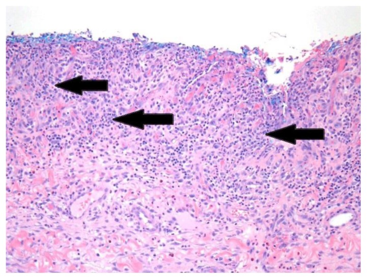
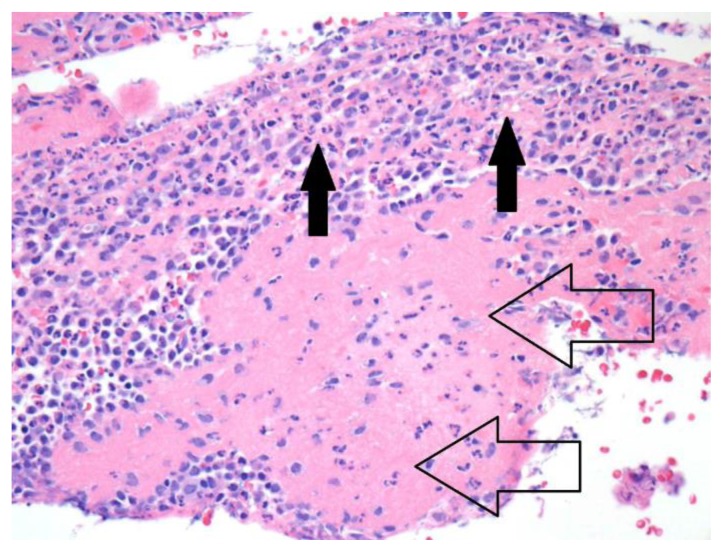
Pathology of the thinned, inflamed skin showed a marked mixed, predominantly acute, inflammation involving the dermis and epidermis with prominent dermal eosinophils (Figure 8). All the pathologic tissue samples of muscle, chest wall and breast confirmed dense fibrotic tissue with acute and chronic inflammation and histiocytic infiltrate, consistent with inflamed implant capsule (Figure 9). Cytology of the peri-implant fluid showed fibrin, macrophages and acute inflammation. No carcinoma was identified in any of the tissue specimens or peri-implant fluid samples. In addition, microbiology of the fluid obtained during surgery demonstrated no anaerobic, aerobic or fungal growth.
Figure 8.
44-year-old male with left lateral chest wall inflamed and thinned skin.
FINDINGS: Histology demonstrates marked mixed, predominantly acute, inflammation (arrows) involving the dermis and epidermis.
TECHNIQUE: Hematoxylin and eosin stain, 20× magnification.
Figure 9.
Histology, 44-year-old male with left peri-implant soft-tissue density.
FINDINGS: Histology demonstrates dense fibrotic tissue (open arrows) with acute and chronic inflammation and histiocytic infiltrate (closed arrows), consistent with inflamed implant capsule.
TECHNIQUE: Hematoxylin and eosin stain, 40× magnification.
DISCUSSION
Etiology & Demographics
Although physicians are more familiar with the procedure and imaging findings associated with female breast augmentation, male chest enhancement with pectoral implants is becoming a more common body implant surgical procedure [1, 4–7]. There are predominately three populations of men who undergo pectoral implant surgery. Originally, the surgery was for men who had Poland Syndrome, congenital absence of the pectoralis muscle on one side, or for men with chest muscle atrophy secondary to injury or other chest deformity, such as pectus excavatum deformity [2–4]. The most common reason today is men who want to improve their body image [4–6].
Clinical & Imaging Findings
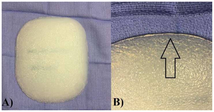
There are several types of commercially available male pectoral implants in the United States. Unlike traditional female breast implants, the male pectoral implants are made of a solid, pliable, silicone polymer [4] or contain a cohesive silicone gel [5, 6]. Consequently, male pectoral implants do not rupture [4–6]. Another benefit of the solid pectoral implant is that it can be modified intra-operatively by the surgeon with a scalpel or scissor if needed [5, 6]. Depending on the cosmetic result desired, the pectoral implant may be rectangular or oval in shape. As seen in Figure 10, the edges of the pectoral implant are tapered and the exterior surface of the implant is textured. Texturing of the pectoral implant surface is supposed to prevent displacement of the implant [6].
Figure 10.
Example of a Pectoral Implant (AART, INC, pectoral implant, style 2, Carson City, NV, USA)
FINDINGS:
A) Photograph demonstrates an example of a rectangular-shaped pectoral implant.
B) Photograph demonstrates the taped, smooth edge of the pectoral implant (open arrow) and the textured exterior surface.
TECHNIQUE:
A) Color photograph, Nikon Coolpix A, digital camera, 16.0 Megapixel.
B) Color photograph, Nikon Coolpix A, digital camera, 16.0 Megapixel.
After the appropriate implant style and size are chosen, the surgical technique involves making an approximately 4–6 cm incision in the axilla followed by identifying the lateral border of the pectoralis muscle [2, 5, 6]. A pocket for the implant is then created behind the muscle with a blunt dissector. The surgeon is careful not to dissect more 1–2 cm below the areola of the breast; otherwise, the implant could be positioned too low and would have a more female appearance [4–6]. Meticulous hemostasis is performed to decrease any risk of complication such as bleeding, hematoma, or malposition of the implant. The implant is placed and oriented to the desired result. Closure is performed in layers followed by application of a compressive chest dressing. Post-operative instructions include wound care, and limiting lifting to less than 10 pounds with no exercise for five weeks.
With the exception of implant rupture, complications of male chest enhancement surgery are the same as female breast augmentation [4–7]. Complications include implant displacement with and without extrusion, hematoma, seroma, infection and capsule formation [2, 3, 5, 6, 8]. When male pectoral implant displacement is severe, as in this case, it is readily appreciated on physical exam. However, if the displacement is subtle it may not be detected clinically. The other reported complications of implant placement are usually detected on physical exam and can be correlated with the appropriate clinical history [2, 3, 5, and 6]. However, if these findings are located deep to the muscles or posterior to the implant, they may not be appreciated clinically.
Although there are many published articles describing the surgical procedure for male pectoral implant placement and its complications [2, 4–6], the imaging features reported are very limited [8]. Our case demonstrates the mammographic, sonographic and CT imaging findings of a male patient with bilateral pectoral silicone implants with associated complications.
Imaging can play a vital role for therapeutic and surgical planning of these patients and especially for those with a clinical concern for carcinoma. On mammographic imaging, the normally positioned silicone pectoral implant should be posterior to the pectoralis muscle and oriented parallel to the chest wall, Figure 3. Similar to a female silicone implant, the pectoral implant appears white on the mammogram since the x-rays are attenuated by the silicone [7]. As with female patients with implants, both the mammographic “implant in-view” and “implant displaced” views can be performed on patients with pectoral implants to evaluate breast tissue [7, 9]. These views are a modified technique to try to image the implant (implant in-view) and the tissue overlying the implant [9]. By displacing the implant posteriorly against the chest wall and pulling breast tissue over and in front of the implant (implant displaced view), marked improvement in compression and visualization of substantially more breast tissue is achieved [9]. As with female breast implants, evaluation of the posterior margin of the male pectoral implant will not be seen mammographically [7, 9]. This is due to its location posterior to the pectoralis major muscle. In addition, if there is subtle implant displacement this may not be appreciated on a standard 2-view mammogram.
Ultrasound is a helpful imaging modality to evaluate for any clinical or mammographically detected breast & chest wall findings in men. It is readily available and does not use radiation [7]. In addition, it can also be used to evaluate for axillary adenopathy. Sonographically, the solid, silicone pectoral implant is anechoic and its anterior margin may be seen as a thin echogenic line. The posterior margin of the implant is not seen due to the attenuation of the ultrasound beam by the silicone [7]. Therefore, a limitation of ultrasound is its inability to evaluate for an abnormality posteriorly to the pectoral implant. In our case, we encountered this limitation since we could not appreciate the posterior peri-implant fluid collection or to distinguish capsular thickening. Another limitation of ultrasound is that it can only demonstrate a small area of the chest at a time. Thus, this can limit the evaluation of subtle implant displacement.
Magnetic resonance imaging (MRI) of the chest is a non-ionizing imaging modality that has superior soft-tissue resolution [10–12]. Compared to other imaging modalities, MRI has the highest sensitivity and specificity in evaluating silicone implant rupture in women [7]. Unlike female silicone implants that have a capsule/shell and are filled with silicone [7], male pectoral implants are solid. Because the pectoral implants are solid, they do not rupture [4–6]. Consequently, there is no clinical indication for MRI for male pectoral implant integrity evaluation. In addition, MRI has limitations. These limitations include increased cost (up to twice the cost of a chest CT), coil coverage over the chest with larger sized patients, increased scanning time (15 to 60 minutes compared to 5 minutes for a CT of the chest) and artifacts [12]. In addition to the above limitations of MRI, CT of the chest is a diagnostic, large field, cross sectional imaging modality that is well tolerated and provides good spatial resolution, including depiction of osseous and soft-tissue structures [10, 11]. CT can also image a large tissue volume in a short acquisition time, reducing the effect of respiratory motion in the thorax [10]. Thus, CT of the chest with contrast is the preferred imaging modality in these patients to evaluate for pectoral implant position, evaluation of anatomic structures, complications and any disease process that effects the chest.
On CT, the pectoral silicone implant is hyperdense compared to muscle but less dense than bone [8]. The implant is normally located between the pectoralis major and minor muscles and the implant’s long axis is parallel to the chest wall [4]. The implant should be covered by the pectoralis major muscle and should not extend more than one to two centimeters inferiorly to the areolar [4–6]. If the implant is not in the appropriate position, then the radiologist should report the implant as displaced and describe its location. CT of the chest with contrast is the preferred imaging modality prior to any definitive surgery to demonstrate anatomy, implant displacement, peri-implant fluid/soft tissue density and potential involvement of the chest by other diseases. In this case, the fluid posterior to the displaced implant and the soft tissue density around the implant were only visualized by CT imaging.
Differential Diagnoses
Physical examination and imaging both demonstrated that there was gross displacement with near extrusion of the left pectoral implant through the skin of the left side of the chest in this patient. Of note, subtle displacement of a pectoral implant can be difficult to appreciate on physical examination, but can be seen with imaging. The patient’s right pectoral implant was normally located and without associated findings. In this HIV+ patient, the findings of a pericapsular fluid collection and pericapsular thickening were concerning for a bacterial or fungal infection. Other possible causes included acute and chronic inflammation from the displaced implant. Furthermore, these findings also raised the possibility for an atypical presentation of carcinoma, anaplastic large cell lymphoma. Anaplastic large cell lymphoma has been reported with peri-implant fluid in females with long-term breast implants [13]. An increase in the incidence of primary breast cancer has not been reported in patients with implants [13].
Treatment & Prognosis
Common treatment of a displaced pectoral implant is surgical revision or removal. In this case, the patient had his implant removed. Unfortunately, his post-operative course was complicated by a methicillin resistant staphylococcus aureus chest wound infection that was successfully treated with antibiotics and sequential packing.
In conclusion, our case illustrates an example of male chest enhancement with silicone pectoral implants. Since male aesthetic surgical procedures are on the rise, and the trend will most likely continue, radiologists need to become familiar with this type of augmentation, its potential complications and imaging features.
TEACHING POINT
Male breast enhancement with silicon pectoral implants are becoming more common in the general population. Although complications of the pectoral implants are infrequent, radiologic imaging provides significant information of implant location and other unanticipated findings prior to definitive surgical therapy.
Table 1.
Comparison between male chest deformity and aesthetic pectoral implant placement patients.
| Chest Deformity | Aesthetic Enhancement | |
|---|---|---|
| Etiology | Congenital, trauma | Body dysmorphic disorder |
| Incidence |
|
Overall incidence unknown |
| Gender Ratio | Male to female 3:1 | Unknown |
| Age Predilection | 20–30’s | 20–40’s |
| Symptoms | Chest/arm decreased strength | None of the chest |
| Complications |
|
|
| Risk Factors | Unknown | Unknown |
| Treatment |
|
|
| Prognosis | Excellent | Excellent |
| Imaging Findings |
|
|
Table 2.
Imaging findings of male pectoral implant positioning.
| Mammography | Ultrasound | Computed Tomography | |
|---|---|---|---|
| Normal Implant Position |
|
|
|
| Implant Displacement |
|
|
|
Table 3.
Differential diagnosis table of chest imaging findings in a male patient with pectoral implants.
| Mammography/Ultrasound/Computed Tomography | |
|---|---|
| Breast Mass | Gynecomastia, findings secondary to trauma, post-surgical change, primary breast cancer |
| Peri-Implant Fluid Collection |
|
| Peri-Implant Soft Tissue Density |
|
| Adenopathy | Metastatic disease from primary or non-breast primary cancer, lymphoma, leukemia, inflammatory/reactive |
ACKNOWLEDGEMENTS
We would like to thank Mr. Jason Hauser at the University of North Carolina for his assistance of article preparation.
ABBREVIATIONS
- CT
Computed tomography
- HIV +
Human immunodeficiency virus positive
- kV
Kilovolts
- mAs
Milliamperage second
- MRI
Magnetic Resonance Imaging
REFERENCES
- 1.American Society of Plastic Surgeons. Cosmetic Plastic Surgery Statistics Cosmetic Procedure Trends 2014. [Accessed March 5, 2015]. http://www.plasticsurgery.org/Documents/news-resources/statistics/2014-statistics/cosmetic-procedure-trends-2014-pdf
- 2.Pereira LH, Sabatovich O, Santana KP, Picanco R, Sterodimas A. Surgical correction of Poland’s syndrome in males – a purposely designed implant. J Plast Reconstr Aesthe Surg. 2008;61:393–9. doi: 10.1016/j.bjps.2006.12.008. [DOI] [PubMed] [Google Scholar]
- 3.Margulis A, Sela M, Neuman R, Buller-Sharon A. Reconstruction of pectus excavatum with silicone implants. J Plast Reconstr Aesthet Surg. 2006;59(10):1082–6. doi: 10.1016/j.bjps.2005.12.035. [DOI] [PubMed] [Google Scholar]
- 4.Aiache A. Male chest correction: pectoral implants. Clin Plast Surg. 1991;18(4):823–8. [PubMed] [Google Scholar]
- 5.Benito-Ruiz J, Raigosa JM, Manzano-Surroca M, Salvador L. Male chest enhancement: pectoral implants. Aesth Plast Surg. 2008;32(1):101–4. doi: 10.1007/s00266-007-9018-5. [DOI] [PubMed] [Google Scholar]
- 6.Horn G. A new concept in male chest reshaping: anatomical pectoral implants and liposculpture. Aesth Plast Surg. 2002;26:23–5. doi: 10.1007/s00266-001-0032-8. [DOI] [PubMed] [Google Scholar]
- 7.Juanpere S, Perez E, Huc O, Motos N, Pont J. Imaging of Breast Implants – A Pictorial Review. Insights Imaging. 2011;2(6):653–70. doi: 10.1007/s13244-011-0122-3. [DOI] [PMC free article] [PubMed] [Google Scholar]
- 8.Yahyavi-Firouz-Abadi N, Menias CO, Bhalla S, Siegel C, Gayer G. Imaging of Cosmetic Plastic Procedures and Implants in the Body and Their Potential Complications. AJR. 2015;151:707–715. doi: 10.2214/AJR.14.13516. [DOI] [PubMed] [Google Scholar]
- 9.Eklund GW, Busby RC, Miller SH, Job JS. Improved imaging of the augmented breast. AJR. 1988;151:469–73. doi: 10.2214/ajr.151.3.469. [DOI] [PubMed] [Google Scholar]
- 10.Mullen CP, Madan R, Trotman-Dickerson B, Qian X, Jacobson FL, Hunsaker A. Radiology of Chest Wall Masses. AJR. 2011;197:W460–470. doi: 10.2214/AJR.10.7259. [DOI] [PubMed] [Google Scholar]
- 11.Jeung MY, Gangi A, Gasser B, et al. Imaging of Chest Wall Disorders. RadioGraphics. 1999;19:617–637. doi: 10.1148/radiographics.19.3.g99ma02617. [DOI] [PubMed] [Google Scholar]
- 12.Lee CU, White DB, Sykes AM. Establishing a Chest MRI Practice and its Clinical Applications: Our Insight and Protocols. Clin Imaging Sci. 2014;4:17. doi: 10.4103/2156-7514.129288. [DOI] [PMC free article] [PubMed] [Google Scholar]
- 13. [Accessed March 5, 2015];Anaplastic Large Cell Lymphoma (ALCL) In Women with Breast Implants: Preliminary FDA Findings and Analyses. http://www.fda.gov/MedicalDevices/ProductsandMedicalProcedures/ImplantsandProsthetics/BreastImplants/ucm239996.htm. [Google Scholar]


