

Abstract
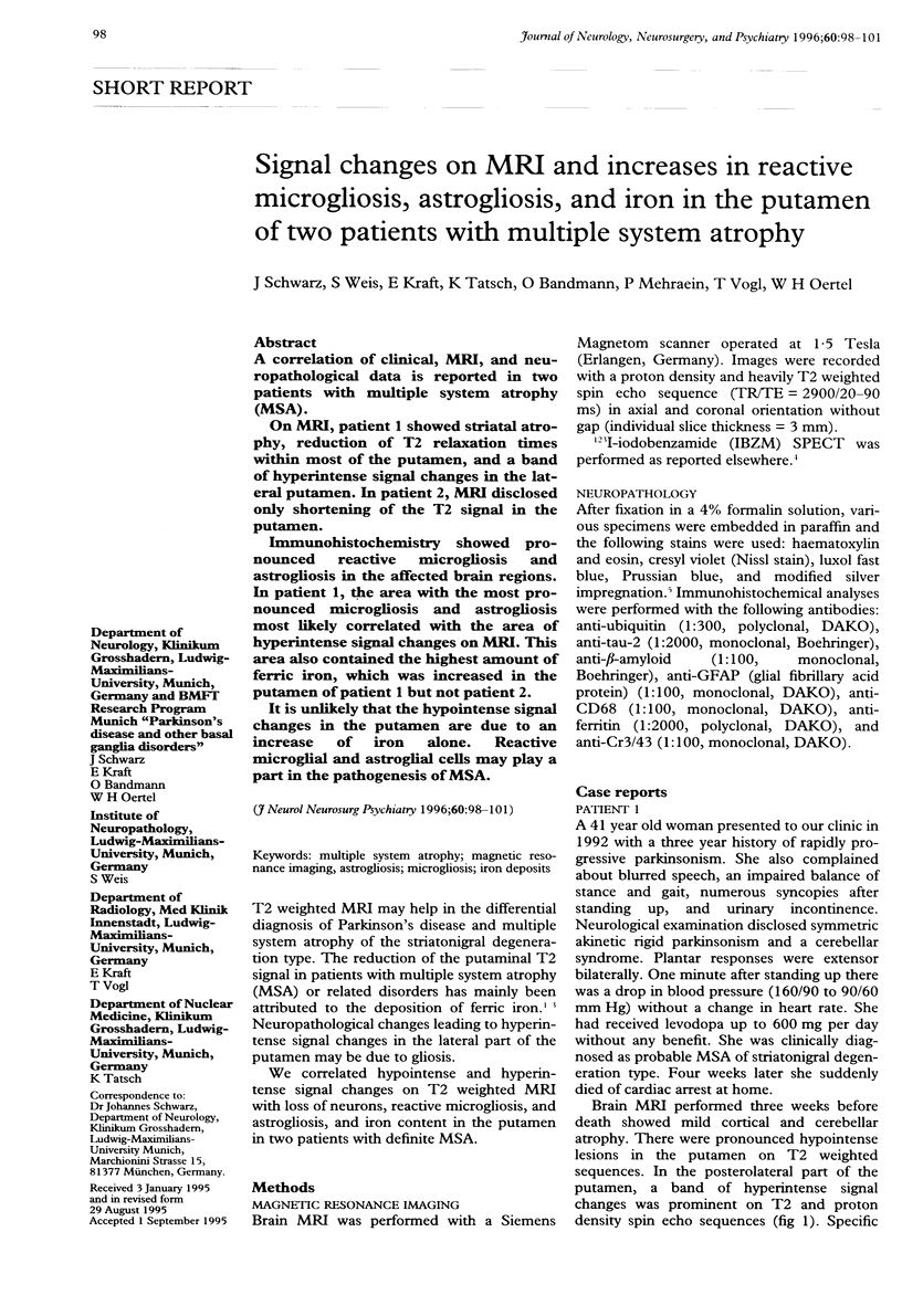
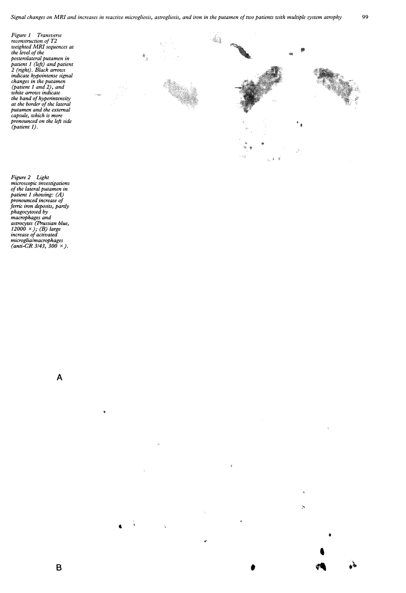
A correlation of clinical, MRI, and neuropathological data is reported in two patients with multiple system atrophy (MSA). On MRI, patient 1 showed striatal atrophy, reduction of T2 relaxation times within most of the putamen, and a band of hyperintense signal changes in the lateral putamen. In patient 2, MRI disclosed only shortening of the T2 signal in the putamen. Immunohistochemistry showed pronounced reactive microgliosis and astrogliosis in the affected brain regions. In patient 1, the area with the most pronounced microgliosis and astrogliosis most likely correlated with the area of hyperintense signal changes on MRI. This area also contained the highest amount of ferric iron, which was increased in the putamen of patient 1 but not patient 2. It is unlikely that the hypointense signal changes in the putamen are due to an increase of iron alone. Reactive microglial and astroglial cells may play a part in the pathogenesis of MSA.
Full text
PDF

Images in this article
Selected References
These references are in PubMed. This may not be the complete list of references from this article.
- Abe H., Mehraein P., Weis S. A modified NOR-silver impregnation technique for amyloid plaques and neurofibrillary tangles: comparative assessment. Neuropathol Appl Neurobiol. 1994 Oct;20(5):478–486. doi: 10.1111/j.1365-2990.1994.tb00999.x. [DOI] [PubMed] [Google Scholar]
- Chen J. C., Hardy P. A., Kucharczyk W., Clauberg M., Joshi J. G., Vourlas A., Dhar M., Henkelman R. M. MR of human postmortem brain tissue: correlative study between T2 and assays of iron and ferritin in Parkinson and Huntington disease. AJNR Am J Neuroradiol. 1993 Mar-Apr;14(2):275–281. [PMC free article] [PubMed] [Google Scholar]
- Daniel S. E., Geddes J. F., Revesz T. Glial cytoplasmic inclusions are not exclusive to multiple system atrophy. J Neurol Neurosurg Psychiatry. 1995 Feb;58(2):262–262. doi: 10.1136/jnnp.58.2.262. [DOI] [PMC free article] [PubMed] [Google Scholar]
- Lang A. E., Curran T., Provias J., Bergeron C. Striatonigral degeneration: iron deposition in putamen correlates with the slit-like void signal of magnetic resonance imaging. Can J Neurol Sci. 1994 Nov;21(4):311–318. doi: 10.1017/s0317167100040889. [DOI] [PubMed] [Google Scholar]
- Lantos P. L., Papp M. I. Cellular pathology of multiple system atrophy: a review. J Neurol Neurosurg Psychiatry. 1994 Feb;57(2):129–133. doi: 10.1136/jnnp.57.2.129. [DOI] [PMC free article] [PubMed] [Google Scholar]
- O'Brien C., Sung J. H., McGeachie R. E., Lee M. C. Striatonigral degeneration: clinical, MRI, and pathologic correlation. Neurology. 1990 Apr;40(4):710–711. doi: 10.1212/wnl.40.4.710. [DOI] [PubMed] [Google Scholar]
- Olanow C. W. Magnetic resonance imaging in parkinsonism. Neurol Clin. 1992 May;10(2):405–420. [PubMed] [Google Scholar]
- Schwarz J., Tatsch K., Vogl T., Kirsch C. M., Trenkwalder C., Arnold G., Gasser T., Oertel W. H. Marked reduction of striatal dopamine D2 receptors as detected by 123IBZM-SPECT in a Wilson's disease patient with generalized dystonia. Mov Disord. 1992;7(1):58–61. doi: 10.1002/mds.870070111. [DOI] [PubMed] [Google Scholar]