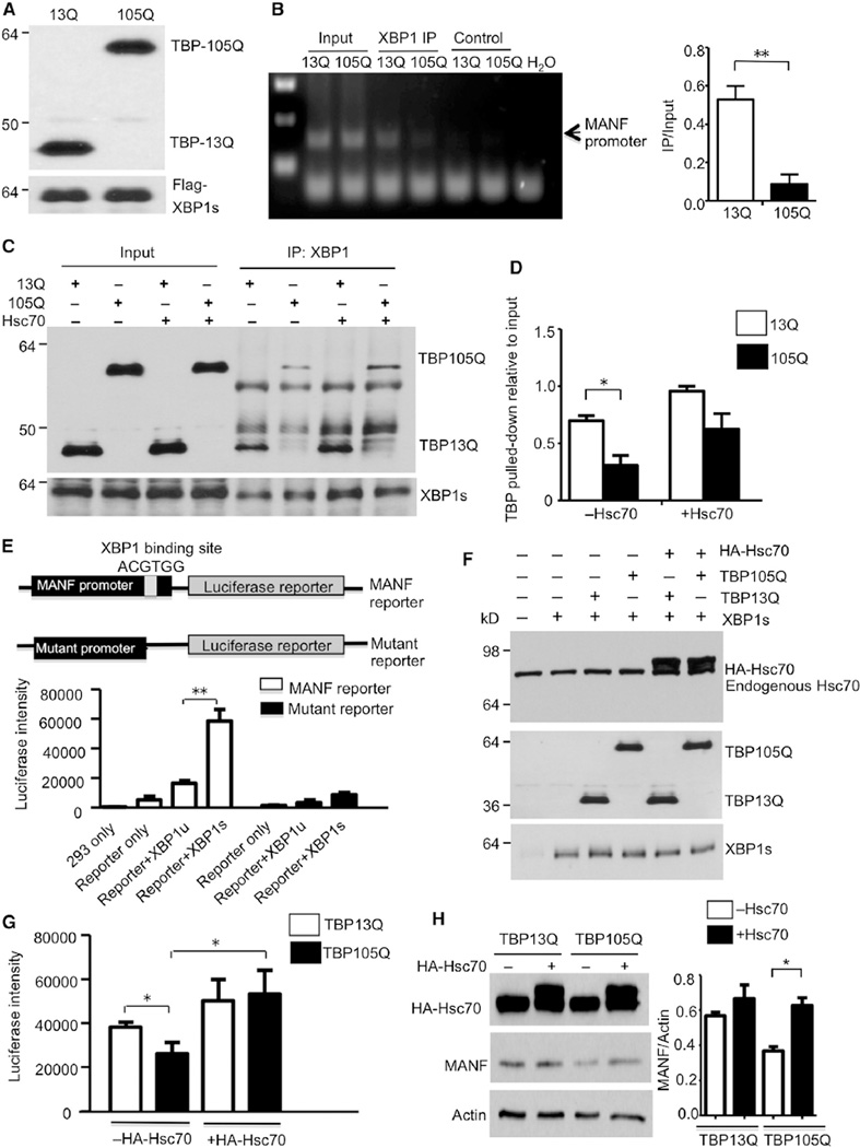
Figure 6. Expanded polyQ Impairs the Binding of TBP with XBP1s and Its Transcriptional Activity for MANF Expression.
(A) Western blotting analysis confirmed the expression of respective proteins (TBP13Q, TBP105Q, and XBP1s) in cells used for ChIP assay.
(B) Semiquantitative PCR result using lysates from chromatin immunoprecipitation with anti-FLAG (ChIP, left). Control was beads only without anti-FLAG. More MANF promoter was pulled down in cells transfected with TBP13Q than with TBP105Q (right, **p < 0.01).
(C) Coimmunoprecipitation of transfected XBP1s with TBP in the presence or absence of Hsc70. XBP1s was pulled down by XBP1 antibody in each sample, and less TBP105Q was pulled down with XBP1s. Overexpression of Hsc70 increased the precipitated amount of TBP (both 13Q and 105Q).
(D) The relative amounts of TBP precipitated with XBP1s (*p < 0.05).
(E) Schematic map of luciferase constructs for reporter assay. Luciferase activity of transfected HEK293 cells showed that XBP1s, but not XBP1u, greatly increased luciferase intensity.
(F) Western blotting verifying the coexpression of TBP, Hsc70, and XBP1s with the MANF promoter reporter.
(G) Overexpression of Hsc70 significantly rescued the impaired luciferase activity by TBP105Q (*p < 0.05).
(H) Western blot analysis of MANF expression levels in TBP stably transfected cells that were transfected with (+) or without (−) HA-tagged Hsc70. The ratios of MANF to actin on the blot are also presented (*p < 0.05). Data are represented as mean ± SEM.

